Abstract
The female hormone estrogen is an important factor in the regulation of airway function and inflammation, and sex differences in the prevalence of asthma are well described. Using an animal model, we determined how sex differences may underlie the development of altered airway function in response to allergen exposure. We compared sex differences in the development of airway hyperresponsiveness (AHR) after allergen exposure exclusively via the airways. Ovalbumin (OVA) was administered by nebulization on 10 consecutive days in BALB/c mice. After methacholine challenge, significant AHR developed in male mice but not in female mice. Ovariectomized female mice showed significant AHR after 10-day OVA inhalation. ICI182,780, an estrogen antagonist, similarly enhanced airway responsiveness even when administered 1 hour before assay. In contrast, 17β-estradiol dose-dependently suppressed AHR in male mice. In all cases, airway responsiveness was inhibited by the administration of a neurokinin 1 receptor antagonist. These results demonstrate that sex differences in 10-day OVA-induced AHR are due to endogenous estrogen, which negatively regulates airway responsiveness in female mice. Cumulatively, the results suggest that endogenous estrogen may regulate the neurokinin 1–dependent prejunctional activation of airway smooth muscle in allergen-exposed mice.
Keywords: estrogen, sex, airway hyperresponsiveness, EFS, neuronal activation
CLINICAL RELEVANCE
Sex differences impact asthma. For the first time, a mechanism demonstrating the ability of estrogen to regulate an NK-1–dependent pathway has been identified. Interventions targeting this pathway may affect asthma outcomes.
Sex differences have been described to play a role in various diseases, including atherosclerosis (1), osteoporosis (2), and certain neurologic disorders (3). Accumulating evidence suggests that hormonal factors are important in the expression of such sex differences at the phenotypic and genotypic levels. A number of epidemiologic and experimental reports suggest that the female hormone estrogen is an important factor in the regulation of airway function and inflammation. Estrogen can prevent cholinergic constriction of asthmatic tracheal rings in vitro (4), and estrogen treatment decreases airway responsiveness to acetylcholine in ovariectomized rats (5). On the other hand, female mice may be more susceptible than male mice to allergen-induced airway inflammation (6–8). The role of estrogen on sex differences in asthma has been the subject of much discussion (9, 10).
There are many animal protocols for investigating allergen-induced airway hyperresponsiveness (AHR) and inflammation, with the majority using initial sensitization to allergen together with an adjuvant such as alum. Using this sensitization approach, some mouse studies have suggested a female superiority in the development of allergic airway inflammation (6–8). We examined the development of AHR after allergen exposure exclusively via the airways for 10 consecutive days in the absence of any adjuvant. Although AHR to inhaled methacholine (MCh) was not detected in female mice after 10-day ovalbumin (OVA) inhalation in vivo, electrical field stimulation (EFS) in vitro detected altered airway responsiveness. In large part, this EFS response has been attributed to muscarinic receptor dysfunction, activation of neurokinin 1 (NK-1)–dependent pathways, and increased acetylcholine (ACh) release (11, 12). In the present study, we identified major differences in the development of MCh-induced alterations in airway resistance (RL) and dynamic compliance (Cdyn) after 10-day OVA inhalation between male and female mice. The aim of the present study was to compare the mechanism(s) underlying this sex difference and demonstrate that one of the sex hormones, estrogen, regulates the development of AHR after allergen exposure in female mice. The data suggest that estrogen may inhibit the activation of NK-1–related prejunctional pathways, which are enhanced by allergen exposure.
MATERIALS AND METHODS
Animals
Female and male BALB/c mice, 8 to 10 weeks of age (Jackson Laboratories, Bar Harbor, ME) were maintained on an OVA-free diet. All experimental animals used in this study were under a protocol approved by the Institutional Animal Care and Use Committee of the National Jewish Medical and Research Center.
Ovariectomy
Female mice at 5 to 6 weeks of age were anesthetized by intraperitoneal injection of 50 mg/kg of pentobarbital sodium and underwent ovariectomy (OVX) using a dorsolateral approach. Sham operation and OVX were performed at least 2 weeks before initiation of the experiments.
OVA Exposure Without Adjuvant
Experimental groups consisted of four mice per group, and each experiment was performed at least twice. Mice were exposed to OVA (albumin from chicken egg white, Grade V; Sigma, St. Louis, MO) as described previously (13). Briefly, a solution of 0.9% saline or endotoxin-depleted 1% OVA solution was delivered by ultrasonic nebulization (Omron, Kyoto, Japan) for 20 minutes daily over 10 consecutive days in a closed chamber. Twenty-four hr after the last airway challenge, airway responsiveness was assessed in vivo and in vitro, and bronchoalveolar lavage fluid (BALF), blood, and lung tissue were collected.
Drugs
ICI182,780, an estrogen receptor antagonist (Tocris Bioscience, Ellisville, MO) and 17β-estradiol (E2; Sigma) were suspended in 0.1% Tween 80 containing phosphate-buffered saline at concentrations of 10 ml/kg and 50 to 100 μg/kg body weight. The highly selective NK-1 receptor antagonist Sendide was obtained from Bachem California (Torrance, CA). The estrogen receptor antagonist ICI182,780 was administered intraperitoneally immediately before each OVA inhalation or 1 hour before determination of AHR to inhaled MCh (Sigma). Sendide was administered intraperitoneally 2 hours before determination of AHR.
Determination of Airway Responsiveness
RL and Cdyn were determined as changes in airway function after aerosolized MCh challenge in vivo. Mice were anesthetized with sodium pentobarbital (90 mg/kg, intraperitoneally), tracheostomized, and mechanically ventilated at a rate of 160 breaths/minute with a constant tidal volume of air (0.2 ml). Lung function was assessed as previously described (14). Aerosolized MCh was administered for eight breaths at a rate of 60 breaths/min, tidal volume of 500 μL by a second ventilator (Model 683; Harvard Apparatus, South Natick, MA) in increasing concentrations (1.6, 3.1, 6.3, and 12.5 mg/ml). After each MCh challenge, the data were continuously collected for 1 to 5 minutes, and maximum values of RL and minimum values for Cdyn were obtained.
For determination of airway responsiveness in vitro, airway responsiveness was assessed by measuring airway smooth muscle (tracheal rings) responsiveness to carbachol (CCh) (Sigma) and EFS 24 hours after the last airway challenge. CCh challenge was performed with increasing doses of the drug from 10−8 to 10−4 M. EFS was administered with an increasing frequency from 0.5 to 30 Hz. We have established earlier (15) that the EFS at the frequency range and mode used stimulates cholinergic fibers causing release of endogenous acetylcholine which mediates airway smooth muscle contraction; the EFS response is abolished in the presence of atropine. The effective concentrations of CCh resulting in 50% of the maximal contraction (EC50) and the electrical frequency resulting in 50% of the maximal contraction (ES50) were calculated from linear plots for each individual animal and were compared between the groups.
Measurement of Total and OVA-Specific Immunoglobulin E
Serum levels of total immunoglobulin (Ig)E and OVA-specific IgE were measured by enzyme-linked immunosorbent assay (ELISA) as described (13). The OVA-specific IgE titers of the samples were related to pooled standards that were generated in the laboratory and expressed as ELISA units per milliliter. Total IgE levels were calculated by comparison with known mouse IgE standards (BD Pharmingen, San Diego, CA). The limit of detection was 100 pg/ml for total IgE.
Determination of Cell Numbers and Cytokine Levels in BALF
Immediately after the assessment of AHR, lungs were lavaged via the tracheal cannula with Hanks' balanced salt solution (1 ml/mouse). Total leukocyte numbers were measured with a Coulter Counter (Coulter Corporation, Hialeah, FL). Differential cell counts were made from cytocentrifuged preparations using a Cytospin 2 (Shandon Ltd., Runcorn, Cheshire, UK) and stained with Leukostat (Fisher Diagnostics, Pittsburgh, PA). At least 200 cells were counted under 400× magnification.
Bronchoalveolar lavage (BAL) supernatants were collected and kept frozen at −80°C until assayed. The levels of cytokine in the supernatants of BALF samples were determined by ELISA. IL-4, IL-5, IL-12 (p70), IFN-γ (all from BD Pharmingen) and IL-13 (R&D Systems, Minneapolis, MN) were measured following the manufacturers' directions. The limits of detection were 4 pg/ml for IL-4 and IL-5, 1.5 pg/ml for IL-13, and 15 pg/ml for IL-12 and IFN-γ.
Histopathologic Examination
Lungs were fixed by inflation (1 ml/whole lung) and immersion in 10% formalin. Cells containing eosinophilic major basic protein were identified by immunohistochemical staining as previously described using rabbit anti-mouse major basic protein (provided by Dr. J. J. Lee, Mayo Clinic, Scottsdale, AZ). The slides were examined in a blinded fashion with a Nikon microscope (Melville, NY) equipped with a fluorescein filter system. For detection of mucus-containing cells in formalin-fixed airway tissue, sections were stained with periodic acid-Schiff and H&E and quantitated as previously described (16).
Data Analysis
All parametric data were compared using Student's t test between two groups and Tukey-Kramer to compare multiple groups. Because measured values may not be normally distributed and due to the small sample sizes, nonparametric analysis using the Mann-Whitney U test was also used to confirm that the statistical differences remained significant. A P value of <0.05 was considered statistically significant. Values for all measurements were expressed as the mean ± SEM.
RESULTS
Sex Differences after OVA Inhalation
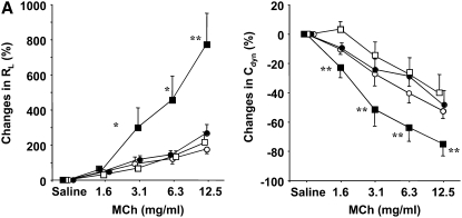
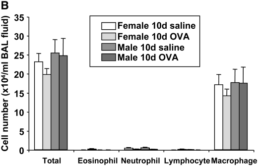
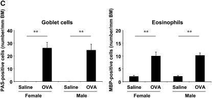
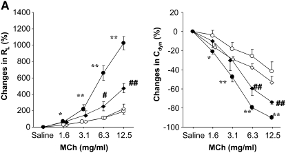
The extent of 10-day OVA-induced AHR to inhaled MCh in female and male BALB/c mice were compared. There were no significant differences in the baseline values between female and male mice. Responses in the 10-day saline-treated groups showed low level MCh dose-responsiveness for RL and Cdyn in male and female mice (Figure 1A). After 10-day OVA inhalation, significant AHR to inhaled MCh was elicited in male mice, but not in female mice (Figure 1A). This 10-day exposure protocol was previously shown to elicit little in the way of BAL inflammatory cell accumulation and there were no differences between female and male mice (Figure 1B). Ten-day OVA inhalation elicited significant increases in numbers of goblet cells and eosinophils in the lung tissue but there were no differences in the numbers of these cells (comparing male and female mice) (Figure 1C). Neither OVA inhalation nor sex differences influenced the levels of IL-4, IL-5, IL-12, IL-13, or IFN-γ in the BALF (data not shown). Serum levels of total IgE were significantly increased in both sexes after 10-day OVA inhalation; the baseline values for total IgE were higher in female than in male mice (Table 1). Serum levels of OVA-specific IgE were increased in both sexes after 10-day OVA inhalation, and no differences between the sexes were detected (Table 1).
Figure 1.
Comparison of airway responsiveness and inflammation in female and male mice after 10-day ovalbumin (OVA) inhalation. Female and male mice were exposed to either a 0.9% saline solution or a 1% OVA solution for 10 days (20 min/d). (A) Airway hyperresponsiveness (AHR) to inhaled methacholine (MCh) after 10-day OVA inhalation. AHR (airway resistance [RL] and lung dynamic compliance [Cdyn]) to inhaled MCh was assayed 24 hours after the last OVA challenge. Circles, female mice; squares, male mice; open symbols, 10-day saline; solid symbols, 10-day OVA. (B) Bronchoalveolar lavage (BAL) cell composition after 10-day OVA inhalation. BAL fluid was recovered immediately after the AHR assay was completed. (C) Numbers of periodic acid-Schiff–positive and major basic protein–positive cells. Results represent the mean ± SEM (n = 8). *P < 0.05 and **P < 0.01 versus 10-day saline exposure in each sex. ND = not detected. The baseline values of RL and Cdyn were as follows: 0.57 ± 0.03 cm H2O/ml/second (n = 16) and 0.069 ± 0.003 ml/cm H2O (n = 16) in female saline-treated mice, respectively; 0.59 ± 0.03 cm H2O/ml/s (n = 12) and 0.070 ± 0.002 ml/cm H2O (n = 12) in female OVA-treated mice, respectively; 0.55 ± 0.02 cm H2O/ml/second (n = 16) and 0.065 ± 0.003 ml/cm H2O (n = 16) in male saline-treated mice, respectively; 0.57 ± 0.02 cm H2O/ml/second (n = 21) and 0.064 ± 0.003 ml/cm H2O (n = 21) in male OVA-treated mice, respectively. BM = basement membrane.
TABLE 1.
COMPARISON OF ENDOGENOUS IgE ANTIBODIES BETWEEN FEMALE AND MALE MICE AFTER 10-d OVALBUMIN INHALATION
| Treatment | Total IgE (ng/ml) | OVA-Specific IgE (EU/ml) |
|---|---|---|
| Female saline | 14.6 ± 1.2 | 0 ± 0 |
| Female OVA | 22.7 ± 1.8* | 27.6 ± 7.9* |
| Male saline | 6.3 ± 0.7 | 0 ± 0 |
| Male OVA | 11.2 ± 1.0* | 27.0 ± 10.1† |
Definition of abbreviation: EU, ELISA units; IgG, immunoglobulin G; OVA, ovalbumin.
P < 0.01, statistically significant compared with saline group in each sex.
P < 0.05, statistically significant compared with saline group in each sex.
Airway Smooth Muscle Contractility
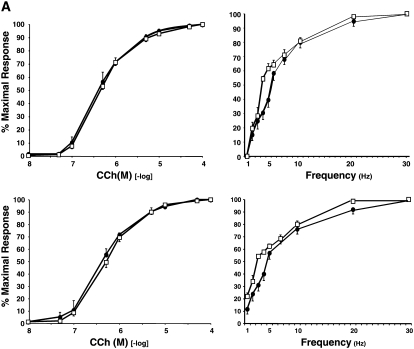
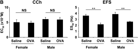
To compare airway (tracheal) smooth muscle contractility, which may contribute to the development of AHR, CCh- and EFS-induced responses were evaluated in vitro and compared. The effective concentration required for EC50 for CCh in saline- and OVA-treated groups were 5.0 ± 0.9 × 10⁻⁷ and 4.8 ± 0.6 × 10⁻⁷ M in female mice, and 6.0 ± 0.7 × 10⁻⁷ and 5.9 ± 0.8 × 10⁻⁷ M in male mice, respectively (Figure 2), indicating no up-regulation of cholinergic airway responsiveness after 10-day OVA inhalation in male or female mice. In contrast, the electrical frequency required for ES50 in the saline- and OVA-treated groups were 3.6 ± 0.2 and 2.1 ± 0.2 Hz in females, and 3.9 ± 0.3 and 2.6 ± 0.1 Hz in males, respectively (Figure 2) demonstrating comparable and significant decreases in ES50 values in both after 10-day OVA inhalation. Maximal contractile responses (Tmax) were also determined in both CCh- and EFS-induced response. However, no significant differences were observed in Tmax (data not shown) as previously reported (17).
Figure 2.
Tracheal smooth muscle responsiveness to carbachol (CCh) and electrical field stimulation (EFS) after 10-day OVA inhalation. (A) Representative CCh and EFS responses. Top panels: Solid circles, female saline; open squares, female OVA. Bottom panels: Solid circles, male saline; open squares, male OVA. (B) Summary of results of CCh- and EFS-induced contractility. Results represent the mean ± SEM (n = 8). **P < 0.01 statistically significant. NS = not significant. 95% confidence intervals overlapped among the groups.
Development of AHR to Inhaled MCh in Ovariectomized Female Mice after 10-Day OVA Inhalation In Vivo
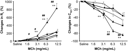
Based on the MCh-induced AHR data distinguishing male and female mice, it appeared that there may be factors related to sex differences which regulate airway responsiveness in vivo, but not in vitro. We therefore evaluated the influence of OVX as a means of reducing endogenous female hormone production (Figure 3). Sham-surgery female mice did not develop AHR after 10-day OVA inhalation and no differences were detected in OVX female mice after 10-day saline inhalation. However, airway responsiveness to inhaled MCh was significantly enhanced after 10-day OVA inhalation in the OVX mice, and this was reversed after E2 administration. These results suggested that hormonal factors derived from the ovary are directly (or indirectly) critical to regulating airway function in vivo after allergen challenge exclusively via the airways. These increases in AHR were not accompanied by any changes in BAL cell composition or cytokine levels (data not shown).
Figure 3.
Development of AHR to inhaled MCh after 10-day OVA inhalation in ovariectomized (OVX) female mice. Sham-operated or OVX female mice were exposed to 0.9% saline or 1% OVA solution for 10 days. AHR was assayed 24 hours after the last OVA challenge. Sham operation or OVX was performed at least 2 weeks before initiation of allergen exposure. 17β-estradiol (E2) at a dose of 100 μg/kg was administered intraperitoneally to OVX mice 1 hour before assay of AHR. Results represent the mean ± SEM (n = 8). **P < 0.01 versus sham/OVA (open triangles) or OVX/saline (solid circles). #P < 0.05 and ##P < 0.01 versus sham/saline (open circles) or OVX/OVA/E2 (squares). Solid triangles, OVX/OVA.
Effect of an Estrogen Antagonist on 10-Day OVA-Induced AHR in Female Mice In Vivo
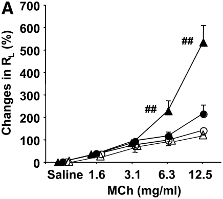
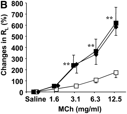
Estrogen is a leading candidate as the female hormonal factor produced and released from the ovaries. To define the differences between male and female mice, effects of an estrogen antagonist were evaluated on 10-day OVA-induced AHR to inhaled MCh in vivo. Ten-day vehicle and 10-day OVA treatment did not elicit significant AHR to MCh in female mice (Figure 4A). After daily administration of the estrogen receptor antagonist ICI182,780 (10 mg/kg/d, intraperitoneally) increases in RL were significantly enhanced in female mice but only in OVA-exposed and not saline-exposed mice (Figure 4A). In saline-exposed mice, administration of the estrogen receptor antagonist had no effect. In male mice, daily administration of this compound did not influence AHR (Figure 4B). Despite the increases in AHR in the estrogen receptor antagonist-treated female mice, no changes in BAL inflammatory cell composition or cytokine levels were detected (data not shown).
Figure 4.
Estrogen receptor antagonist alters AHR in female mice. (A) Effect of daily administration of an estrogen antagonist on AHR in female mice. Open circles, 10 days saline/10 days vehicle; solid circles, 10 days OVA/10 days vehicle; open triangles, 10 days saline/10 days ICI (10 mg/kg/d); solid triangles, 10 days OVA/10 days ICI (10 mg/kg/d). (B) Effect of daily administration of the antagonist in male mice after 10-day OVA inhalation in vivo. Open squares, 10 days saline/10 days vehicle; solid squares, 10 days OVA/10 days vehicle; diamonds, 10 days OVA/10 days ICI (10 mg/kg/d). AHR was assayed 24 hours after the last OVA challenge. ICI182,780 at a dose of 10 mg/kg/d was administered intraperitoneally immediately before each OVA challenge. Results represent the mean ± SEM (n = 8). **P < 0.01 and ##P < 0.01 comparing 10-day saline/10-day vehicle and 10-day OVA/10-day vehicle.
Effect of Exogenous Estrogen on 10-Day OVA-Induced AHR in Male Mice In Vivo
As significant AHR in vivo developed in male mice after 10-day OVA inhalation, we evaluated the effects of exogenous estrogen administration on this response. In male mice, administration of E2 suppressed the development of AHR after 10-day OVA inhalation in a dose-dependent manner, with complete attenuation of AHR at a dose of 100 μg/kg/d of E2 (Figure 5). Suppression of AHR was not accompanied by any changes in BAL cell accumulation or cytokine levels (data not shown). This suppression was reversed with daily coadministration of the estrogen receptor antagonist (10 mg/kg/d of ICI182,780 for 10 days), establishing the specificity of the exogenous estrogen effect (Figure 5). Together, these results demonstrate the negative regulatory effects of endogenous and exogenous estrogen on AHR in female and male mice, respectively.
Figure 5.
Effect of daily administration of E2 on 10-day OVA-induced AHR in male mice in vivo. AHR was assayed 24 hours after the last OVA challenge. E2 at doses of 50 and 100 μg/kg was administered intraperitoneally immediately before each OVA challenge. Results represent the mean ± SEM (n = 8). #P < 0.05 and ##P < 0.01 comparing 10-day OVA/10-day vehicle (squares); *P < 0.05 and **P < 0.01 comparing 10-day OVA/10-day E2 100 μg/kg/day (circles). Diamonds, 10-day OVA/10-day E2 (50 μg/kg/d); triangles, 10-day OVA/10-day E2 (μg/kg/d) + 10-day ICI.
Acute Effects of an Estrogen Antagonist and Exogenous Estrogen on AHR to MCh after 10-Day OVA Inhalation In Vivo
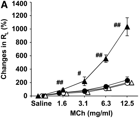
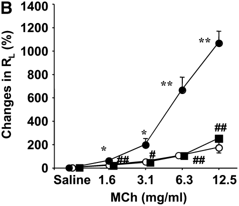
The results reported above were obtained in a protocol which saw the compounds administered on 10 consecutive days. To focus on the response targeted by estrogen, airway constriction, the estrogen antagonist or exogenous estrogen were administered 1 h before the AHR assay, and 24 hours after the last OVA inhalation. AHR to inhaled MCh did not appear in female mice after 10-day OVA inhalation with vehicle treatment (Figure 6A). ICI182,780 at a dose of 10 mg/kg administered 1 hour before assay, significantly enhanced airway responsiveness in female mice after 10-day OVA exposure (but not saline) (Figure 6A). In parallel, the increases in the response to inhaled MCh which developed in male mice after 10-day OVA inhalation, were totally suppressed when estrogen was given 1 hour before assay (Figure 6B). Based on these results and the absence of changes in BAL cell composition or cytokine levels, it appeared that endogenous or exogenous estrogen given after allergen exposure was completed, regulated airway contractility directly. In no case did the estrogen receptor antagonist or estrogen itself impact AHR in the absence of allergen exposure.
Figure 6.
Rapid effect of (A) an estrogen antagonist and (B) exogenous E2 on airway responsiveness to MCh in vivo. AHR was assayed 24 hours after the last OVA challenge. ICI182,780 (ICI) at a dose of 10 mg/kg to female mice (A) or E2 at a dose of 100 μg/kg to male mice (B) was administered intraperitoneally 1 hour before the AHR assay. Results represent the mean ± SEM (n = 8). #P < 0.05 and ##P < 0.01 comparing 10-day OVA/vehicle. *P < 0.05 and **P < 0.01 comparing 10-day saline/vehicle. Open circles, 10-day saline/vehicle; solid circles, 10-day OVA/vehicle; open triangles, 10-day saline/ICI; solid triangles, 10-day OVA ICI. Squares, 10-day OVA/E2.
Effect of Exogenous Estrogen on CCh- and EFS-Induced Airway Constriction after 10-Day OVA Inhalation
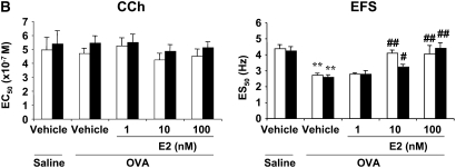
To directly investigate whether estrogen influences airway constriction directly, we examined the effects of estrogen on CCh- and EFS-induced airway constriction in 10-day saline- or OVA-exposed mice in vitro. Estrogen at a dose of 100 nM did not affect CCh- or EFS-induced constriction of trachea smooth muscle from mice exposed to 10-day saline inhalation, and no differences were observed between males and females (Figure 7A). However, ES50 values were decreased in male and female mice after 10-day OVA exposure, and E2 dose-dependently normalized ES50 values (Figure 7B). These results imply that estrogen does not affect cholinergic airway constriction directly but may inhibit prejunctional activation by factors generated during the 10 days of allergen exposure.
Figure 7.
Effect of exogenous E2 on in vitro airway responsiveness to CCh or EFS after (A) 10-day saline or (B) 10-day OVA exposure. Tracheas from female (open bars) and male (solid bars) mice were isolated from 10-day saline- or OVA-treated mice. The tracheas were preincubated with vehicle or E2 (1–100 nM) for 30 minutes. After incubation, airway contractility to CCh or EFS was determined. Results represent the mean ± SEM (n = 8). **P < 0.01 comparing 10-day saline/vehicle group in either sex. #P < 0.05 and ##P < 0.01 comparing 10-day OVA/vehicle group in either sex. In the CCh and EFS groups, the 95% confidence intervals overlapped.
In Vivo Effects of an NK-1 Receptor Antagonist
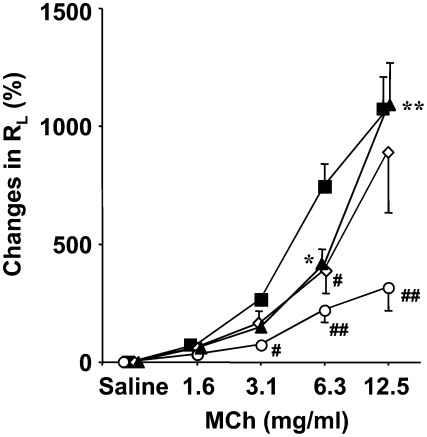
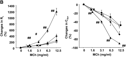
Neuropeptides, such as substance P, are critical regulators of airway responsiveness. Activation of NK-1 receptors enhances ACh release by EFS (11). We evaluated the effects of Sendide, an NK-1 receptor antagonist, on 10-day OVA-induced AHR in male mice and on the enhancement of AHR by ICI182,780 in female mice. Sendide at a dose of 40 nmol/kg, given 2 hours before assay, significantly suppressed AHR (RL and Cdyn) in 10-day OVA-exposed male mice but not in 10-day saline-exposed mice (Figure 8A). ICI182,780, given 1 hour before assay, significantly enhanced airway responsiveness in female mice after 10-day OVA exposure (Figure 8B). Sendide, at a dose of 40 nmol/kg, fully suppressed this enhancement of RL and decrease in Cdyn (Figure 8B). These data suggest that NK-1 is critical to the development of AHR after 10-day OVA inhalation in vivo, and that endogenous estrogen regulates the development of AHR in female mice exposed to OVA exclusively via the airways via an NK-1–dependent pathway.
Figure 8.
Effect of a neurokinin-1 receptor antagonist (Sendide) on (A) 10-day OVA-induced AHR in male mice and (B) enhancement of AHR by an estrogen antagonist in 10-day OVA-exposed female mice. AHR was assayed 24 hours after the last OVA challenge. Sendide at a dose of 40 nmol/kg and/or ICI182,780 at a dose of 10 mg/kg was administered intraperitoneally 2 hours and/or 1 hour, respectively, before the AHR assay. Results represent the mean ± SEM (n = 8). #P < 0.05 and ##P < 0.01 comparing 10-day OVA/vehicle or 10-day saline/Sendide. *P < 0.05 and **P < 0.01 comparing 10-day saline/vehicle or 10-day saline/Sendide. †P < 0.05 and ††P < 0.01 comparing 10-day OVA/ICI/PBS. PBS was a vehicle for Sendide. ICI = ICI182,780. A: open circles, 10-day saline/vehicle; solid circles, 10-day OVA/vehicle; open diamonds, 10-day saline/Sendide; solid diamonds, 10-day OVA/Sendide. B: open circles, 10-day saline/vehicle/PBS; solid circles, 10-day OVA/vehicle/PBS; triangles, 10-day OVA/ICI/PBS; diamonds, 10-day OVA/ICI/Sendide.
In Vitro Effects of an NK-1 Receptor Antagonist
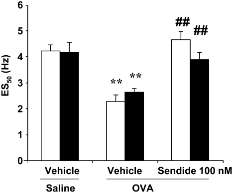
Together, the data suggested that endogenous estrogen may regulate an NK-1–dependent pathway leading to AHR in mice exposed to 10-day OVA. Furthermore, exogenous estrogen suppressed the 10-day OVA-induced AHR detected by EFS in vitro (see Figure 7B). We next confirmed the contribution of the NK-1 pathway to 10-d OVA-induced AHR detected by EFS in vitro. Pretreatment of tracheal smooth muscle preparations with Sendide (100 nM) significantly suppressed the decrease in ES50 in female and male mice exposed to OVA for 10 days, and there were no sex differences in the degree of suppression (Figure 9). Thus, NK-1 seems to be critical to development of AHR after 10-day OVA inhalation.
Figure 9.
Involvement of neurokinin-1 in 10-day OVA-induced AHR detected by EFS in vitro. Tracheas from female (open bars) and male (solid bars) mice were isolated from 10-day saline- or OVA-exposed mice. The tracheas were preincubated with vehicle or Sendide (100 nM) for 30 minutes. After the preincubation, airway contractility to EFS was assayed. Results represent the mean ± SEM (n = 8). ††P < 0.01 comparing 10-day saline/vehicle. ##P < 0.01 comparing 10-day OVA/vehicle. ES50 = 50% of maximal contraction.
DISCUSSION
There is substantial evidence that female sex hormones play a regulatory role in asthma pathophysiology, and in particular, development of AHR. Asthma prevalence rates are higher in women than men with a switch occurring at the time of puberty (18–20). There are impressive menstrual cycle–associated variations in pulmonary function with asthma worsening during low hormone levels premenstrually (21, 22). Although these clinical findings suggest that sex hormones may influence the prevalence and severity of asthma, the role of specific hormones in dictating these changes is unclear. A number of studies in this area have been reported without consensus, likely reflecting the complexity of the pathways involved and undefined regulatory factors at various times in established disease. In particular, the effects of estrogen in asthma have been discordant, with both beneficial and negative effects described (7, 8, 23). In the former, supplemental estrogens were used as a steroid-sparing agent in females with asthma; in the latter, post-menopausal estrogen therapy increased the subsequent risk of asthma. A number of papers describe a worsening of asthma during the premenstrual and intramenstrual periods in a subgroup of women with asthma (21, 24–27), although the mechanism underlying this susceptibility remains unclear. Ensom and colleagues demonstrated the beneficial effects of estradiol in a severely asthmatic woman with premenstrual worsening of asthma (24).
The present study examined sex differences in a model of allergen exposure that was exclusively delivered via the airways and in the absence of systemic sensitization and adjuvant. This approach has been shown to be dependent on IgE, FcɛRI expression, and mast cells, and differs from the requirements when systemic sensitization with adjuvant are employed (13). In some studies which use systemic sensitization to allergen with adjuvant followed by allergen challenge via the airways, female mice developed greater AHR, with estrogen enhancing airway inflammation (6–8). In contrast, in naive mice or following less robust sensitization and challenge, as performed here, the inhibitory effects of estrogen on development of AHR may be more obvious (4, 5, 23). Carey et al also showed that deficiency of the estrogen receptor resulted in higher AHR. In our study, OVA exposure exclusively via the airways resulted in little inflammatory cell accumulation in the BALF but did elicit a modest eosinophilic inflammatory response in lung tissue as well as goblet cell metaplasia (23). In female mice, 10 days of OVA exposure failed to alter lung function in vivo when assessed after MCh inhalation. Increased airway responsiveness was detected only by the altered response to electrical field stimulation, a decrease in ES50. Such decreases have been linked to muscarinic receptor dysfunction, endogenous substance P, and increased acetylcholine release (13, 15), perhaps the result of eosinophilic inflammation (28). In contrast to female mice, male mice subjected to 10 days of OVA exposure developed a similar decrease in ES50 but also exhibited a robust MCh dose-dependent increase in RL and a decrease in Cdyn in vivo. These differences in MCh-induced AHR between male and female mice did not seem to be related to differences in airway inflammation, BAL cytokine levels, or numbers of goblet cells. Together, these data pointed to sex differences in the response of airway smooth muscle to inhaled MCh after allergen exposure as opposed to differences in the immune/inflammatory response often associated with increased MCh reactivity (29, 30).
In different models, estrogen has been shown to recapitulate sex differences. As examples, estrogen pretreatment of male mice prevented endotoxin-induced hepatic injury and reduced poly-ADP-ribosylation in vivo (31). Down-modulation of E-selectin and IL-6 gene expression by estrogen may have contributed to a reduction in the cellular infiltration seen in a model of acute anterior uveitis (32). Several reports of risk factors associated with AHR have noted a higher level of responsiveness in female compared with male subjects (6, 23, 33–35), implicating that sex hormones play a role in altered airway responsiveness. However, the specific mechanisms responsible for the development of AHR were unclear.
In female mice, ovariectomy enabled development of MCh-induced changes in RL (increased) and Cdyn (decreased). Confirmation of the role of estrogen as an important regulator of airway function was demonstrated by the administration of an estrogen receptor antagonist, which similarly enhanced MCh-induced changes in airway function in female mice. Because administration of the estrogen receptor antagonist 1 hour before assay of airway function showed similar enhancement of AHR, this suggested direct effects on airway smooth muscle, but only in the allergen-exposed group. In male mice, administration of estrogen reduced the 10-day OVA-induced changes in MCh-dependent reactivity, and this could be prevented by coadministration of an estrogen receptor antagonist. Together, these data identified estrogen as a critical regulator of the development of AHR after exposure to allergen exclusively via the airways. Furthermore, because manipulation of airway reactivity to MCh with estrogen, the estrogen receptor antagonist, or ovariectomy was accomplished without concomitant changes in airway inflammation, BAL cytokine levels, or numbers of goblet cells, this was consistent with estrogen regulating airway function at the level of airway smooth muscle. This notion was also supported by the in vivo findings that these manipulations (other than OVX) could be performed after full completion of allergen exposure, just 1 hour before measuring AHR to inhaled MCh, with very similar results.
These in vivo results were paralleled by examining the effects of estrogen in vitro on CCh- or EFS-induced contractility of tracheal smooth muscle. Estrogen has been reported to inhibit CCh-induced airway constriction after passive sensitization with serum from patients with asthma (4). In the present study, no enhancement of CCh-induced contractility was detected in vitro after 10-day OVA inhalation. Exogenous estrogen did not influence CCh-induced tracheal smooth muscle responses in male or female mice after OVA exposure. In contrast, exogenous estrogen added to the muscle preparation suppressed EFS-induced contractility in tracheal smooth muscle from male and female mice after OVA exposure but not after saline exposure. These data demonstrate that the effects of estrogen on airway smooth muscle contractility are restricted to those triggered by EFS and not CCh. To extend these findings, we showed using a specific NK-1 receptor antagonist that OVA exposure activated an NK-1–dependent pathway resulting in altered airway function in vitro and in vivo. Moreover, the NK-1 receptor antagonist attenuated the effects of the estrogen receptor antagonist in enhancing AHR in female mice in vivo. NK1 receptor stimulation has been shown to augment cholinergic neurotransmission in mouse trachea when in vitro airway responsiveness was detected by EFS (36). Thus, these data support the notion that the rapid onset of estrogen regulation of smooth muscle contractility is NK-1–dependent and involves prejunctional activation but does not interfere with cholinergic airway smooth muscle constriction. The 10-day OVA inhalation leads to a cascade of immunologic/inflammatory responses (13). The assumption is that these events trigger and up-regulate an NK-1–dependent pathway, leading to increases in ACh release. The role of NK receptors and their agonistic tachykinins in allergic airway inflammation is somewhat controversial (37). In a recent study, Boot and colleagues demonstrated that a NK1/NK2 dual antagonist failed to modulate AHR and airway inflammation after allergen challenge in patients with asthma (38). However, the role of NK receptors cannot be discounted because it is difficult to confirm blocking of all available NK receptors in the airways within such a clinical design.
In this study, male–female differences in the response to allergen exposure were revealed when MCh-induced changes in airway function were monitored. Investigation of these differences identified estrogen as an important regulator of airway function at the prejunctional level in vitro and in vivo. Manipulation of estrogen production or estrogen receptor antagonism converted female mice from nonresponders to responders and male mice from responders to nonresponders in an NK-1–dependent manner. These findings identify potential approaches to modify altered airway function in patients with asthma, especially at critical stages of their disease.
Acknowledgments
The authors thank Dr. J. J. Lee (Mayo Clinic, Scottsdale, AZ) for providing the anti–major basic protein antibodies and L. N. Cunningham and D. Nabighian (National Jewish Medical and Research Center, Denver, CO) for their assistance.
This work was supported by National Institutes of Health grants HL-36577 and HL-61005 and by Environmental Protection Agency grant R825702. The content is solely the responsibility of the authors and does not necessarily represent the official views of the NHLBI or the NIH.
Originally Published in Press as DOI: 10.1165/rcmb.2007-0298OC on December 6, 2007
Conflict of Interest Statement: None of the authors has a financial relationship with a commercial entity that has an interest in the subject of this manuscript.
References
- 1.Meyer MR, Haas E, Barton M. Sex differences of cardiovascular disease: new perspectives for estrogen receptor signaling. Hypertension 2006;47:1019–1026. [DOI] [PubMed] [Google Scholar]
- 2.Most W, van der Wee-Pals L, Ederveen A, Papapoulos S, Lowik C. Ovariectomy and orchidectomy induce a transient increase in the osteoclastogenic potential of bone marrow cells in the mouse. Bone 1997;20:27–30. [DOI] [PubMed] [Google Scholar]
- 3.Gillies GE, Murray HE, Dexter D, McArthur S. Sex dimorphisms in the neuroprotective effects of estrogen in an animal model of Parkinson's disease. Pharmacol Biochem Behav 2004;78:513–522. [DOI] [PubMed] [Google Scholar]
- 4.Dimitropoulou C, White RE, Ownby DR, Catravas JD. Estrogen reduces carbachol-induced constriction of asthmatic airways by stimulating large-conductance voltage and calcium-dependent potassium channels. Am J Respir Cell Mol Biol 2005;32:239–247. [DOI] [PubMed] [Google Scholar]
- 5.Degano B, Prevost MC, Berger P, Molimard M, Pontier S, Rami J, Escamilla R. Estradiol decreases the acetylcholine-elicited airway reactivity in ovariectomized rats through an increase in epithelial acetylcholinesterase activity. Am J Respir Crit Care Med 2001;164:1849–1854. [DOI] [PubMed] [Google Scholar]
- 6.Melgert BN, Postma DS, Kuipers I, Geerlings M, Luinge MA, van der Strate BW, Kerstjens HA, Timens W, Hylkema MN. Female mice are more susceptible to the development of allergic airway inflammation than male mice. Clin Exp Allergy 2005;35:1496–1503. [DOI] [PubMed] [Google Scholar]
- 7.Corteling R, Trifilieff A. Sex comparison in a murine model of allergen-driven airway inflammation and the response to budesonide treatment. BMC Pharmacol 2004;4:4. [DOI] [PMC free article] [PubMed] [Google Scholar]
- 8.Riffo-Vasquez Y, Ligeiro de Oliveira AP, Page CP, Spina D, Tavares-de-Lima W. Role of sex hormones in allergic inflammation in mice. Clin Exp Allergy 2007;37:459–470. [DOI] [PubMed] [Google Scholar]
- 9.Dijkstra A, Howard TD, Vonk JM, Ampleford EJ, Lange LA, Bleecker ER, Meyers DA, Postma DS. Estrogen receptor 1 polymorphisms are associated with airway hyperresponsiveness and lung function decline, particularly in female subjects with asthma. J Allergy Clin Immunol 2006;117:604–611. [DOI] [PubMed] [Google Scholar]
- 10.Schatz M, Clark S, Camargo CA Jr. Sex differences in the presentation and course of asthma hospitalizations. Chest 2006;129:50–55. [DOI] [PubMed] [Google Scholar]
- 11.Colasurdo GN, Loader JE, Graves JP, Larsen GL. Modulation of acetylcholine release in rabbit airways in vitro. Am J Physiol 1995;268:L432–L437. [DOI] [PubMed] [Google Scholar]
- 12.Larsen GL, Loader JE, Nguyen DD, Fratelli C, Dakhama A, Colasurdo GN. Mechanisms determining cholinergic neural responses in airways of young and mature rabbits. Pediatr Pulmonol 2004;38:97–106. [DOI] [PubMed] [Google Scholar]
- 13.Taube C, Wei X, Swasey CH, Joetham A, Zarini S, Lively T, Takeda K, Loader J, Miyahara N, Kodama T, et al. Mast cells, Fc epsilon RI, and IL-13 are required for development of airway hyperresponsiveness after aerosolized allergen exposure in the absence of adjuvant. J Immunol 2004;172:6398–6406. [DOI] [PubMed] [Google Scholar]
- 14.Takeda K, Hamelmann E, Joetham A, Shultz LD, Larsen GL, Irvin CG, Gelfand EW. Development of eosinophilic airway inflammation and airway hyperresponsiveness in mast cell-deficient mice. J Exp Med 1997;186:449–454. [DOI] [PMC free article] [PubMed] [Google Scholar]
- 15.Larsen GL, Fame TM, Renz H, Loader JE, Graves J, Hill M, Gelfand EW. Increased acetylcholine release in tracheas from allergen-exposed IgE-immune mice. Am J Physiol 1994;266:L263–L270. [DOI] [PubMed] [Google Scholar]
- 16.Tomkinson A, Duez C, Cieslewicz G, Pratt JC, Joetham A, Shanafelt MC, Gundel R, Gelfand EW. A murine IL-4 receptor antagonist that inhibits IL-4- and IL-13-induced responses prevents antigen-induced airway eosinophilia and airway hyperresponsiveness. J Immunol 2001;166:5792–5800. [DOI] [PubMed] [Google Scholar]
- 17.Larsen GL, Renz H, Loader JE, Bradley KL, Gelfand EW. Airway response to electrical field stimulation in sensitized inbred mice: passive transfer of increased responsiveness with peribronchial lymph nodes. J Clin Invest 1992;89:747–752. [DOI] [PMC free article] [PubMed] [Google Scholar]
- 18.Skobeloff EM, Spivey WH, St Clair SS, Schoffstall JM. The influence of age and sex on asthma admissions. JAMA 1992;268:3437–3440. [PubMed] [Google Scholar]
- 19.Hyndman SJ, Williams DR, Merrill SL, Lipscombe JM, Palmer CR. Rates of admission to hospital for asthma. BMJ 1994;308:1596–1600. [DOI] [PMC free article] [PubMed] [Google Scholar]
- 20.Bloomberg GR, Trinkaus KM, Fisher EB Jr, Musick JR, Strunk RC. Hospital readmissions for childhood asthma: a 10-year metropolitan study. Am J Respir Crit Care Med 2003;167:1068–1076. [DOI] [PubMed] [Google Scholar]
- 21.Gibbs CJ, Coutts II, Lock R, Finnegan OC, White RJ. Premenstrual exacerbation of asthma. Thorax 1984;39:833–836. [DOI] [PMC free article] [PubMed] [Google Scholar]
- 22.Pauli BD, Reid RL, Munt PW, Wigle RD, Forkert L. Influence of the menstrual cycle on airway function in asthmatic and normal subjects. Am Rev Respir Dis 1989;140:358–362. [DOI] [PubMed] [Google Scholar]
- 23.Carey MA, Bradbury JA, Card JW, Noorman MP, Haykal-Coates N, Gavett SH, Graves JP, Walker VR, Zhu D, Jacobs ER, et al. Spontaneous airway hyperresponsiveness in estrogen receptor-alpha deficient mice. Am J Respir Crit Care Med 2007;175:126–135. [DOI] [PMC free article] [PubMed] [Google Scholar]
- 24.Ensom MH, Chong G, Beaudin B, Bai TR. Estradiol in severe asthma with premenstrual worsening. Ann Pharmacother 2003;37:1610–1613. [DOI] [PubMed] [Google Scholar]
- 25.Ensom MH, Chong E, Carter D. Premenstrual symptoms in women with premenstrual asthma. Pharmacotherapy 1999;19:374–382. [DOI] [PubMed] [Google Scholar]
- 26.Oguzulgen IK, Turktas H, Erbas D. Airway inflammation in premenstrual asthma. J Asthma 2002;39:517–522. [DOI] [PubMed] [Google Scholar]
- 27.Tan KS. Premenstrual asthma: epidemiology, pathogenesis and treatment. Drugs 2001;61:2079–2086. [DOI] [PubMed] [Google Scholar]
- 28.Fryer AD, Stein LH, Nie Z, Curtis DE, Evans CM, Hodgson ST, Jose PJ, Belmonte KE, Fitch E, Jacoby DB. Neuronal eotaxin and the effects of CCR3 antagonist on airway hyperreactivity and M2 receptor dysfunction. J Clin Invest 2006;116:228–236. [DOI] [PMC free article] [PubMed] [Google Scholar]
- 29.Tomkinson A, Duez C, Cieslewicz G, Gelfand EW. Eotaxin-1-deficient mice develop airway eosinophilia and airway hyperresponsiveness. Int Arch Allergy Immunol 2001;126:119–125. [DOI] [PubMed] [Google Scholar]
- 30.Wolyniec WW, De Sanctis GT, Nabozny G, Torcellini C, Haynes N, Joetham A, Gelfand EW, Drazen JM, Noonan TC. Reduction of antigen-induced airway hyperreactivity and eosinophilia in ICAM-1-deficient mice. Am J Respir Cell Mol Biol 1998;18:777–785. [DOI] [PubMed] [Google Scholar]
- 31.Mabley JG, Horvath EM, Murthy KG, Zsengeller Z, Vaslin A, Benko R, Kollai M, Szabo C. Sex differences in the endotoxin-induced inflammatory and vascular responses: potential role of poly(ADP-ribose) polymerase activation. J Pharmacol Exp Ther 2005;315:812–820. [DOI] [PubMed] [Google Scholar]
- 32.Miyamoto N, Mandai M, Suzuma I, Suzuma K, Kobayashi K, Honda Y. Estrogen protects against cellular infiltration by reducing the expressions of E-selectin and IL-6 in endotoxin-induced uveitis. J Immunol 1999;163:374–379. [PubMed] [Google Scholar]
- 33.Whitacre CC, Reingold SC, O'Looney PA. A sex gap in autoimmunity. Science 1999;283:1277–1278. [DOI] [PubMed] [Google Scholar]
- 34.Da Silva JA, Pinto A, Cutolo M, Porto A. Sex differences in adrenal and gonadal responses to inflammatory aggression. Ann N Y Acad Sci 1999;876:148–151. [DOI] [PubMed] [Google Scholar]
- 35.Amadori A, Zamarchi R, De Silvestro G, Forza G, Cavatton G, Danieli GA, Clementi M, Chieco-Bianchi L. Genetic control of the CD4/CD8 T-cell ratio in humans. Nat Med 1995;1:1279–1283. [DOI] [PubMed] [Google Scholar]
- 36.Tournoy KG, De Swert KO, Leclere PG, Lefebvre RA, Pauwels RA, Joos GF. Modulatory role of tachykinin NK1 receptor in cholinergic contraction of mouse trachea. Eur Respir J 2003;21:3–10. [DOI] [PubMed] [Google Scholar]
- 37.Joos GF, De Swert KO, Schelfhout V, Pauwels RA. The role of neural inflammation in asthma and chronic obstructive pulmonary disease. Ann N Y Acad Sci 2003;992:218–230. [DOI] [PubMed] [Google Scholar]
- 38.Boot JD, de Haas S, Tarasevych S, Roy C, Wang L, Amin D, Cohen J, Sterk PJ, Miller B, Paccaly A, et al. Effect of an NK1/NK2 receptor antagonist on airway responses and inflammation to allergen in asthma. Am J Respir Crit Care Med 2007;175:450–457. [DOI] [PubMed] [Google Scholar]


