

Abstract
The objectives of augmentation of the nucleus pulposus following disc removal are to prevent disc height loss and the associated biomechanical and biochemical changes. Flowable materials may be injected via a small incision, allowing minimally invasive access to the disc space. Fluids can interdigitate with the irregular surgical defects and may even physically bond to the adjacent tissue. Injectable biomaterials allow for incorporation and uniform dispersion of cells and/or therapeutic agents. Injectable biomaterials have been developed that may act as a substitute for the disc nucleus pulposus. Our work has focused on the evaluation of a recombinant protein copolymer consisting of amino acid sequence blocks derived from silk and elastin structural proteins as an injectable biomaterial for augmentation of the nucleus pulposus. This implant, NuCore™ Injectable Nucleus is being developed by Spine Wave (Shelton, CT). The NuCore™ material is comprised of a solution of the protein polymer and a polyfunctional cross-linking agent. The material closely mimics the protein content, water content, pH and complex modulus of the natural nucleus pulposus. Extensive mechanical testing, biocompatibility and toxicology testing have been performed on the material. Characterization studies indicate that the NuCore™ Injectable Nucleus is able to restore the biomechanics of the disc following a microdiscectomy. Extensive biomaterial characterization shows the material to be non-toxic and biocompatible. The mechanical properties of the material mimic those of the natural nucleus pulposus. Thus NuCore™ Injectable Nucleus is suitable to replace the natural nucleus pulposus following a discectomy procedure. Human clinical evaluation is underway in a multi center clinical study on the use of the material as an adjunct to microdiscectomy. Further clinical studies of the use of NuCore™ Injectable Nucleus for treatment of early stage degenerative disc disease are planned in the near future. On-going efforts are characterizing the use of the material as a cell delivery vehicle for disc repair and reconstruction. Related development efforts are exploring methods for repair and regeneration of the cartilage endplate that are implemented to enhance the host-implant interface. Prior to the introduction of the above-mentioned biomaterial, our work proposes to utilize a process for the treatment of the vertebral endplates. The goal of this process is to restore the endplates as closely as possible to their natural state prior to disease or degeneration. The nature of the treatment will depend upon the form of the endplate degeneration and on the type of scaffolding that is intended to be introduced in the nuclear cavity. Endplate therapy is a potential means of enhancing biomaterial integration and cell survival, but remains a long-term and currently untested methodology.
Keywords: Intervertebral disc repair, Disc prosthesis, Injectable biomaterial, Vertebral endplate, Minimally invasive surgery
Part 1: Injectable biomaterials
Introduction
Discectomy is the most common spinal surgical treatment and typically performed in a relatively young patient population (25–40 years) and the impact of altered biomechanics and long-term sequelae in this young patient population may be significant [29, 40, 67]. Substantial disc height reduction following discectomy may occur and is evident soon following the discectomy procedure. Disc height loss has been found to be proportional to the amount of nucleus removed in an in vitro study [11]. Clinically, the operated disc spaces of patients post-operatively are significantly more narrow following discectomy than controls [26]. Scoville and Corkill [55] found a 50% incidence of narrowing following surgery at the 3 month follow-up. Tibrewal and Pearcy [57] found disc space narrowing evident within 3 months following surgery as compared to non-operated-controls.
Proper disc height is necessary to ensure proper functioning of the intervertebral disc and spinal column. Changes in disc height can have both local and global effects. On the local (or cellular) level decreased disc height and volume results in increased load on the remaining nucleus pulposus, which can lead to a decrease in cell matrix synthesis and an increase in cell necrosis and apoptosis. It has been shown in other cartilaginous tissues that increased static loading decreases matrix protein biosynthesis [13, 20, 46]. Animal models have shown that overloading of the intervertebral disc can initiate disc degeneration [32, 36]. In addition, increases in intradiscal pressure create an unfavorable environment for fluid transfer into the disc, which can cause a further decrease in disc height.
Decreased disc height also results in significant changes in the global mechanical stability of the spine, which may result in further degeneration of the spinal segment. With decreasing height of the disc, the facet joints bear increasing loads and may undergo hypertrophy and degeneration, which may act as a source of pain over time [27]. Decreased stiffness of the spinal column and increased range of motion resulting from loss of disc height can lead to further instability of the spine [49]. Excessive motion can manifest itself in abnormal muscle, ligament and tendon loading, which can ultimately be a source of back pain.
Radicular pain may result from a decrease in foraminal volume caused by decreased disc height. Specifically, as disc height decreases, the volume of the foraminal canal, through which the spinal nerve roots pass, decreases. This decrease may lead to spinal nerve impingement, with associated radiating pain and dysfunction. Finally, adjacent segment loading increases as the disc height decreases at a given level [44]. The discs that must bear additional loading are now susceptible to accelerated degeneration and compromise, which may eventually propagate along the destabilized spinal column. A further issue with microdiscectomy surgery is the occurrence of reherniation, which may lead to the need to reoperate. Atlas et al. [3] reported a reoperation rate of 25% at 10 year follow up in a study of patients with lumbar disc herniation, with the median time to reoperation 24 months. Carraggee et al. [18] reported an 11.5% reherniation rate, with 6.5% re-operation at 5 years.
Material and surgical objectives
The objectives of augmentation of the nucleus pulposus following disc removal are to prevent disc height loss and the associated biomechanical and biochemical changes resulting from reduced disc height and volume. The use of an injectable material for nucleus replacement gives the flexibility of allowing treatment of partial nucleotomies following microdiscectomy, as well as indications such as early stage degenerative disc disease (DDD), where more complete nucleus removal and replacement may be required. An injectable biomaterial is ideal for restoration of disc volume removed during discectomy and for preventing loss of disc height. Flowable materials may be injected via a small incision, allowing minimally invasive access to the disc space where appropriate. Fluids can interdigitate with the irregular surgical defects and may, depending on the material used, physically bond to the adjacent tissue. Injectable biomaterials allow for incorporation and uniform dispersion of cells and/or therapeutic agents. Growth factors, such as members of the BMP, TGF and IGF families, may be valuable in enhancing the repair process. Inhibitors of inflammatory cytokines (e.g., interleukins, tumor necrosis factors) and proteases (e.g., matrix metalloproteases) may act to retard matrix degradation and the potential effects of these cytokines on surrounding tissue and neural (especially nocciceptive) structures.
The primary considerations for injectable scaffolds for disc repair include mechanical strength and durability, promotion of tissue formation, biodegradability, biocompatibility, sterilizability, minimal setting time and temperature change, low viscosity for easy injection, as well as ease in accessing the disc space. The scaffold must exhibit the necessary mechanical properties as well as provide physical support. Due to the large number of loading cycles experienced in the spine, it is important that the scaffold be able to withstand significant physical loading. In addition, physical support should be provided by the injectable scaffold through restoration of disc height. Preferably, the scaffold would promote matrix formation while degrading over time. The biocompatibility of the material is also of great importance. Neither the initial material nor its degradation products should elicit an unresolved immune response, promote immunotoxicity, or express cytotoxicity. To minimize infections and related immune responses, the implanted material must be easily sterilized while retaining the original bioactivity and chemical composition.
Generally, the candidate biomaterials are injected as viscous fluids and then cured through methods such as thermosensitive crosslinking, pH-sensitive crosslinking, photopolymerization, or addition of a solidifying agent to form a gel-like substance. It is important to consider the amount of time it takes for the material to set. The setting time should be long enough to allow for accurate placement during the procedure yet be short enough so as to not prolong the length of the surgical procedure. If the material experiences a temperature change while hardening, the increase in temperature should be small. Heat generated during this process should not cause harm to surrounding tissue. The viscosity of the material should balance the need for the substance to remain at the site of its introduction into the disc and ability of the surgeon to manipulate its placement, with the need to assure complete filling of the intradiscal space or voids. Ease in accessing the disc space also needs to be considered. Polymers that cure through a photopolymerization procedure could pose a problem due to a limited ability to access the small cavities of the disc space with light needed to initiate cross-linking.
Injectable biomaterial options
Injectable biomaterials have been considered as an augment to a discectomy for over 40 years. As early as 1962, Alf Nachemson [42] suggested the injection of room temperature vulcanizing silicone into a degenerated disc using an ordinary syringe. In 1974, Schneider and Oyen [53, 54] studied the use of silicone elastomer in the intervertebral disc. Since then, injectable biomaterials or scaffolds have been developed that may act as a substitute for the disc nucleus pulposus, such as hyaluronic acid, fibrin glue, alginate, elastin-like polypeptides, collagen type I gel and others. A number of patents have been issued concerning various injectable biomaterials that may have utility for nucleus augmentation including: cross-linkable silk elastin copolymer [17, 25, 56], polyurethane-filled balloons [4, 24], aldehyde cross-linked bovine serum albumin [68], collagen-PEG [52]; chitosan [21]; various injectable synthetic polymers [39]; recombinant bioelastic materials [61]; light-curable PEG polymers and other multicomponent precursor systems [30, 31]. Several groups are actively pursuing the development of an injectable biomaterial for use in the intervertebral disc.
Our efforts have focused on the evaluation of a recombinant protein copolymer consisting of amino acid sequence blocks derived from silk and elastin structural proteins as an injectable biomaterial for augmentation of the nucleus pulposus. These proteins have been well-characterized over more than a decade of intensive research and development. The material appears to have ideal characteristics for the augmentation of the nucleus pulposus following discectomy procedures. This implant, the NuCore™ Injectable Nucleus is being developed by Spine Wave (Shelton, CT, USA). Cappello et al. [15, 16] have reported on the development and characterization of structural protein polymers, especially those derived from the structural proteins silk and elastin. They have developed a technology for the production of synthetically designed protein polymers consisting of repeated blocks of amino acid sequence. Through a combination of biological and chemical methods, block polymers are produced using gene template directed synthesis. Using this method, the design and polymerization of a new polymer occurs once during the synthesis of the gene template. Through the construction of synthetic genes, it is possible to specify the sequence of protein blocks (the unit of repetition of a protein polymer) several hundred amino acids in length, many fold greater than the limit of sequence control of chemical synthesis.
Injectable disc nucleus
The protein polymer used in the NuCore™ material is a copolymer of silk and elastin, with two silk blocks and eight elastin blocks per polymer repeat. One of the elastin blocks is modified to provide for chemical cross-linking. The protein polymer is synthesized using recombinant DNA techniques via Escherichia coli strain K12, a nonpathogenic strain of bacterium and the workhorse of recombinant protein expression. Following batch fermentation, the cells are ruptured by homogenization and the protein polymer is purified from the lysate using precipitation and a series of filtration and adsorption purification steps. The identity and purity of the polymer is confirmed using amino acid composition, amino acid terminal sequencing, mass spectroscopy and other biochemical tests.
The material is comprised of a solution of the protein polymer and a polyfunctional cross-linking agent. The material is formulated to closely match the properties of the human nucleus pulposus as shown in Table 1.
Table 1.
Comparison of properties between NuCore™ material and nucleus pulposus
| Property | NuCore™ material | Nucleus pulposus |
|---|---|---|
| Protein content | 19.4% | 13.6–21.9%a |
| Water content | 79.1% | 74–81% |
| PH | 7.1 | 6.7–7.1 |
| Complex shear modulus (G*) | 26 kPa | 7–21 kPab |
The material closely mimics the protein content, water content, pH and complex modulus of the natural nucleus pulposus. The material is injected following mixing with a very low concentration of a diisocyanate-based cross-linking agent and has approximately a ninety-second working time following addition of the cross-linking agent before it becomes a viscous gel. The material is essentially cured within 5 min and reaches near final mechanical strength approximately 30 min following addition of the crosslinker.
Testing of cadaver functional spinal units was done to determine how well the NuCore™ material restores stability and function to a spinal unit [37]. Anterior column units were tested in compression, first in the intact condition, then with annulotomy, then with a partial nucleotomy, and finally with NuCore™ material injected. Statistical analysis using repeated measures ANOVA and post hoc Tukey comparisons showed that the discectomy caused a significant loss of height during the test (P < 0.05). However, the NuCore™ material injection caused a restoration such that there was no significant difference between the displacement of the intact condition (1.28 mm) and the NuCore™ material treated condition (1.22 mm).
Specimens were also tested in flexion/extension, right and left lateral bending, and right and left axial rotation [63]. Testing evaluated first the intact condition, then after a partial nucleotomy, and finally after NuCore™ material injection. Statistical analysis using repeated measures ANOVA and post hoc Tukey pairwise comparisons showed that in all degrees of freedom, the discectomy condition caused a destabilization of the unit (P < 0.05) whereas once the NuCore™ material was injected, in no cases were there significant differences between the intact or NuCore™ material conditions (P > 0.05). This analysis indicates the NuCore™ Injectable Nucleus restores function and stability to the spine after a destabilizing discectomy procedure.
To simulate dynamic loading over long periods of time, testing has been performed in a synthetic disc model. The annulus fibrosus of the disc was simulated with a molded silicone elastomer. This silicone annulus was injected with NuCore™ material, and the model was subjected to cyclic loading up to 10 million cycles with no failure of the NuCore™ material or test model. The sinusoidal applied load in axial compression had a maximum of 1,000 N and a minimum of 100 N, applied at a frequency of 3 Hz for 10 million cycles. In a separate test in axial torsion, with a compressive pre-load of 250 N, a torque of ± 2 Nm was applied using a sinusoidal waveform at a frequency of 3 Hz for 10 million cycles. This testing indicates the NuCore™ material to be fatigue resistant and durable, capable of withstanding in vivo loads for an extended period of time.
Testing of cadaver anterior column units was done to determine how well the NuCore™ material resists forces to cause extrusion from the IVD space. Segments were tested in axial compression in both a neutral posture and in hyperflexion. In all cases, there was no extrusion prior to bony failure and/or endplate failure. The average failure load of the spinal segments was 3,555 N in the neutral position, and 2,637 N in the hyperflexed position, well above physiologic levels. Results demonstrated that the NuCore material integrated extensively with the surrounding disc tissue and did not extrude during any of the testing. This is shown in Fig. 1 where the NuCore™ material is pigmented to aid visualization.
Fig. 1.
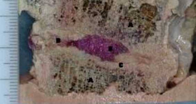
Lateral cross-section of cadaveric spinal motion segment with NuCore™ material filling nuclear cavity (red dye added to material for visualization)
Extensive biocompatibility and toxicology testing following the ISO 10993 guidelines has been performed on the NuCore™ material. Acute tests include cytotoxicity, sensitization (guinea pig), intracutaneous reactivity (rabbit), systemic toxicity (mouse), pyrogenicity, muscle implant evaluation and genotoxicity testing. The material is non-cytotoxic, non irritating and non-toxic in all of these test evaluations. Chronic toxicity testing has been conducted in a rat model, with material placed subcutaneously and evaluated at time points to 1 year and beyond, with no toxicity seen. Neurofunctional testing in a rat showed no neurotoxicity of the material when placed adjacent to spinal nerve roots.
Part 2: vertebral endplate therapy
Background to the concept
The intervertebral disc is the largest avascular structure in the body and the endplate serves as the main route for metabolites in and out of the disc nucleus [58, 59]. Its permeability to solutes depends upon the capillary bed and the presence of vascular contacts between marrow spaces of the vertebral body and the hyaline cartilage of the endplate [22, 23, 28, 47, 65]. Nutrients transported by the contact vessels are required for cellular metabolism and biosynthesis of extracellular matrix. Metabolic waste products (e.g., lactic acid) must be removed to prevent their accumulation within the disc [7]. Water can also diffuse through the endplates to maintain a proper intra-discal pressure, which ultimately results in an appropriate disc height for mechanical function [2, 48]. Poor supply of essential nutrients and build up of waste products are often cited as significant contributors to degeneration of the intervertebral disc [1, 12, 38, 60, 64, 65].
There appears to be a strong link between degeneration of the intervertebral disc and changes to the adjacent endplate. Nachemson et al. [43] found a correlation between impermeability of the endplate and disc degeneration. Degenerative changes in the disc appear to be preceded by histological evidence of endplate lesions and microfracture [8, 50, 62]. There is evidence of increasing calcification of endplate cartilage and occluded nutrient canals with increasing age [6, 19]. Signal intensity changes in vertebral marrow and endplate changes on MRI have been strongly associated with disc degeneration [35, 41]. Density of endplate openings has been found to significantly correlate with morphological degeneration grade [5].
As discussed above, treatment of disc degeneration can proceed as outlined previously, i.e., a discectomy followed by the introduction of some form of scaffold into the intradiscal space. In some cases, the scaffold is a solid implant or spinal fusion that does not preserve any of the mechanical properties of the disc. However, where the scaffold is of the injectable biomaterial type that seeks to restore normal disc function, patency of the endplates is of critical concern. If the disc has sclerotic or thickened endplates, no restorative scaffold will work in its intended way because minimal fluid/nutrient diffusion is permitted. In other words, if the foundation is deficient, the entire treatment of the disc will fall short of its goal.
Objective of EP treatment and proposed methodology
Our current work in this area involves developing methods for endplate treatment that are implemented following removal of a portion of the nucleus pulposus. As discussed above, disc repair or regeneration may be achieved by the introduction of a biomaterial into the disc cavity that is capable of restoring disc height and substantially normal disc function. Prior to the introduction of the above-mentioned biomaterial, our work proposes to implement a process for the pretreatment of the vertebral endplates. The goal of this process is to restore the endplates as closely as possible to their natural state prior to disease or degeneration. The nature of the treatment will depend upon the form of the endplate degeneration and on the type of scaffolding that is intended to be introduced in the nuclear cavity.
One possible method involves the decalcification of sclerotic, thickened and impermeable vertebral endplates. Calcification of the articular cartilage of the endplates impedes fluid diffusion and may be addressed by injection of compounds capable of binding calcium. The decalcifying agent is maintained in contact with the endplates over a suitable incubation duration, after which the agent, along with the byproducts of its operation, can be flushed from the disc space. In addition, large proteoglycans, such as aggrecan, can also hinder the passage of certain solutes through the endplates. The removal of these proteoglycans via an appropriate enzyme, such as trypsin, can further enhance the permeability of the endplate. This treatment could accompany treatment for calcification.
Another focus area is enhancing the vascularity of the endplate region. An established network of blood vessels at the endplate is, of course, critical to nutrient transport and proper biological functioning of the disc. Chemical treatments are known that increase vascularity in tissues, such as cytokines and growth factors capable of inducing endothelial cell growth (e.g., VEGF, vascular endothelial growth factor). Growth factors and cytokines can become absorbed into the endplate cells and extracellular matrix so only a minimal incubation time may be necessary, and no flushing would be required after application of the treatment. Besides direct delivery of cytokines, known methods of gene therapy can be used to transfect resident cells (e.g., endothelial cells) on a transient basis to secrete the cytokine of interest [45, 66]. Since this cell growth stimulating material is intended to remain, this treatment can be reserved to the end of the endplate treatment process.
Another area of application for endplate therapy involves methods to enhance the integration between the injectable biomaterial and surrounding tissues. Solid or injectable in situ curable biomaterials require that the prosthesis stay well fixed within the disc space. Anchorage and integration at the graft-host interface (i.e., between the prosthesis and the endplates) is essential to the long-term viability of the prosthesis. However, the cartilaginous endplates and the annulus fibrosus contain abundant large and small proteoglycans that may impede integration between the tissue and the prosthesis. For articular cartilage repair, researchers have proposed enzymatic digestion of cartilage adjacent to the graft site to denude it of proteoglycans and allow for interdigitation with the newly synthesized matrix components [9, 10, 51]. It has been found that a protease treated matrix has much less structured water and matrix, and is more easily infiltrated with newly synthesized matrix from repair tissue. Pre-treatment of affected endplates with trypsin, hyaluronidase or other enzymes may allow cartilaginous tissue buds to extend from adjacent tissue into a pre-treated graft to create an undulating, well-integrated surface at the graft-tissue interface. It has been demonstrated that a sequential digestion of the cartilaginous matrix with hyaluronidase followed by trypsin effectively extracts proteoglycan without any significant disruption of the underlying collagen fiber network [14]. Endplate pretreatment may involve treatment with trypsin, or with the sequential application of hyaluronidase and trypsin, followed by a suitable incubation period. Once the incubation time has expired, the solution and its byproducts are flushed from the disc space and the cavity is well irrigated, such as with a saline solution. Other components of the extracellular matrix may also be targeted by appropriate materials, such as proteolytic enzymes. A proteolytic inhibitor may be necessary following incubation with the protease or enzyme.
Finally, cell migration into the injected scaffold may also be enhanced by vertebral endplate pretreatment. Tissue engineering involves the use of systems of cells, scaffolds and growth factors/cytokines to reconstruct and regenerate tissues and organs. Certain disc prostheses may be populated with cells from the patient’s own natural disc prior to injection. Cells may also be harvested from other cartilaginous tissues of the body, such as the non-articulating areas of hyaline cartilage in the knee. Adult mesenchymal stem cells may be isolated and purified from bone marrow or adipose tissue. However, accessing, isolating and purifying cells in this manner can pose a host of problems. One way to avoid these technical challenges is by implanting an acellular scaffold. In this approach, cells are not actually placed in/on the scaffold, but instead resident cells are allowed to migrate into and populate the scaffold. It has been found that the trypsin pre-treatment of cartilage explants allows chondrocytes to proliferate and rapidly replenish matrix lost during the trypsin treatment [14].
Summary and future directions
In conclusion, characterization studies indicate that the NuCore™ Injectable Nucleus is able to restore the biomechanics of the disc following a microdiscectomy. Extensive biomaterial characterization shows the material to be non-toxic and biocompatible. The mechanical properties of the material mimic those of the natural nucleus pulposus. Thus NuCore™ Injectable Nucleus is suitable to replace the natural nucleus pulposus following a discectomy procedure. Human clinical evaluation is underway in a multi center clinical study on the use of the material as an adjunct to microdiscectomy. Further clinical studies of the use of NuCore™ Injectable Nucleus for treatment of early stage degenerative disc disease are planned in the near future. On-going efforts are characterizing the use of the material as a cell delivery vehicle for disc repair and reconstruction. Finally, endplate therapy is a potential means of enhancing biomaterial integration and cell survival, but remains a long-term and currently untested methodology.
References
- 1.Adams MA, Bogduk N, Burton K, Dolan P. Summary: spinal ageing, degeneration and pain. In: Adams MA, Bogduk N, Burton K, Dolan P, editors. The biomechanics of back pain. New York: Churchill Livingstone; 2002. pp. 197–203. [Google Scholar]
- 2.Adams MA, Hutton WC. The effects of posture on the fluid content of lumbar intervertebral discs. Spine. 1983;8:665–671. doi: 10.1097/00007632-198309000-00013. [DOI] [PubMed] [Google Scholar]
- 3.Atlas SJ, Deyo RA, Ancker Mvd, Singer DE, Keller RB, Patrick DL. The Maine-Seattle back questionnaire: A 12-item disability questionnaire for evaluating patients with lumbar sciatica or stenosis. Spine. 2003;28:1869–1876. doi: 10.1097/01.BRS.0000083205.82614.01. [DOI] [PubMed] [Google Scholar]
- 4.Bao Q-BH, Yuan A (2001) Implantable tissue repair device. Patent 6,224,630
- 5.Benneker LM, Heini PF, Anderson SE, Ito K. Young investigator award winner: vertebral endplate marrow contact channel occlusions and intervertebral disc degeneration. Spine. 2005;7:97–102. doi: 10.1097/01.brs.0000150833.93248.09. [DOI] [PubMed] [Google Scholar]
- 6.Bernick S, Cailliet R. Vertebral end-plate changes with aging of human vertebrae. Spine. 1982;7:97–102. doi: 10.1097/00007632-198203000-00002. [DOI] [PubMed] [Google Scholar]
- 7.Bibby SRS, Jones DA, Ribley RM, Urban JPG. Metabolism of the intervertebral disc: effects of low levels of oxygen, glucose, and pH on rates of energy metabolism of bovine nucleus pulposus cells. Spine. 2005;30:487–496. doi: 10.1097/01.brs.0000154619.38122.47. [DOI] [PubMed] [Google Scholar]
- 8.Boos N, Weissbach S, Rohrbach H, Weiler C, Spratt KF, Nerlich AG. Classification of age-related changes in lumbar intervertebral discs. Spine. 2002;27:2631–2644. doi: 10.1097/00007632-200212010-00002. [DOI] [PubMed] [Google Scholar]
- 9.Bos PK, DeGroot J, Budde M, Verhaar JA, Osch GJv. Specific enzymatic treatment of bovine and human articular cartilage: Implications for integrative cartilage repair. Arthritis Rheum. 2002;46:976–985. doi: 10.1002/art.10208. [DOI] [PubMed] [Google Scholar]
- 10.Bravenboer JvdB, Maur CDId, Bos PK, Feenstra L, Verhaar JA, Weinans H, van Osch GJVM. Improved cartilage integration and interfacial strength after enzymatic treatment in a cartilage transplantation model. Arthritis Res Therapy. 2005;6:469–476. doi: 10.1186/ar1216. [DOI] [PMC free article] [PubMed] [Google Scholar]
- 11.Brinckmann P, Grootenboer H. Change of disc height, radial disc bulge, and intradiscal pressure from discectomy: an in vitro investigation of human lumbar discs. Spine. 1991;16:641–646. doi: 10.1097/00007632-199106000-00008. [DOI] [PubMed] [Google Scholar]
- 12.Brodin H. Paths of nutrition in articular cartilage and intervertebral discs. Acta Orthop Scand. 1955;24:177–183. doi: 10.3109/17453675408988561. [DOI] [PubMed] [Google Scholar]
- 13.Buschmann MD, Gluzband YA, Grodzinsky AJ, Huziker EB. Mechanical compression modulates matrix biosynthesis in chondrocyte/agarose culture. J Cell Sci. 1995;108:1497–1508. doi: 10.1242/jcs.108.4.1497. [DOI] [PubMed] [Google Scholar]
- 14.Caplan AI, Elyaderani M, Mochizuki Y, Wakatani S, Goldberg VM. Overview: Principles of cartilage repair and regeneration. Clin Orthop Rel Res. 1997;342:254–269. doi: 10.1097/00003086-199709000-00033. [DOI] [PubMed] [Google Scholar]
- 15.Cappello J. Genetically engineered protein polymers. In: Domb AJ, Kost J, Wiseman D, editors. Handbook of degradable polymers. Amsterdam: Harwood Academic Publishers; 1996. pp. 387–414. [Google Scholar]
- 16.Cappello Ferrari J F. Microbial production of structural protein polymers. In: Mobley, editor. Plastics from microbes. Munich: Carl Hanser Verlag; 1994. pp. 35–92. [Google Scholar]
- 17.Cappello JE, Stedronsky R (2002) Synthetic proteins for in vivo drug delivery and tissue augmentation, U.S. Patent 6,380,154
- 18.Carragee EJ, Han MY, Yang B, Kim DH, Kraemer H, Billys J. Activity restrictions after posterior lumbar discectomy: a prospective study of outcomes in 152 cases with no postoperative restrictions. Spine. 1999;24:2346–2351. doi: 10.1097/00007632-199911150-00010. [DOI] [PubMed] [Google Scholar]
- 19.Chandraraj S, Briggs CA, Opeskin K. Disc herniations in the young and end-plate vascularity. Clin Anat. 1998;11:171–176. doi: 10.1002/(SICI)1098-2353(1998)11:3<171::AID-CA4>3.0.CO;2-W. [DOI] [PubMed] [Google Scholar]
- 20.Chen J, Yan W, Setton LA. Static compression induces zonal-specific changes in gene expression for extracellular matrix and cytoskeletal proteins in intervertebral disc cells in vitro. Matrix Biol. 2004;22:573–583. doi: 10.1016/j.matbio.2003.11.008. [DOI] [PubMed] [Google Scholar]
- 21.Chenite A, Chaput C, Wang D, Combes C, Buschmann MD, Hoemann CD, Leroux JC, Atkinson BL, Binette F, Selmani A. Novel injectable neutral solutions of chitosan form biodegradable gels in situ. Biomaterials. 2000;21:2155–2161. doi: 10.1016/S0142-9612(00)00116-2. [DOI] [PubMed] [Google Scholar]
- 22.Crock H, Goldwasser VM. Anatomic studies of the circulation in the region of the vertebral end-plate in adult greyhound dogs. Spine. 1984;9:702–706. doi: 10.1097/00007632-198410000-00009. [DOI] [PubMed] [Google Scholar]
- 23.Donisch E, Trapp WW. The cartilage endplates of the human vertebral column (some considerations of postnatal development). Anat Rec. 1971;169:705–716. doi: 10.1002/ar.1091690409. [DOI] [PubMed] [Google Scholar]
- 24.Felt JC, Rydell MA, Zdrahala RJ, Arsenyev A (2001) Biomaterial for in situ tissue repair, U.S. Patent 6,306,177
- 25.Ferrari FA, Richardson C, Chambers J, Causey S, Pollock TJ, Cappello J, Crissmann JW (2002) Peptides comprising repetitive units of amino acids and DNA sequences encoding the same, U.S. Patent 6,355,776
- 26.Frymoyer JW, Hanley EN, Howe J, Kuhlmann D, Matteri RE. A comparison of radiographic findings in fusion and nonfusion patients ten and more years following disc surgery. Spine. 1979;4:435–440. doi: 10.1097/00007632-197909000-00008. [DOI] [PubMed] [Google Scholar]
- 27.Gotfried Y, Bradford DS, Oegena TR., Jr Facet joint changes after chemonucleolysis-induced disc space narrowing. Spine. 1986;11:944–950. doi: 10.1097/00007632-198611000-00016. [DOI] [PubMed] [Google Scholar]
- 28.Hassler O. The human intervertebral disc: a micro-angiographical study of its vascular supply at various ages. Acta Orthop Scand. 1970;40:765–772. doi: 10.3109/17453676908989540. [DOI] [PubMed] [Google Scholar]
- 29.Hermantin FU, Peters T, Quartararo L, Kambin P. A prospective, randomized study comparing the results of open discectomy with those of video-assisted arthroscopic microdiscectomy. J Bone Jt Surg. 1999;81-A:958–965. doi: 10.2106/00004623-199907000-00008. [DOI] [PubMed] [Google Scholar]
- 30.Hubbell JA, Pathak CP, Sawhney AS, Desai NP, Hill JL (1997) Photopolymerizable biodegradable hydrogels as tissue contacting materials and controlled-release microcarriers, U.S. Patent 5,626,863
- 31.Hubbell JA, Wetering Pvd, Cowling DSP (2002) Novel polymer compounds, Patent 2002/0177680
- 32.Iatridis JC, Mente PL, Stokes IAF, Aronsson DD, Alini M. Compression -induced changes in intervertebral disc properties in a rat tail model. Spine. 1999;24:996–1002. doi: 10.1097/00007632-199905150-00013. [DOI] [PubMed] [Google Scholar]
- 33.Iatridis JC, Weidenbaum M, Setton LA, Mow VC. Is the nucleus pulposus a solid or fluid? Mechanical behavior of the nucleus pulposus of the human intervertebral disc. Spine. 1996;21:1174–1184. doi: 10.1097/00007632-199605150-00009. [DOI] [PubMed] [Google Scholar]
- 34.Kitano T, Zerwekh J, Usui Y, Edwards M, Flickere P, Mooney V. Biochemical changes associated with the symptomatic human intervertebral disc. Clin Orthop Rel Res. 1993;293:372–377. [PubMed] [Google Scholar]
- 35.Kokkonen SM, Kurunlahti M, Tervonen O, Iikko E, Vanharanta H. Endplate degeneration observed on magnetic resonance imaging of the lumbar spine: correlation with pain provocation and disc changes observed on computed tomography diskography. Spine. 2002;27:2274–2278. doi: 10.1097/00007632-200210150-00017. [DOI] [PubMed] [Google Scholar]
- 36.Lotz JC, Colliou OK, Chin JR, Duncan NA, Liebenberg E. Compression-induced degeneration of the intervertebral disc: an in vivo mouse model and finite-element study. Spine. 1998;23:2493–2506. doi: 10.1097/00007632-199812010-00004. [DOI] [PubMed] [Google Scholar]
- 37.Mahar AT, Oka R, Whitledge J, Cappello JR, Powell J, McArthur T (2002) Biomechanical efficacy of a protein polymer hydrogel for inter-vertebral nucleus augmentation and replacement. World Congress on Biomechanics, Calgary, Canada, p 356
- 38.Maroudas A, Stockwell RA, Nachemson A, Urban J. Factors involved in the nutrition of the human lumbar intervertebral disc: cellularity and diffusion of glucose in vitro. J Anat. 1975;120:113–130. [PMC free article] [PubMed] [Google Scholar]
- 39.Milner R, Arrowsmith P, Millan EJ (2001) Intervertebral disc implant, U.S. Patent 6,187,048
- 40.Mochida J, Toh E, Nomura T, Nishimura K. The risks and benefits of percutaneous nucleotomy for lumbar disc herniation: a ten-year longitudinal study. J Bone Jt Surg. 2001;83-B:501–505. doi: 10.1302/0301-620X.83B4.11508. [DOI] [PubMed] [Google Scholar]
- 41.Modic MT, Steinberg PM, Ross JS, Masaryk TJ, Carter JR. Degenerative disc disease: assessment of changes in vertebral body marrow with mr imaging. Radiology. 1988;166:193–199. doi: 10.1148/radiology.166.1.3336678. [DOI] [PubMed] [Google Scholar]
- 42.Nachemson A. Some mechanical properties of the lumbar intervertebral disc. Bull Hosp Joint Dis. 1962;23:130–132. [PubMed] [Google Scholar]
- 43.Nachemson A, Lewin T, Maroudas A, Freeman MAR. In vitro diffusion of dye through the end-plates and the annulus fibrosus of human lumbar inter-vertebral discs. Acta Orthop Scand. 1970;41:589–607. doi: 10.3109/17453677008991550. [DOI] [PubMed] [Google Scholar]
- 44.Natarajan RN, Ke JH, Andersson GBJ. A model to study the disc degeneration process. Spine. 1994;19:259–265. doi: 10.1097/00007632-199402000-00001. [DOI] [PubMed] [Google Scholar]
- 45.Nishida K, Gilbertson LG, Evans CH, Kang JD. Spine update: Potential applications of gene therapy to the treatment of spinal disorders. Spine. 2000;25:1308–1314. doi: 10.1097/00007632-200005150-00021. [DOI] [PubMed] [Google Scholar]
- 46.Ohshima H, Urban JPG, Bergel DH. Effect of static load on matrix synthesis rates in the intervertebral disc measured in vitro by a new perfusion technique. J Orthop Res. 1995;13:22–29. doi: 10.1002/jor.1100130106. [DOI] [PubMed] [Google Scholar]
- 47.Oki S, Matsuda Y, Shibata T, Okumura H, Desaki J. Morphologic differences of the vascular buds in the vertebral endplate: scanning electron microscipic study. Spine. 1996;21:174–177. doi: 10.1097/00007632-199601150-00003. [DOI] [PubMed] [Google Scholar]
- 48.Paajanen H, Lehto I, Alanen A, Erkintalo M, Komu M. Diurnal fluid changes of lumbar discs measured indirectly by magnetic resonance imaging. J Orthop Res. 1994;12:509–514. doi: 10.1002/jor.1100120407. [DOI] [PubMed] [Google Scholar]
- 49.Panjabi MM, Krag MH, Chung TQ. Effects of disc injury on mechanical behavior of the human spine. Spine. 1984;9:707–713. doi: 10.1097/00007632-198410000-00010. [DOI] [PubMed] [Google Scholar]
- 50.Pritzker KPH. Aging and degeneration in the lumbar intervertebral disc. Orthop Clin North Am. 1977;8:65–77. [PubMed] [Google Scholar]
- 51.Quinn T, Hunziker B. Controlled enzymatic matrix degradation for integrative cartilage repair: effects on viable cell density and proteoglycan deposition. Tissue Eng. 2002;8:799–806. doi: 10.1089/10763270260424150. [DOI] [PubMed] [Google Scholar]
- 52.Rhee WM, DeLustro FA, Berg RA (2001) Method of making crosslinked polymer matrices in tissue treatment applications, U.S. Patent 6,323,278
- 53.Schneider P, Oyen GR. Intervertebral disc replacement, experimental studies, clinical consequences. Z Orthop Ihre Grenzgeb. 1974a;112:791–792. [PubMed] [Google Scholar]
- 54.Schneider P, Oyen GR. Plastic surgery on intervertebral disc: Part i intervertebral disc replacement in the lumbar regions with silicone rubber. Theoretical and experimental studies. Z Orthop Ihre Grenzgeb. 1974b;112:1078–1086. [PubMed] [Google Scholar]
- 55.Scoville W, Corkill BG. Lumbar disc surgery: technique of radical removal and early mobilization. J Neurosurg. 1973;39:265–269. doi: 10.3171/jns.1973.39.2.0265. [DOI] [PubMed] [Google Scholar]
- 56.Stedronsky E, Cappello RJ (2002) Sealing or filling tissue defects using polyfunctional crosslinking agents and protein polymers, U.S. Patent 6,423,333
- 57.Tibrewal SB, Pearcy MJ. Lumbar intervertebral disc heights in normal subjects and patients with disc herniation. Spine. 1985;10:452–454. doi: 10.1097/00007632-198506000-00009. [DOI] [PubMed] [Google Scholar]
- 58.Urban JPG, Holm S, Maroudas A. Diffusion of small solutes into the intervertebral disc: an in vivo study. Biorheology. 1978;15:203–223. doi: 10.3233/bir-1978-153-409. [DOI] [PubMed] [Google Scholar]
- 59.Urban JP, Maroudas GA. The measurement of fixed charge density in the intervertebral disc. Biochim Biophys Acta. 1979;586:166–178. [Google Scholar]
- 60.Urban JPG, Smith S, Fairbank JCT. Nutrition of the intervertebral disc. Spine. 2004;29:2700–2709. doi: 10.1097/01.brs.0000146499.97948.52. [DOI] [PubMed] [Google Scholar]
- 61.Urry DW (1991) Polynanopeptide bioelastomers having an increased elastic modulus, U.S. Patent 5,064,430
- 62.Vernon-Roberts BC, Pirie J. Healing trabecular microfractures in the bodies of lumbar vertebrae. Ann Rheum Dis. 1973;32:406–412. doi: 10.1136/ard.32.Suppl.46. [DOI] [PMC free article] [PubMed] [Google Scholar]
- 63.Walkenhorst J, Kitchel S, Spenciner D (2004) Effect of injectable disc nucleus on function of human cadaver spine. In: 11th International meeting on advanced spine techniques (IMAST) Bermuda
- 64.Wallace AL, Wyatt BC, McCarthy ID, Hughes SPF. Humoral regulation of blood flow in the vertebral endplates. Spine. 1994;19:1324–1328. doi: 10.1097/00007632-199406000-00004. [DOI] [PubMed] [Google Scholar]
- 65.Whalen JL, Parke WW, Mazur JM, Stauffer ES. The intrinsic vasculature of developing vertebral end plates and its nutritive significance to the intervertebral discs. J Pediatric Orthop. 1985;5:403–410. doi: 10.1097/01241398-198507000-00003. [DOI] [PubMed] [Google Scholar]
- 66.Yoon ST. The potential for gene therapy for the treatment of disc degeneration. Orthop Clin North Am. 2004;35:95–100. doi: 10.1016/S0030-5898(03)00097-X. [DOI] [PubMed] [Google Scholar]
- 67.Yorimitsu E, Chiba K, Toyama Y, Hirabayashi K. Long-term outcomes of standard discectomy for lumbar disc herniation: a follow-up study of more than 10 years. Spine. 2001;26:652–657. doi: 10.1097/00007632-200103150-00019. [DOI] [PubMed] [Google Scholar]
- 68.Yuksel KU, Walsh SP, Black KS (2005) In situ bioprosthetic filler and methods, particularily for the in situ formation of vertebral disc bioprosthetics, U.S. Patent 6,921,412