Abstract
The replicative properties of influenza virus hemagglutinin (HA) mutants with altered receptor binding characteristics were analyzed following intranasal inoculation of mice. Among the mutants examined was a virus containing a Y98F substitution at a conserved position in the receptor binding site that leads to a 20-fold reduction in binding. This mutant can replicate as well as wild-type (WT) virus in MDCK cells and in embryonated chicken eggs but is highly attenuated in mice, exhibiting titers in lungs more than 1,000-fold lower than those of the WT. The capacity of the Y98F mutant to induce antibody responses and the structural locations of HA reversion mutations are examined.
Influenza A viruses attach to host cells due to interactions between the hemagglutinin (HA) and cell surface glycoconjugates containing terminal sialic acids (17). A shallow depression of conserved amino acids at the membrane-distal tip of each monomer of the HA trimer serves as the receptor binding site, and this has been characterized in detail based on the structures of HA-receptor analog complexes (9, 11, 30, 32). In all HA-receptor complexes, the location of sialic acid in the binding site is virtually superimposable in the binding pocket, while the structural configuration of the other sugars of receptor analogs can vary considerably.
The type of linkage by which sialic acid is attached to the penultimate galactose sugar of influenza virus receptors can be associated with species specificity, with avian viruses preferring α2,3-linked receptors and human viruses favoring receptors containing α2,6 linkages (5, 27, 29). Numerous reports have related changes in linkage specificity to HA residues 226 and 228 (H3 numbering) at the left side of the binding site (5, 14, 23, 28, 29, 34-37, 39). However, studies on HA mutations associated with growth of human clinical isolates in eggs (7, 8, 16, 24, 25), variability among viruses isolated from different species (5, 19, 21, 22, 26, 28), and mutations of avian virus HAs that affect the binding specificity (20, 31, 34, 35, 39, 40) demonstrate that a range of residues in the vicinity of the binding site may affect receptor recognition properties. Depending on the host cells and replication environment, as well as the virus strain or subtype, any number of these residues could be relevant for adaptation.
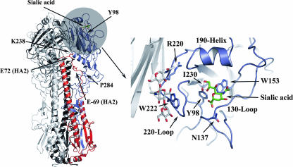
The structure of the HA of A/Aichi/2/68 virus (H3 subtype) and that of its receptor binding region are shown in Fig. 1. In a previous study, several HA mutants with substitutions at conserved positions were analyzed quantitatively for binding to human erythrocytes and a range of phenotypes was detected (18). Despite the abundance of data on receptor binding mutants, receptor specificity, and the relationship to host species for influenza viruses, there is little information that directly relates receptor binding activity that has been quantitatively determined to efficiency of influenza virus infection in vivo. Here we investigate a selection of the mutant viruses generated by reverse genetics for their ability to infect and replicate in BALB/c mice following intranasal (i.n.) inoculation. The mutant with the poorest binding activity (Y98F mutant) was severely attenuated in mice, whereas replication was not compromised for mutants more moderately inhibited for binding. Antibody responses and mutants selected in Y98F mutant-infected mice were also analyzed. These findings enhance our understanding of the relationship between receptor binding and virus fitness in a susceptible host.
FIG. 1.
Left panel, ribbon diagram showing the HA trimer. One of the three monomers is colored to show the HA1 subunit in blue and the HA2 subunit in red. The region encompassing the receptor binding domain is shaded, and the location of bound sialic acid is indicated in green. The location of HA1 Tyr-98 at the base of the binding pocket is also indicated, as are some of the positions identified in reversion mutants that may influence trimer stability. Right panel, enlarged view of the receptor binding region. The binding site is a shallow depression bordered by a short α-helix (the 190 helix) at the membrane-distal edge, the 130 loop at the front of the site, and the 220 loop at the left side. Conserved residues Y98, W153, H183, and Y195 form the base of the site, and the positions of Y98 and W153 are indicated. Sialic acid is shown in green, and the location of residues identified in reversion mutants are indicated (as they appear in WT HA). The sialic acid binding pocket is located in the monomer colored in blue, but residues from the adjacent monomer (shown in gray) can influence binding, particularly if carbohydrate attachments are involved. This is illustrated by the location of the carbohydrate chain originating from HA1 Asn-165 of the neighboring monomer, shown here in gray and red at the left side of the binding site.
Results and discussion.
The mouse is an established model for studying the pathogenesis of susceptible strains of influenza virus (38). Certain H3-subtype human virus strains have been shown to be capable of infecting mice following intranasal inoculation even though α2,6-linked receptors are expressed poorly or are not detected in the respiratory tract using specific lectin staining (10, 12). We used BALB/c mice to evaluate the abilities of our mutant receptor binding site viruses to infect, replicate, and cause disease. Based on previous results (18), four viruses with mutant Aichi HAs were chosen for analysis: one with binding activity similar to that of the wild type (WT) (S228G mutant), two with moderate inhibition of binding (S136T and G225D mutants), and one for which erythrocyte binding was reduced by 20-fold relative to WT levels (Y98F mutant). Three mice per group were infected i.n. with 106 PFU of WT or mutant viruses. At three days postinfection, all except those infected with the Y98F virus displayed characteristic signs of illness, such as shivering, ruffled fur, hunched posture, and weight loss. Mice were sacrificed, and titers of virus in lungs were determined by plaque assay on MDCK cells. These are shown in Table 1, along with previous results on erythrocyte binding and viral titers in MDCK cells (18). The titers of virus in lungs from mice infected with WT, S136T, G225D, or S228G virus were similar (approximately 106 PFU/ml), while the lung viral titers of Y98F mutant-infected mice were reduced by three logs. These results reflect those observed with MDCKRR cells deficient in sialic acid receptors (2), in which only the Y98F virus is significantly restricted for growth (18). We have also done infectivity studies of WT and mutant viruses on WT MDCK cells treated with various concentrations of Clostridium perfringens neuraminidase (ranging from 0 to 100 mU/ml) to specifically cleave sialic acid from the cell surface in an incremental fashion. These showed the Y98F mutant to be the most severely restricted, followed by the mutants with the intermediate binding phenotype, and the effect was particularly apparent at lower concentrations of neuraminidase (data not shown). WT MDCK cells and human erythrocytes have been reported to express abundant levels of both α2,3- and α2,6-linked cell surface sialosides (13, 15), indicating that the Y98F mutant is compromised for binding to each receptor type. The results suggest that the polyvalent binding interactions between the virus and host cell receptors involve a functional interplay between binding affinity and receptor density and that each plays a role in the outcome of infection.
TABLE 1.
Infectivity and growth characteristics of HA receptor binding site mutants
| Mutation | Binding (% WT level)a | HA titer (turkey RBCsb) | Titer in MDCK cells (log10 PFU/ml) | Lung viral titer (log10 PFU/ml) on day 3c |
|---|---|---|---|---|
| None | 100 | 256 | 7.0 | 6.3 |
| Y98F | 5 | <2 | 7.0 | 3.4 |
| S136T | 45 | 128a | 6.8 | 6.6 |
| S225D | 53 | 512a | 6.8 | 6.1 |
| S228G | 112 | 512a | 7.0 | 6.1 |
| W222R | 64 | 5.6 | 6.7 | |
| K238N | 512 | 6.6 | 5.2 |
Results from reference 18.
RBCs, red blood cells.
Mice were infected i.n. with 106 PFU of WT or mutant H3N1 viruses generated as described previously (18) or with 7 × 10³ PFU for the experiment with pseudorevertant viruses. Lungs from three mice per group were collected and pooled, and titers were determined on MDCK cells.
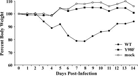
In another set of experiments, mice were infected with a range (106 to 101 PFU) of WT or Y98F virus doses (Table 2). A group infected with 106 PFU of the WT virus exhibited greater than 20% weight loss by day 8, whereas mice infected with the same dose of Y98F virus exhibited insignificant weight loss through day 4 before gaining weight (Fig. 2). Another group of mice was used to determine day 3 lung titers, which again showed titers in Y98F mutant-infected mice to be three to four logs lower than those of the WT with any inoculation dose. Furthermore, serial titration of the virus inocula from 106 to 101 PFU established that the 50% mouse infectious dose for Y98F virus was 1,000-fold higher than that for WT virus (104.5 PFU versus 101.5 PFU), confirming that the Y98F mutant is highly attenuated in BALB/c mice.
TABLE 2.
Infectivity and virulence of Y98F mutant virus in mice
| Virus | 50% mouse infectious dosea (log10 PFU) | Inoculum (PFU) | Lung viral titer (log10 PFU/ml) at day 3b | Mean maximum % wt lossc |
|---|---|---|---|---|
| WT | 1.5 | 106 | 6.4 | 21.4 |
| 105 | 6.2 | |||
| 104 | 6.3 | |||
| Y98F | 4.5 | 106 | 3.5 | 1.2 |
| 105 | 1.8 | |||
| 104 | 2.1 |
Mice (n = 2) were infected i.n. with serial dilutions of virus from 106 to 101 PFU. Three days later, lungs were harvested and titers were determined individually in MDCK cells. The 50% mouse infectious dose was determined as the highest dilution of virus which infected mice.
Mice were infected i.n. with 106 to 104 PFU of WT or mutant virus. Lungs from three mice per inoculum group were collected and pooled, and titers were determined on MDCK cells.
A separate group of four mice was infected with 106 PFU of virus (each mouse) and was monitored for weight loss daily for 14 days. Mice infected with WT virus and Y98F virus exhibited maximum weight loss on days 8 and 4 postinfection, respectively. One mouse infected with WT virus succumbed on day 8.
FIG. 2.
Time course of weight loss in mice infected with WT or Y98F mutant viruses.
To evaluate the ability of the attenuated Y98F virus to induce protective antibody responses, groups of four mice were inoculated intranasally with 106 PFU of WT virus, Y98F virus, or virus diluent. Three months after infection, sera were collected and tested for the presence of hemagglutinin inhibition (HI) antibodies to the WT virus. Both WT and Y98F virus induced substantial serum HI antibody, and the respective geometric mean titers were similar (Table 3). Mice were then challenged with 106 PFU of WT virus and sacrificed on day 3 postinfection, and the lung viral titers were determined (Table 3). More than 5 logs of virus were detected in the lungs of control mice, whereas no virus was detected in mice initially infected with either WT or Y98F virus. Thus, the protective antibody responses conferred by Y98F virus were comparable with those of mice that survived an initial disease-inducing infection with WT virus.
TABLE 3.
Y98F mutant induces protective immune response equivalent to that for WT virus
| Virus inoculuma | Postinfection serum HI antibody GMTb | Lung viral titer (log10 PFU/ml) at day 3 postchallengec |
|---|---|---|
| WT virus | 126 | <1.3 |
| Y98F virus | 112 | <1.3 |
| None | <10 | 5.8 |
Groups of mice were infected i.n. with 106 PFU of WT or Y98F mutant virus or were mock infected with virus diluent.
Three months postinfection, sera from three or four mice per group were collected and tested individually for the presence of HI antibody. Data are expressed as the geometric mean titer (GMT). A value of <10 represents the lower limit of antibody detection.
Three months postinfection, mice were challenged with 106 PFU of WT virus; 3 days later, animals were euthanized, lungs from three or four mice per group were collected and pooled, and titers were determined to assess the presence of virus on MDCK cells. The lower limit of virus detection was 101.3 PFU/ml.
Viruses were isolated from the lungs of Y98F-infected mice at day 3 and plaque purified on MDCK cells, and the HAs of several were sequenced. Among the 18 plaques analyzed, 5 had no sequence changes, retaining the Y98F mutation (two displayed P284H heterogeneity). Six were true WT revertants to Y98, and the others had pseudoreversion mutations with changes at alternative positions. Three of these had an R220L mutation, and the others contained the change W222R, K238N, I230M, or N137S. It is not possible to discriminate between mutations that may have been present in the quasispecies of MDCK-grown virus inoculum and amplified in the mouse respiratory tract versus those that were generated and selected in vivo. Regardless, the fact that these mutations clustered primarily in the receptor binding region indicates that the amino acid substitutions are likely to convey a selective advantage over Y98F virus based on the binding phenotype. The observation that mice infected with Y98F virus stocks do not display disease symptoms suggests that early innate immune responses may be induced before variants of greater fitness reach significant titers.
Figure 1 shows the location of Y98 in WT HA. The hydroxyl of its phenol side chain forms hydrogen bonds to the 8- and 9-hydroxyls of the sialic acid glycerol moiety and NE2 of the H183 imidazole side chain. In WT HA, I230 packs against Y98 and W153. Mutation to methionine would allow more-intimate hydrophobic packing with both Y98 and W153, which could introduce added stability to the site and compensate for the loss of the H bond between Y98 and H183. In WT HA, an H bond is formed between the main chain of N137 and the carboxylate of sialic acid, and this is unlikely to be altered by mutation to serine. However, an S-to-A substitution at this position has recently been reported to influence binding specificity of an H5-subtype HA (40).
Tryptophan 222 is located on the left side of the binding domain. In many H1-subtype HAs, lysine is present at this position, where it forms hydrogen bonds to Gal-2 of both the human and avian receptors (9). However, W222 of the H3 subtype packs against the carbohydrate attached to N165 of a neighboring monomer. Mutation from W to R at this position could have two effects. First, it would introduce a side chain capable of forming hydrogen bond interactions with Gal-2 of the receptor, similar to that observed in H1 HAs. Second, the conformation of the carbohydrate at N165 of the neighboring monomer could be altered and influence receptor binding.
Arginine 220 lies at a monomer-monomer interface of HA adjacent to the binding site. Substitutions at this position could change the conformation of the 220 loop and could also influence HA trimer stability by disrupting monomer-monomer contacts. Residues 238 and 284 are located approximately 40 Å from the binding site but could also be relevant for trimer stability, since they are situated at interfaces of domains that relocate relative to one another during membrane fusion. K238 is involved in an ion pair with E72 of HA2 from a neighboring monomer that would be eliminated by mutation to N. Proline 284 lies in an HA1-HA2 interface within a monomer, and a histidine at this position could form an ion pair with E69 of HA2. In each example, the changes could lead to altered stability of subunit interfaces, as noted for the R220L mutant above. Mutations at position 218, which resides in proximity to 220 at a monomer-monomer interface, are known to cause both elevated fusion pH and changes in receptor specificity (6). In some situations, changes at subunit interfaces may affect the location and interactions of carbohydrates originating from neighboring monomers.
Pseudorevertant virus plaques with the W222R, I230M, K238N, and P284H mutations isolated during the experiments summarized in Table 2 were amplified on MDCK cells for further analysis. Interestingly, during the two passages required to amplify stocks of these viruses in MDCK cells, the consensus sequence of both the I230M and P284H (which was heterogeneous) virus stocks reverted back to the WT sequence at these positions while retaining the Y98F mutation. In addition, the yield of the W222R mutant in MDCK cells was reduced by at least a log relative to that of the WT or the other mutants (Table 1). Both the W222R virus and the K238N mutant, which was observed to grow reasonably well on MDCK cells, were able to agglutinate erythrocytes despite the continued presence of the Y98F mutation (Table 1). This provides evidence for a direct effect of these mutations on receptor binding properties. Furthermore, the W222R and K238N pseudorevertant viruses and the WT were used to reinfect mice intranasally, and 3-day lung titers of these showed the mutations to either completely (W222R) or partially (K238N) revert the attenuated phenotype conferred by the Y98F mutation (Table 1).
These results support the interpretation that the Y98F mutant is attenuated in mice due to its binding properties, that mutations located at various positions in and around the HA receptor binding site can be selected during replication of this mutant in the mouse respiratory tract, and that at least a subset of these can restore binding and reverse the attenuated phenotype. In addition, they clearly demonstrate that the selective pressure operating on HA binding is different in MDCK cells and the mouse respiratory tract. This comes as no surprise, but it serves as a warning for studies on natural isolates or biological mutants that require amplification prior to the analysis of their binding properties. Our current study does not address the effects of the pseudoreversion mutations on α2,3 or α2,6 linkage specificity or the length of the glycan chains to which the mutant HAs bind (4), but these will be interesting to investigate.
The Y98F mutant is attenuated for replication in the respiratory tracts of mice, but we did not look for the presence of virus elsewhere. However, it should be noted that mutants with reduced binding capacity have the potential to disseminate more efficiently to become more pathogenic. Such examples have been reported for murine polyomavirus strains with enhanced tumorigenicity in mice (33) and with mutants of the Sindbis virus E2 glycoprotein that reduce binding to heparin sulfate receptors but cause increased viremia and delay virus clearance in infected mice (3).
Although viruses such as the Y98F mutant have certain properties that are desirable in an attenuated vaccine, the scale and scope of reversion as highlighted here are likely to mitigate against this. However, since receptor binding is not required for fusion (1), such mutants may be useful as surrogate fusogenic proteins in viral vectors designed for gene therapy or targeted delivery purposes.
Acknowledgments
This work was supported by NIH Public Health Service grant AI66870 to D.A.S. and by contract HHSN266200700006C from NIH/NIAID.
The findings and conclusions in this report are those of the author(s) and do not necessarily represent the views of the funding agency.
Footnotes
Published ahead of print on 19 March 2008.
REFERENCES
- 1.Bosch, V., B. Kramer, T. Pfeiffer, L. Starck, and D. A. Steinhauer. 2001. Inhibition of release of lentivirus particles with incorporated human influenza virus haemagglutinin by binding to sialic acid-containing cellular receptors. J. Gen. Virol. 822485-2494. [DOI] [PubMed] [Google Scholar]
- 2.Brandli, A. W., G. C. Hansson, E. Rodriguez-Boulan, and K. Simons. 1988. A polarized epithelial cell mutant deficient in translocation of UDP-galactose into the Golgi complex. J. Biol. Chem. 26316283-16290. [PubMed] [Google Scholar]
- 3.Byrnes, A. P., and D. E. Griffin. 2000. Large-plaque mutants of Sindbis virus show reduced binding to heparan sulfate, heightened viremia, and slower clearance from the circulation. J. Virol. 74644-651. [DOI] [PMC free article] [PubMed] [Google Scholar]
- 4.Chandrasekaran, A., A. Srinivasan, R. Raman, K. Viswanathan, S. Raguram, T. M. Tumpey, V. Sasisekharan, and R. Sasisekharan. 2008. Glycan topology determines human adaptation of avian H5N1 virus hemagglutinin. Nat. Biotechnol. 26107-113. [DOI] [PubMed] [Google Scholar]
- 5.Connor, R. J., Y. Kawaoka, R. G. Webster, and J. C. Paulson. 1994. Receptor specificity in human, avian, and equine H2 and H3 influenza virus isolates. Virology 20517-23. [DOI] [PubMed] [Google Scholar]
- 6.Daniels, P. S., S. Jeffries, P. Yates, G. C. Schild, G. N. Rogers, J. C. Paulson, S. A. Wharton, A. R. Douglas, J. J. Skehel, and D. C. Wiley. 1987. The receptor-binding and membrane-fusion properties of influenza virus variants selected using anti-haemagglutinin monoclonal antibodies. EMBO J. 61459-1465. [DOI] [PMC free article] [PubMed] [Google Scholar]
- 7.Gambaryan, A. S., J. S. Robertson, and M. N. Matrosovich. 1999. Effects of egg-adaptation on the receptor-binding properties of human influenza A and B viruses. Virology 258232-239. [DOI] [PubMed] [Google Scholar]
- 8.Gambaryan, A. S., A. B. Tuzikov, V. E. Piskarev, S. S. Yamnikova, D. K. Lvov, J. S. Robertson, N. V. Bovin, and M. N. Matrosovich. 1997. Specification of receptor-binding phenotypes of influenza virus isolates from different hosts using synthetic sialylglycopolymers: non-egg-adapted human H1 and H3 influenza A and influenza B viruses share a common high binding affinity for 6′-sialyl(N-acetyllactosamine). Virology 232345-350. [DOI] [PubMed] [Google Scholar]
- 9.Gamblin, S. J., L. F. Haire, R. J. Russell, D. J. Stevens, B. Xiao, Y. Ha, N. Vasisht, D. A. Steinhauer, R. S. Daniels, A. Elliot, D. C. Wiley, and J. J. Skehel. 2004. The structure and receptor binding properties of the 1918 influenza hemagglutinin. Science 3031838-1842. [DOI] [PubMed] [Google Scholar]
- 10.Glaser, L., G. Conenello, J. Paulson, and P. Palese. 2007. Effective replication of human influenza viruses in mice lacking a major alpha2,6 sialyltransferase. Virus Res. 1269-18. [DOI] [PubMed] [Google Scholar]
- 11.Ha, Y., D. J. Stevens, J. J. Skehel, and D. C. Wiley. 2001. X-ray structures of H5 avian and H9 swine influenza virus hemagglutinins bound to avian and human receptor analogs. Proc. Natl. Acad. Sci. USA 9811181-11186. [DOI] [PMC free article] [PubMed] [Google Scholar]
- 12.Ibricevic, A., A. Pekosz, M. J. Walter, C. Newby, J. T. Battaile, E. G. Brown, M. J. Holtzman, and S. L. Brody. 2006. Influenza virus receptor specificity and cell tropism in mouse and human airway epithelial cells. J. Virol. 807469-7480. [DOI] [PMC free article] [PubMed] [Google Scholar]
- 13.Ito, T., Y. Suzuki, L. Mitnaul, A. Vines, H. Kida, and Y. Kawaoka. 1997. Receptor specificity of influenza A viruses correlates with the agglutination of erythrocytes from different animal species. Virology 227493-499. [DOI] [PubMed] [Google Scholar]
- 14.Ito, T., Y. Suzuki, T. Suzuki, A. Takada, T. Horimoto, K. Wells, H. Kida, K. Otsuki, M. Kiso, H. Ishida, and Y. Kawaoka. 2000. Recognition of N-glycolylneuraminic acid linked to galactose by the α2,3 linkage is associated with intestinal replication of influenza A virus in ducks. J. Virol. 749300-9305. [DOI] [PMC free article] [PubMed] [Google Scholar]
- 15.Ito, T., Y. Suzuki, A. Takada, A. Kawamoto, K. Otsuki, H. Masuda, M. Yamada, T. Suzuki, H. Kida, and Y. Kawaoka. 1997. Differences in sialic acid-galactose linkages in the chicken egg amnion and allantois influence human influenza virus receptor specificity and variant selection. J. Virol. 713357-3362. [DOI] [PMC free article] [PubMed] [Google Scholar]
- 16.Katz, J. M., C. W. Naeve, and R. G. Webster. 1987. Host cell-mediated variation in H3N2 influenza viruses. Virology 156386-395. [DOI] [PubMed] [Google Scholar]
- 17.Klenk, E., H. Faillard, and H. Lempfrid. 1955. Uber die enzymatische wirkung von influenzaviren. Hoppe-Seylerias Z. Physiol. Chem. 301235-246. [PubMed] [Google Scholar]
- 18.Martin, J., S. A. Wharton, Y. P. Lin, D. K. Takemoto, J. J. Skehel, D. C. Wiley, and D. A. Steinhauer. 1998. Studies of the binding properties of influenza hemagglutinin receptor-site mutants. Virology 241101-111. [DOI] [PubMed] [Google Scholar]
- 19.Matrosovich, M., P. Gao, and Y. Kawaoka. 1998. Molecular mechanisms of serum resistance of human influenza H3N2 virus and their involvement in virus adaptation in a new host. J. Virol. 726373-6380. [DOI] [PMC free article] [PubMed] [Google Scholar]
- 20.Matrosovich, M., A. Tuzikov, N. Bovin, A. Gambaryan, A. Klimov, M. R. Castrucci, I. Donatelli, and Y. Kawaoka. 2000. Early alterations of the receptor-binding properties of H1, H2, and H3 avian influenza virus hemagglutinins after their introduction into mammals. J. Virol. 748502-8512. [DOI] [PMC free article] [PubMed] [Google Scholar]
- 21.Matrosovich, M., N. Zhou, Y. Kawaoka, and R. Webster. 1999. The surface glycoproteins of H5 influenza viruses isolated from humans, chickens, and wild aquatic birds have distinguishable properties. J. Virol. 731146-1155. [DOI] [PMC free article] [PubMed] [Google Scholar]
- 22.Matrosovich, M. N., A. S. Gambaryan, S. Teneberg, V. E. Piskarev, S. S. Yamnikova, D. K. Lvov, J. S. Robertson, and K. A. Karlsson. 1997. Avian influenza A viruses differ from human viruses by recognition of sialyloligosaccharides and gangliosides and by a higher conservation of the HA receptor-binding site. Virology 233224-234. [DOI] [PubMed] [Google Scholar]
- 23.Naeve, C. W., V. S. Hinshaw, and R. G. Webster. 1984. Mutations in the hemagglutinin receptor-binding site can change the biological properties of an influenza virus. J. Virol. 51567-569. [DOI] [PMC free article] [PubMed] [Google Scholar]
- 24.Robertson, J. S. 1993. Clinical influenza virus and the embryonated hen's egg. Rev. Med. Virol. 397-106. [Google Scholar]
- 25.Robertson, J. S., J. S. Bootman, R. Newman, J. S. Oxford, R. S. Daniels, R. G. Webster, and G. C. Schild. 1987. Structural changes in the haemagglutinin which accompany egg adaptation of an influenza A(H1N1) virus. Virology 16031-37. [DOI] [PubMed] [Google Scholar]
- 26.Rogers, G. N., and B. L. D'Souza. 1989. Receptor binding properties of human and animal H1 influenza virus isolates. Virology 173317-322. [DOI] [PubMed] [Google Scholar]
- 27.Rogers, G. N., R. S. Daniels, J. J. Skehel, D. C. Wiley, X. F. Wang, H. H. Higa, and J. C. Paulson. 1985. Host-mediated selection of influenza virus receptor variants. Sialic acid-alpha 2,6Gal-specific clones of A/duck/Ukraine/1/63 revert to sialic acid-alpha 2,3Gal-specific wild type in ovo. J. Biol. Chem. 2607362-7367. [PubMed] [Google Scholar]
- 28.Rogers, G. N., and J. C. Paulson. 1983. Receptor determinants of human and animal influenza virus isolates: differences in receptor specificity of the H3 hemagglutinin based on species of origin. Virology 127361-373. [DOI] [PubMed] [Google Scholar]
- 29.Rogers, G. N., J. C. Paulson, R. S. Daniels, J. J. Skehel, I. A. Wilson, and D. C. Wiley. 1983. Single amino acid substitutions in influenza haemagglutinin change receptor binding specificity. Nature 30476-78. [DOI] [PubMed] [Google Scholar]
- 30.Russell, R. J., S. J. Gamblin, L. F. Haire, D. J. Stevens, B. Xiao, Y. Ha, and J. J. Skehel. 2004. H1 and H7 influenza haemagglutinin structures extend a structural classification of haemagglutinin subtypes. Virology 325287-296. [DOI] [PubMed] [Google Scholar]
- 31.Ryan-Poirier, K., Y. Suzuki, W. J. Bean, D. Kobasa, A. Takada, T. Ito, and Y. Kawaoka. 1998. Changes in H3 influenza A virus receptor specificity during replication in humans. Virus Res. 56169-176. [DOI] [PubMed] [Google Scholar]
- 32.Skehel, J. J., and D. C. Wiley. 2000. Receptor binding and membrane fusion in virus entry: the influenza hemagglutinin. Annu. Rev. Biochem. 69531-569. [DOI] [PubMed] [Google Scholar]
- 33.Stehle, T., and S. C. Harrison. 1996. Crystal structures of murine polyomavirus in complex with straight-chain and branched-chain sialyloligosaccharide receptor fragments. Structure 4183-194. [DOI] [PubMed] [Google Scholar]
- 34.Stevens, J., O. Blixt, T. M. Tumpey, J. K. Taubenberger, J. C. Paulson, and I. A. Wilson. 2006. Structure and receptor specificity of the hemagglutinin from an H5N1 influenza virus. Science 312404-410. [DOI] [PubMed] [Google Scholar]
- 35.Tumpey, T. M., T. R. Maines, N. Van Hoeven, L. Glaser, A. Solorzano, C. Pappas, N. J. Cox, D. E. Swayne, P. Palese, J. M. Katz, and A. Garcia-Sastre. 2007. A two-amino acid change in the hemagglutinin of the 1918 influenza virus abolishes transmission. Science 315655-659. [DOI] [PubMed] [Google Scholar]
- 36.Vines, A., K. Wells, M. Matrosovich, M. R. Castrucci, T. Ito, and Y. Kawaoka. 1998. The role of influenza A virus hemagglutinin residues 226 and 228 in receptor specificity and host range restriction. J. Virol. 727626-7631. [DOI] [PMC free article] [PubMed] [Google Scholar]
- 37.Wan, H., and D. R. Perez. 2007. Amino acid 226 in the hemagglutinin of H9N2 influenza viruses determines cell tropism and replication in human airway epithelial cells. J. Virol. 815181-5191. [DOI] [PMC free article] [PubMed] [Google Scholar]
- 38.Ward, A. C. 1997. Virulence of influenza A virus for mouse lung. Virus Genes 14187-194. [DOI] [PubMed] [Google Scholar]
- 39.Yamada, S., Y. Suzuki, T. Suzuki, M. Q. Le, C. A. Nidom, Y. Sakai-Tagawa, Y. Muramoto, M. Ito, M. Kiso, T. Horimoto, K. Shinya, T. Sawada, M. Kiso, T. Usui, T. Murata, Y. Lin, A. Hay, L. F. Haire, D. J. Stevens, R. J. Russell, S. J. Gamblin, J. J. Skehel, and Y. Kawaoka. 2006. Haemagglutinin mutations responsible for the binding of H5N1 influenza A viruses to human-type receptors. Nature 444378-382. [DOI] [PubMed] [Google Scholar]
- 40.Yang, Z. Y., C. J. Wei, W. P. Kong, L. Wu, L. Xu, D. F. Smith, and G. J. Nabel. 2007. Immunization by avian H5 influenza hemagglutinin mutants with altered receptor binding specificity. Science 317825-828. [DOI] [PMC free article] [PubMed] [Google Scholar]


