Abstract
Growth of Coxiella burnetii, the agent of Q fever, is strictly limited to colonization of a viable eukaryotic host cell. Following infection, the pathogen replicates exclusively in an acidified (pH 4.5 to 5) phagolysosome-like parasitophorous vacuole. Axenic (host cell free) buffers have been described that activate C. burnetii metabolism in vitro, but metabolism is short-lived, with bacterial protein synthesis halting after a few hours. Here, we describe a complex axenic medium that supports sustained (>24 h) C. burnetii metabolic activity. As an initial step in medium development, several biological buffers (pH 4.5) were screened for C. burnetii metabolic permissiveness. Based on [35S]Cys-Met incorporation, C. burnetii displayed optimal metabolic activity in citrate buffer. To compensate for C. burnetii auxotrophies and other potential metabolic deficiencies, we developed a citrate buffer-based medium termed complex Coxiella medium (CCM) that contains a mixture of three complex nutrient sources (neopeptone, fetal bovine serum, and RPMI cell culture medium). Optimal C. burnetii metabolism occurred in CCM with a high chloride concentration (140 mM) while the concentrations of sodium and potassium had little effect on metabolism. CCM supported prolonged de novo protein and ATP synthesis by C. burnetii (>24 h). Moreover, C. burnetii morphological differentiation was induced in CCM as determined by the transition from small-cell variant to large-cell variant. The sustained in vitro metabolic activity of C. burnetii in CCM provides an important tool to investigate the physiology of this organism including developmental transitions and responses to antimicrobial factors associated with the host cell.
Coxiella burnetii is a bacterial obligate intracellular parasite and the causative agent of Q fever in humans (38). The host range of C. burnetii is broad and includes domesticated animals, wildlife, avian species, and arthropods (25). Q fever is primarily a zoonotic infection that is transmitted via inhalation of contaminated aerosols associated with domestic livestock operations. Acute Q fever is generally a self-limiting infection with influenza-like symptoms (25). Rare endocarditis resulting from chronic infection represents the most severe clinical manifestation of Q fever (25). Although the reported annual incidence of Q fever in the United States is low (approximately 50 cases/year) (25, 27), serologic surveys indicate widespread exposure to C. burnetii (27).
Following infection of a host cell, C. burnetii becomes enclosed in a membranous compartment that transits through the endolysosomal pathway (19). C. burnetii protein synthesis promotes development of this compartment into the organism's replicative niche: the lysosome-like parasitophorous vacuole (PV) (23). Indeed, the mature PV contains a variety of lysosomal markers (38), and studies using pH-sensitive ratiometric probes indicate that the PV lumen is moderately acidic (pH 4.5 to 5.3) (1, 11, 24). In a Vero cell infection model, bacterial replication and expansion of the PV occur following differentiation of the environmentally stable small-cell variant (SCV) to the more fragile large-cell variant (LCV) (6). Condensed chromatin is the most distinguishing feature of the SCV and likely contributes to its low metabolic activity (6, 26). In contrast, the chromatin of the LCV is relaxed, and the cell form has enhanced metabolic and replicative activity relative to the SCV (26). Although information is lacking on factors that regulate C. burnetii morphological transition, developmental regulation of genes by the alternative sigma factor RpoS (33) and nutritional status (6) have been implicated.
Early studies of the axenic (host cell free) metabolic capabilities of C. burnetii used buffers adjusted to neutral pH and suggested that the organism had minimal metabolic capacity (29). The landmark discovery by Hackstadt et al. (14) that C. burnetii axenic metabolic activity is significantly enhanced in an acidic buffer (approximately pH 4.5) allowed detailed analyses of the parameters that are required for transport and utilization of nutrients (14, 15), maintenance of the ATP pool (16), and membrane energization (13). A biochemical stratagem was proposed for C. burnetii (14) whereby the neutral pH of the extracellular environment restricts the organism's metabolism, conferring prolonged viability without replication, whereas the acidic environment of the PV triggers activation of metabolism and replication.
The physiological reason for acid activation of C. burnetii metabolism is still not understood. The proton gradient established by the low luminal pH of the PV may provide a proton motive force that supports transport of metabolites (15). Other possibilities include pH-dependent activation of transporters or sensory systems that affect gene regulation. A pH of 4.5 is reported as optimal for C. burnetii axenic metabolism (14). However, maximal transport and incorporation of individual substrates occur over a wider pH range (14, 20), indicating that the metabolic impact of pH on C. burnetii metabolism may vary according to medium nutrient composition. Energy parasitism has been characterized for Chlamydiae and was once proposed for C. burnetii (40). However, the ATP pool of C. burnetii is stable during axenic metabolism in the presence of a carbon source, consistent with active ATP synthesis (16), and C. burnetii lacks ATP/ADP exchangers (32). Despite active maintenance of the ATP pool (16), protein synthesis by C. burnetii purified from host cells is transient in simple phosphate buffers (pH 4.5) supplemented with amino acids and glucose and declines within 4 h (43). Similarly, DNA synthesis by C. burnetii exhibits a quantitative yield of only 23% over 12 h (4), indicating a lack of replication.
The obligate intracellular nature of C. burnetii imposes considerable limitations on physiologic and biochemical studies of the organism. The limited metabolic activity of C. burnetii in previously established axenic buffers (14, 16, 43) has hampered studies of how the organism responds to environmental conditions associated with its unique lifestyle. Conditions that support prolonged extracellular activity would aid our ability to study C. burnetii biochemistry and potentially lead to a method of axenic cultivation of the organism. To this end, we developed a medium termed complex Coxiella medium (CCM) that supports C. burnetii metabolic fitness for at least 24 h outside the host cell.
MATERIALS AND METHODS
Cultivation and purification of C. burnetii.
The C. burnetii Nine Mile RSA439 phase II isolate (clone 4) was propagated in African green monkey kidney (Vero) fibroblasts (CCL-81; American Type Culture Collection) grown in RPMI medium (Invitrogen Corp., CA) supplemented with 2% fetal bovine serum (FBS). At 7 days postinfection, host cells were disrupted by sonication, and C. burnetii was recovered by centrifugation through a 30% RenoCal-76 (Bracco Diagnostics Inc., NJ) cushion as described previously (5, 34). At this time point postinfection, infected Vero cells contain roughly equal numbers of SCV and LCV forms (6). To achieve highly pure bacterial preparations, pelleted bacteria were additionally fractionated by centrifugation through a 40, 44, and 54% RenoCal-76 step gradient (5, 34). C. burnetii SCV forms were generated by prolonged culture in Vero cells as previously reported (6) and purified as described above. Coxiella genome equivalents were calculated as previously described (6). Purified bacteria were resuspended in K-36 buffer (0.05 M K2HPO4, 0.05 M KH2PO4, 0.1 M KCl, 0.15 M NaCl, pH 7.0) (39) and stored at −80°C until further use. Previous work in our laboratory has revealed no significant difference in axenic metabolic activity of freshly purified, unfrozen C. burnetii and organisms subjected to a single freeze-thaw cycle (A. Omsland and R. A. Heinzen, unpublished data). Therefore, previously frozen C. burnetii was used throughout this study.
Radiolabeling with [35S]cysteine-methionine.
Radiolabeling of C. burnetii proteins was conducted using 2.5 × 10⁹ genome equivalents of freshly thawed organisms in 500 μl of buffer or medium. In comparisons of different buffers or media that did not differ in amino acid concentrations, 25 to 50 μCi of [35S]Cys-Met protein labeling mix (Perkin Elmer, MA) was added directly to the buffers/media. Incubations were conducted for 3 h at 37°C in a screw-cap, 1.0-ml microcentrifuge tube to allow transport and incorporation of radiolabel. The ability of C. burnetii to incorporate radiolabel following preincubation in complex nutrient medium containing different concentrations of amino acids was also assessed. Complex nutrient sources included neopeptone (Bacto neopeptone; Becton Dickinson catalog no. 211681), FBS (Defined Fetal Bovine Serum; HyClone catalog no. SH30070), and RPMI 1640 medium (Invitrogen Corp. catalog no. 61870). Bacteria were preincubated in 500 μl of nutrient medium in 24-well plates, harvested by centrifugation at 20,000 × g for 5 min, and then washed in a citrate labeling buffer (22) (26.5 mM citric acid, 32.2 mM tribasic sodium citrate, 50 mM glycine, 5 mM glutamate, 1 mM glucose, 42.8 mM NaCl, 3.7 mM KH2PO4, 2.7 mM KCl, 1.0 mM MgCl2, 0.1 mM CaCl2, 10 μM FeSO4, pH 4.5) to remove excess nutrients. Bacteria were then resuspended in 500 μl of labeling buffer containing 25 to 50 μCi of [35S]Cys-Met protein labeling mix and incubated as indicated in the figure legends to allow incorporation of the radionuclide. Rifampin, dissolved in dimethyl sulfoxide, was used at final concentrations of 10, 50, or 100 μM. Following radiolabeling of bacterial proteins, bacteria were pelleted for 5 min at 20,000 × g and washed in 200 μl of phosphate-buffered saline (10 mM Na2HPO4, 10 mM NaH2PO4, 150 mM NaCl, pH 7.8) to remove unincorporated [35S]Cys-Met. Bacterial pellets were lysed in equal volumes of a sodium dodecyl sulfate-polyacrylamide gel electrophoresis (SDS-PAGE) sample buffer (87.5 mM Tris-HCl [pH 6.8], 89.7 mM SDS, 350 mM β-mercaptoethanol, 38 μM bromophenol blue, 9% glycerol) and boiled for 10 min. Equal volumes of each sample were analyzed by scintillation counting to determine counts per minute. Twelve percent SDS-PAGE followed by autoradiography using Hyperfilm ECL (Amersham Biosciences, NJ) or CL-Xposure (Pierce, Rockford, IL) film was employed to visualize radiolabeled proteins. Autoradiography was conducted for 24 h. Precision Plus Protein Dual Color Standards (Bio-Rad, Hercules, CA) were used as molecular mass markers.
Analysis of bacterial ATP pool.
Measurement of total C. burnetii ATP levels was conducted with an ATP Determination kit (Invitrogen Corp.) according to the manufacturer's instructions. Briefly, 2.5 × 10⁹ genome equivalents of C. burnetii were incubated in 500 μl of buffer/medium (in 5% CO2 at 37°C) in 24-well plates. At the time of analysis, bacteria were collected by centrifugation at 20,000 × g for 5 min, resuspended in 500 μl of ice-cold phosphate-buffered saline, and mechanically disrupted to release cellular ATP using a FastPrep homogenizer (Q-Biogene Inc., CA) and 0.1-mm zerconia/silica beads (Biospec Products Inc., OK) as a lysing matrix. Samples were centrifuged for 1 min at 20,000 × g to pellet the lysing matrix and loaded in duplicate onto Microlite 2 microtiter plates (Thermo Electron Corporation, MA); luminometric analysis was conducted using a MicroLumat Plus luminometer (Berthold Technologies, TN). An ATP standard curve was established for each experiment.
Transmission electron microscopy.
C. burnetii SCVs were incubated in axenic buffer/medium in 35- by 10-mm dishes for 24 h, pelleted by centrifugation at 20,000 × g for 5 min, and then processed for ultrastructural analysis. Samples were fixed for 2 h in 2.5% glutaraldehyde-4% paraformaldehyde in 100 mM sodium cacodylate buffer (pH 7.2). Cells were postfixed with 0.5% osmium tetroxide-0.8% potassium ferricyanide in 100 mM sodium cacodylate buffer followed by 1% tannic acid in distilled water. Samples were stained overnight with 1% uranyl acetate, washed with distilled water, dehydrated with a graded ethanol series, and embedded in Spurr's resin. Thin sections were cut with an RMC MT-7000 ultramicrotome (Ventana, Tucson, AZ) and stained with 1% uranyl acetate and Reynold's lead citrate. Sections were viewed at 80 kV on a Philips CM-10 transmission electron microscope (FEI, OR). Digital images were acquired with an AMT digital camera (AMT, NY) and processed with Adobe Photoshop (version 7.0) (Adobe Systems, CA).
Immunoblotting.
C. burnetii was pelleted by centrifugation at 20,000 × g for 5 min and lysed by boiling in a solution of 1% SDS. The protein concentration of each sample was determined using a detergent-compatible protein assay kit (Bio-Rad). Samples were diluted in SDS-PAGE sample buffer, and 10 μg of total protein was separated by 17% SDS-PAGE. Proteins were transferred to an Immobilon-P membrane (Millipore, Bedford, MA) that was blocked overnight at 4°C in Tris-buffered saline (50 mM NaCl, 20 mM Tris-HCl, pH 8.0) containing 0.1% Tween-20 and 3% nonfat milk (TBST). Membranes were then incubated for 1 h at room temperature in TBST containing a 1:500 dilution of anti-ScvA rabbit polyclonal antibody (18) and a 1:1,000 dilution of anti-Com1 mouse monoclonal antibody (clone 11B24) (17, 21). Membranes were washed and then incubated for 1 h at room temperature in TBST containing anti-mouse immunoglobulin G and anti-rabbit immunoglobulin G secondary antibodies conjugated to horseradish peroxidase (Pierce, Rockford, IL). Reacting proteins were detected via enhanced chemiluminescence using ECL Pico reagent (Pierce, Rockford, IL) and CL-XPosure film (Pierce, Rockford, IL). Densitometric analysis of protein bands was conducted using ImageJ software (W. Rasband, National Institutes of Health).
Statistical analysis.
Statistical analyses were performed by analysis of variance with a Bonferroni posttest or unpaired Student's t test using Prism software (GraphPad Software Inc., CA). Differences between data sets with P values of <0.05 were considered statistically significant. Depicted data are the mean of three independent experiments ± standard error of the mean.
RESULTS
C. burnetii metabolic activity in biological buffers.
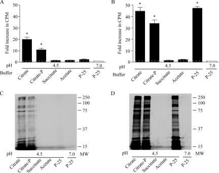
As a first step in establishing a nutrient medium that supports robust and prolonged C. burnetii metabolic activity, we evaluated the metabolic permissiveness of biological buffers with high buffering capacity in the pH range of 4 to 5. The capacity of C. burnetii to metabolize in different buffers was determined by assessing the ability of purified organisms to incorporate [35S]cysteine-methionine. As a late-stage anabolic process, bacterial protein synthesis provides a measure of metabolic activity that requires high activity levels of other central metabolic processes including energy generation, metabolite transport, and transcription/translation. Radiolabel incorporation by C. burnetii was measured following incubation in 59 mM citrate, 27 mM citrate-phosphate, 50 mM succinate, and 100 mM acetate buffers (pH 4.5) (10) supplemented with salts (42.8 mM NaCl, 3.7 mM KH2PO4, 2.7 mM KCl, 1.0 mM MgCl2, 0.1 mM CaCl2, 10 μM FeSO4). Incorporation of radiolabel in P-25 buffer (50 mM KH2PO4, 152.5 mM KCl, 15 mM NaCl, 100 mM glycine, pH 4.5), a phosphate buffer used in previous studies of C. burnetii axenic metabolic activity (13-15), was also measured, with incubations in P-25 (pH 7.0) conducted as a negative control (14). As measured by scintillation counting, citrate and citrate-phosphate buffers supported (20.0 ± 2.1)-fold and (11.0 ± 1.4)-fold increases, respectively, in C. burnetii [35S]Cys-Met incorporation, relative to the negative control, over a 3-h incubation period (Fig. 1A). Negligible levels of [35S]Cys-Met incorporation were observed following incubation in succinate, acetate, and P-25 (pH 4.5) buffers with relative increases of (1.6 ± 0.1)-fold, (1.8 ± 0.1)-fold, and (2.4 ± 0.4)-fold, respectively (Fig. 1A). These results suggest that C. burnetii utilizes citrate as a carbon source. When supplemented with 1.0 mM glutamate, a readily utilized carbon source of C. burnetii (15, 16), citrate, citrate-phosphate, and P-25 buffers all supported substantial radiolabel incorporation with increases of (44.8 ± 3.1)-fold, (33.9 ± 3.0)-fold, and (47.4 ± 1.3)-fold, respectively, in comparison to the negative control. Radiolabel incorporation in succinate and acetate buffers remained negligible (Fig. 1B). SDS-PAGE and autoradiography confirmed that radiolabel was incorporated into C. burnetii de novo synthesized protein (Fig. 1C and D). Citrate buffer was chosen over P-25 buffer for C. burnetii axenic medium development because of its stronger buffering capacity at pH 4.5 to 5.
FIG. 1.
Permissiveness of acidic buffers for C. burnetii metabolism. The metabolic activity of purified C. burnetii in acidic (pH 4.5) buffers was determined by measuring [35S]Cys-Met radiolabel incorporation by bacteria after a 3-h incubation in the indicated buffers without (A and C), or with (B and D) 1 mM glutamate. The level of de novo synthesized protein was determined by scintillation counting (A and B) and SDS-PAGE with autoradiography (C and D). The magnitude of radiolabel incorporation is expressed as the relative increase over incorporation observed in P-25 buffer (pH 7.0) (negative control). Asterisks indicate statistically significant differences (P < 0.05) between the indicated sample and P-25 buffer (pH 7.0). The broken line represents the level of radiolabel incorporation in P-25 buffer (pH 7.0) normalized to 1. Representative autoradiograms are shown. citrate-P, citrate-phosphate; MW, molecular weight (in thousands).
Development of CCM.
In silico metabolic pathway reconstruction of C. burnetii revealed auxotrophies for several amino acids as well as potential deficiencies in the biosynthesis of some vitamins and cofactors. Thus, C. burnetii likely has complex nutritional requirements. Consequently, we tested various complex nutrient mixtures in citrate buffer for the ability to sustain C. burnetii metabolic activity. Because the nutrient mixtures differed in their amino acid composition, [35S]Cys-Met incorporation by C. burnetii could not be measured directly during bacterial incubations in these media. Alternatively, bacteria were preincubated in the test medium and then transferred to a labeling buffer for a 3-h radiolabel pulse. The amount of radiolabel incorporation is thus a measure of C. burnetii “metabolic fitness” following incubation in media differing in nutrient composition. As a reference, comparisons were made to the translational activity in labeling buffer of immediately thawed freezer stocks of C. burnetii that had not undergone previous axenic metabolic activation. These “directly labeled” organisms provide a baseline readout of the metabolic capability of C. burnetii after purification from Vero cells. Initially, three complex nutrient sources (neopeptone, FBS, and RPMI cell culture medium) were individually titrated in citrate-salts buffer (CSB), i.e., citrate buffer with physiologic ion concentrations (Table 1), to establish levels that sustained optimal C. burnetii metabolic fitness following a 24-h incubation (data not shown). CCM was formulated based on these experiments (Table 1).
TABLE 1.
Components of CSB and CCM
| Component | Final concna |
|---|---|
| Ions | |
| Na+ | 190 |
| SO4− | 0.06 |
| Fe2+ | 0.01 |
| Cl− | 140 |
| Ca+ | 0.15 |
| Mg2+ | 1.0 |
| K+ | 4.3 |
| PO4− | 4.5 |
| NO3− | 0.1 |
| HCO3− | 3.0 |
| Nutrients | |
| Neopeptoneb | 0.1 mg/ml |
| FBSb | 1% |
| RPMI 1640b | 1/8× |
| Citrate | 29 |
Concentrations are in mM unless otherwise specified. Values for ions exclude contributions from neopeptone and FBS.
Present only in CCM.
Comparison of C. burnetii metabolic fitness following incubation in CCM and supplemented P-25 buffer.
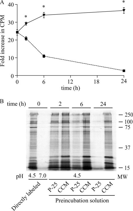
We next directly compared the ability of P-25 buffer supplemented with 5.0 mM glutamate and CCM to sustain C. burnetii metabolic fitness (Fig. 2). C. burnetii was preincubated in CCM or supplemented P-25 buffer for 2, 6, and 24 h and then transferred to labeling buffer for 3 h to allow incorporation of [35S]Cys-Met. Incorporation of radiolabel was quantified by scintillation counting (Fig. 2A). Compared to the translational activity of C. burnetii cells that were directly labeled in labeling buffer (pH 4.5), incorporation of radiolabel increased (4.8 ± 0.8)-fold and (12.2 ± 1.6)-fold following 2- and 24-h preincubations, respectively, in CCM. Radiolabel incorporation in CCM was inhibited when the transcriptional inhibitor rifampin was added to the labeling buffer, indicating that C. burnetii protein synthesis is dependent on de novo transcription (see Fig. S1 in the supplemental material). Radiolabel incorporation decreased (3.5 ± 1.4)-fold and (21.6 ± 0.2)-fold following 2- and 24-h preincubations, respectively, in P-25 buffer supplemented with 5 mM glutamate. As assessed by SDS-PAGE and autoradiography, the degree of de novo protein synthesis during the 3-h labeling period directly correlated with results obtained by scintillation counting (Fig. 2B). Thus, CCM is significantly better than P-25 buffer supplemented with glutamate, a preferred carbon source, at sustaining in vitro C. burnetii metabolic fitness.
FIG. 2.
C. burnetii metabolic fitness in supplemented P-25 buffer and CCM. The ability of P-25 buffer supplemented with 5 mM glutamate (broken line) and CCM (solid line) to sustain C. burnetii metabolic fitness was directly compared by examining de novo protein synthesis in labeling buffer following preincubation in these solutions for 2, 6, and 24 h. Following preincubations, C. burnetii was labeled with [35S]Cys-Met in labeling buffer (pH 4.5) for 3 h and then subjected to scintillation counting (A) and SDS-PAGE and autoradiography (B). In panel A, results are expressed as the relative increase in radiolabel incorporation compared to bacteria that were directly labeled in labeling buffer (pH 7.0) (negative control). The 0-h time point represents the relative increase in radiolabel incorporation by C. burnetii cells that were directly labeled in labeling buffer (pH 4.5) without preincubation. Asterisks indicate statistically significant differences (P < 0.05) between CCM and supplemented P-25 buffer. A representative autoradiogram is shown. MW, molecular weight (in thousands).
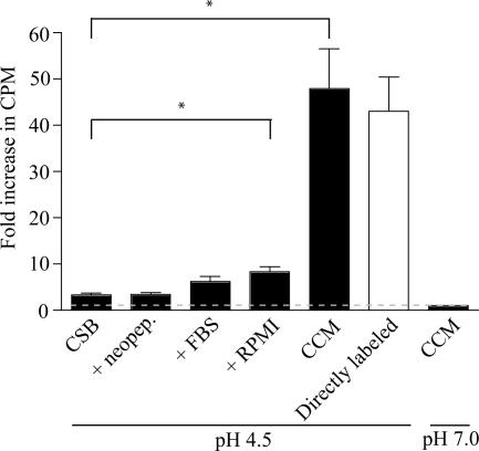
To compare the individual contributions of neopeptone, FBS, and RPMI medium in maintaining C. burnetii metabolic fitness, organisms were preincubated for 24 h in CSB (Table 1) or CSB supplemented with each nutrient source and then labeled for 3 h in labeling buffer supplemented with [35S]Cys-Met (Fig. 3). Radiolabel incorporation of C. burnetii preincubated in CSB supplemented with neopeptone or FBS was not significantly different from the incorporation of organisms preincubated in CSB alone. Addition of RPMI medium to CSB did result in a statistically significant (4.8 ± 1.2)-fold increase in radiolabel incorporation compared to CSB. Moreover, radiolabel incorporation of organisms preincubated in complete CCM containing neopeptone, FBS, and RPMI medium increased by (44.7 ± 8.4)-fold relative to incorporation of organisms preincubated in CSB. This level of protein synthesis was comparable to that of directly labeled organisms.
FIG. 3.
Effect of CCM nutrient supplements on C. burnetii metabolic fitness. The relative impact of CCM nutrient supplements on C. burnetii metabolic fitness was determined by incubating organisms in CSB supplemented with neopeptone (neopep), FBS, or RPMI medium. Bacteria were preincubated in the respective medium formulations for 24 h and then labeled with [35S]Cys-Met in labeling buffer (pH 4.5) for 3 h. De novo protein synthesis by C. burnetii was measured by quantification of radiolabel incorporation by scintillation counting. Results are expressed as the relative increase in radiolabel incorporation compared to the incorporation of bacteria labeled in labeling buffer (pH 7.0) (negative control). Asterisks indicate statistically significant (P < 0.05) differences compared to CSB. The broken line represents the level of radiolabel incorporation in CCM (pH 7.0) normalized to 1.
Effects of CCM ion concentrations, pH, and individual carbon source concentrations on C. burnetii protein synthesis.
We next evaluated the effects of CCM ion concentrations, individual carbon source levels, and pH on C. burnetii protein synthesis. With the exception of experiments involving different glutamate levels, CCM amino acid levels were identical in these experiments. Therefore, [35S]Cys-Met was added directly to CCM formulations, and C. burnetii incorporation of radiolabel was measured after a 3-h incubation.
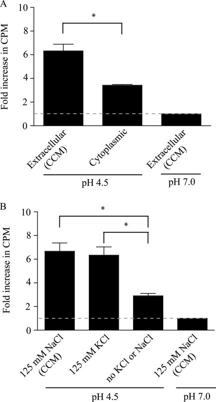
The Coxiella PV interacts with the host cell cytoplasm and the extracellular environment via endocytic fluid phase trafficking. To determine whether C. burnetii metabolic activity is sensitive to the orientation of Cl−, K+, and Na+ gradients associated with the host environment, [35S]Cys-Met incorporation was measured following incubation in CCM modified to mimic either host intracellular (cytoplasmic) ion levels (26 mM Cl−, 110 mM K+, and 17 mM Na+), or host extracellular (serum or interstitial fluid) ion levels (140 mM Cl−, 4.3 mM K+, and 190 mM Na+). Compared to the negative control (i.e., CCM at pH 7.0), radiolabel incorporation of C. burnetii incubated in CCM (pH 4.5) with extracellular ion concentrations increased by (6.3 ± 0.5)-fold. This was significantly greater than the (3.4 ± 0.1)-fold increase of C. burnetii incubated in CCM with intracellular ion concentrations (Fig. 4A). To determine whether C. burnetii metabolic activity in CCM was affected by the source of chloride ion, 125 mM NaCl was replaced with 125 mM KCl. Similar levels of radiolabel incorporation were observed following a 3-h incubation in medium containing either 125 mM NaCl or KCl, with relative increases in incorporation of (6.6 ± 0.7)-fold and (6.3 ± 0.7)-fold, respectively, compared to the negative control. Without the addition of 125 mM chloride, radiolabel incorporation was significantly reduced to only a (2.9 ± 0.2)-fold increase relative to the negative control (Fig. 4B). Thus, C. burnetii requires a high concentration of Cl− ions for optimal axenic metabolic activity that is independent of whether Na+ and K+ serves as the counter ion. It should be noted that we could not precisely determine the contributions of neopeptone and FBS to the ion levels of CCM. However, based on the product information provided by the manufacturer (see Materials and Methods), these nutrient sources should contribute less than 1 mM Cl−, K+, and Na+ to CCM.
FIG. 4.
Effect of CCM ion concentrations on C. burnetii metabolic activity. (A) The effect of medium ion levels on C. burnetii metabolic activity was assessed by measuring incorporation of [35S]Cys-Met into C. burnetii de novo synthesized protein following a 3-h incubation in CCM with Cl−, K+, and Na+ concentrations similar to the host cell cytoplasm (26 mM Cl−, 110 mM K+, and 17 mM Na+) or extracellular (140 mM Cl−, 4.3 mM K+, and 190 mM Na+) environment. Incorporation of radiolabel was quantified by scintillation counting and is expressed as the increase relative to the incorporation of C. burnetii incubated in CCM (pH 7.0) with extracellular levels of Cl−, K+, and Na+ (negative control). Asterisks indicate statistically significant differences (P < 0.05) between media with extracellular or intracellular ion levels. The broken line represents the level of radiolabel incorporation in medium with extracellular ion levels at pH 7.0 normalized to 1. (B) The importance of the source of chloride ion on C. burnetii metabolic activity was examined by measuring [35S]Cys-Met incorporation of organisms incubated for 3 h in CCM containing 125 mM KCl or NaCl or in CCM without added KCl or NaCl. Incorporation of radiolabel was quantified by scintillation counting and is expressed as the relative increase over the incorporation of C. burnetii incubated in CCM (pH 7.0) containing 125 mM NaCl (negative control). Asterisks indicate statistically significant differences (P < 0.05) compared to CCM without KCl or NaCl. The broken line represents the level of radiolabel incorporation in medium containing 125 mM NaCl at pH 7.0 normalized to 1.
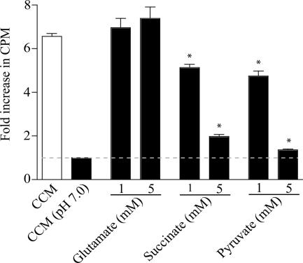
The nature and concentration of specific carbon sources can significantly impact bacterial metabolism. Therefore, the effect of supplementing CCM with glutamate, succinate, or pyruvate on C. burnetii protein synthesis was assessed. These carbon sources have been previously demonstrated to be readily catabolized and incorporated by C. burnetii (15, 16). Relative to nonsupplemented CCM, statistically significant changes in C. burnetii [35S]Cys-Met incorporation were not observed following a 3-h incubation in CCM supplemented with 1 or 5 mM glutamate (Fig. 5). However, significant concentration-dependent declines in radiolabel incorporation were observed following incubation of C. burnetii in CCM supplemented with succinate and pyruvate (Fig. 5). Compared to nonsupplemented CCM, the addition of 1 and 5 mM succinate resulted in (1.4 ± 0.1)-fold and (4.6 ± 0.1)-fold reductions in [35S]Cys-Met incorporation, respectively, while the addition of 1 and 5 mM pyruvate resulted in (1.8 ± 0.2)-fold and (5.2 ± 0.1)-fold reductions, respectively.
FIG. 5.
Effects of individual carbon source levels on C. burnetii metabolic activity. The effect of supplementing CCM with efficiently oxidized carbon sources (16) on C. burnetii de novo protein synthesis was measured. Organisms were incubated for 3 h in CCM containing [35S]Cys-Met and supplemented with 1 or 5 mM glutamate, succinate, or pyruvate. Incorporation of radiolabel was quantified by scintillation counting and is expressed as the increase relative to the incorporation of C. burnetii incubated in CCM (pH 7.0) (negative control). Asterisks indicate statistically significant (P < 0.05) differences between radiolabel incorporation by C. burnetii incubated in CCM and in CCM supplemented with a 1 or 5 mM concentration of the carbon source. The broken line represents the level of radiolabel incorporation in CCM (pH 7.0) normalized to 1.
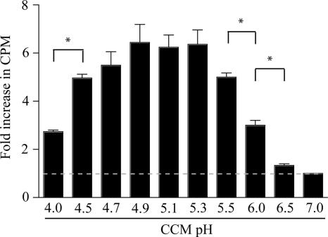
Based on the use of pH-sensitive fluorescent probes, three independent studies indicate that the pH of the C. burnetii PV is mildly acidic (pH 4.5 to 5.3) (1, 11, 24). However, maximal rates of C. burnetii metabolite transport and incorporation into cellular products in vitro exhibit a wider range of pH optima (14, 15, 20). For example, in P-25 buffer maximal transport and catabolism of glutamate occur at a pH of 3.5 (14, 15), with declines in these activities within 1 pH unit of this optimum. To determine the pH of CCM that supports optimal C. burnetii metabolic activity, incorporation of [35S]Cys-Met was measured after a 3-h incubation in this medium adjusted over a pH range of 4.0 to 7.0 (Fig. 6). Radiolabel incorporation of C. burnetii incubated in CCM (pH 4.0) increased (2.7 ± 0.1)-fold relative to incorporation of organisms incubated in CCM at pH 7.0 (negative control). Elevation of CCM pH to 4.5 and 4.9 resulted in increases in radiolabel incorporation of (4.9 ± 0.1)-fold and (6.4 ± 0.7)-fold, respectively, relative to the negative control. Approximately equal levels of radiolabel were incorporated during incubation in CCM at pH 4.9, 5.1, and 5.3, with decreasing incorporation at higher pH values. The high metabolic activity of C. burnetii between pH 4.5 and 5.5 is consistent with the organism's use of the diverse substrates present in CCM that differ moderately in their pH optima for transport and incorporation into cellular material.
FIG. 6.
Effect of CCM pH on C. burnetii metabolic activity. The pH for optimal C. burnetii metabolism in CCM was determined by incubating bacteria for 3 h in CCM adjusted to different pH values in the presence of [35S]Cys-Met. Radiolabel incorporation was measured by scintillation counting. C. burnetii incorporation of [35S]Cys-Met is expressed as the increase relative to the incorporation of organisms incubated in CCM (pH 7.0) (negative control). Asterisks indicate statistically significant (P < 0.05) differences between the indicated samples. The broken line represents the level of radiolabel incorporation in CCM (pH 7.0) normalized to 1.
ATP pool synthesis and stability during incubation in CCM.
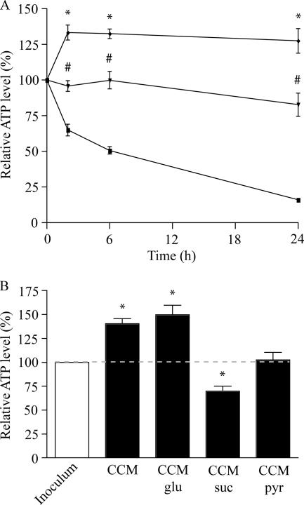
As a second method of evaluating the metabolic state of C. burnetii during prolonged incubation in CCM, the bacterial ATP pool was measured following 2-, 6-, and 24-h incubations in CCM, CSB, or P-25 buffer (Fig. 7A). Following incubation in CCM (pH 4.5) for 2 h, the C. burnetii ATP pool increased 33.5% ± 5.2% over that of the inoculum and remained elevated throughout the 24-h incubation period. Conversely, incubation in CSB for 2 and 24 h resulted in ATP pool decreases of 4.2% ± 3.8% and 17.1% ± 8.1%, respectively. More dramatic declines of 34.9% ± 4.0% and 83.8% ± 1.2% occurred following 2 and 24 h of incubation, respectively, in P-25 buffer. Collectively, these data indicate that incubation of C. burnetii in CCM stimulates ATP synthesis and that the elevated ATP pool remains stable for at least 24 h.
FIG. 7.
Analysis of C. burnetii ATP pool during incubation in CCM. (A) C. burnetii ATP pools were compared following incubation in CCM (⧫), CSB (▾), or P-25 buffer (▪) for 2, 6, and 24 h. Data are expressed as the percentage of the total ATP pool of the inoculum (time zero). *, statistically significant differences (P < 0.05) between CCM and P-25 buffer; #, statistically significant differences (P < 0.05) between P-25 buffer and CSB. (B) The stability of the C. burnetii ATP pool during incubation in CCM supplemented with an efficiently oxidized carbon source was assessed. ATP was measured following a 3-h incubation in CCM or in CCM supplemented with 5 mM glutamate (glu), succinate (suc), or pyruvate (pyr). Asterisks indicate statistically significant differences between the inoculum and indicated sample. The broken line represents the ATP level of the inoculum normalized to 100%.
To determine whether levels of de novo protein synthesis by C. burnetii incubated in CCM supplemented with glutamate, succinate, or pyruvate (Fig. 5) correlated with ATP levels, bacterial ATP pools were measured following a 3-h incubation in CCM with or without 5 mM supplements of these nutrients (Fig. 7B). Compared to the inoculum, the ATP pool of C. burnetii incubated in CCM and in CCM supplemented with 5 mM glutamate increased 40.4% ± 5.3% and 49.7% ± 9.9%, respectively. Conversely, the ATP pool decreased 30.2% ± 5.4% following incubation in CCM supplemented with 5 mM succinate, and a minimal 2.6% ± 6.7% increase was observed after incubation in CCM with 5 mM pyruvate. Together with data depicted in Fig. 5, these results show that improved C. burnetii protein synthesis in CCM correlates with significant increases in the ATP pool. Moreover, C. burnetii axenic metabolism appears sensitive to the concentration of certain carbon sources even during metabolism of a broad spectrum of nutrients.
Morphogenesis of C. burnetii SCV to LCV during incubation in CCM.
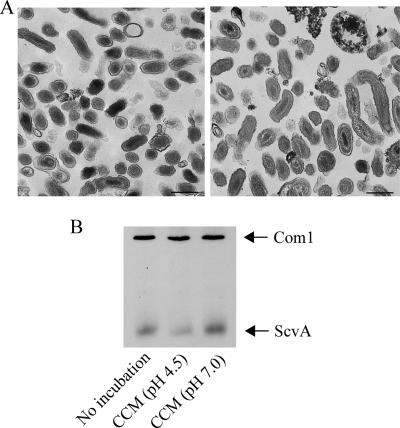
The C. burnetii LCV form is considered more replicatively and metabolically active than the SCV form (6). Thus, maintenance of the LCV is likely important for C. burnetii to optimally metabolize in an axenic medium. To assess the capacity of CCM to promote morphogenesis of the SCV to LCV and to maintain the LCV form, purified C. burnetii SCVs (6) were incubated in CCM for 24 h, and C. burnetii ultrastructure was assessed by transmission electron microscopy. SCVs prior to incubation in CCM had ultrastructural characteristics consistent with this cell form including size (≤0.5 μm) and electron-dense condensed chromatin (18) (Fig. 8A, left panel). Following incubation of SCVs in CCM for 24 h, most C. burnetii cells appeared ultrastructurally similar to the LCVs with an increase in cell size (>0.5 μm) and dispersed chromatin (Fig. 8A, right panel). To further examine SCV-to-LCV transitions in CCM, immunoblotting was performed to determine the amount of ScvA in C. burnetii before and after incubation in CCM (Fig. 8B). ScvA is an SCV-specific protein (18), and its degradation is considered a molecular marker of SCV-to-LCV morphogenesis (6, 18). By scanning densitometry, the level of ScvA in SCVs incubated in CCM (pH 4.5) for 24 h declined approximately 50% relative to SCVs that did not undergo axenic incubation (Fig. 8B), a result also observed by quantitative immunogold transmission electron microscopy (data not shown). This level of reduction in SCV ScvA content is similar to previously observed reductions following SCV infection of Vero cells for 8 to 12 h (6, 18). The level of Com1, an outer membrane protein that is equally present in the SCV and the LCV (17, 21), did not change. Neither ScvA nor Com1 levels were significantly altered by incubation for 24 h in CCM (pH 7.0). Taken together, these data suggest that C. burnetii SCVs undergo morphological differentiation to the LCVs in CCM in a process that is reminiscent of differentiation following in vivo infection and that the LCV form is maintained in this medium.
FIG. 8.
Transition of C. burnetii SCV to LCV during incubation in CCM. Purified C. burnetii SCVs were analyzed by transmission electron microscopy before and after a 24-h incubation in CCM to assess potential morphological transitions. (A) SCVs prior to axenic incubation in CCM showed ultrastructural characteristics of this cell form (left panel). Following SCV incubation in CCM, organisms displayed an ultrastructure more characteristic of the LCV form (e.g., size of >0.5 μm and relaxed chromatin; right panel). Bar, 0.5 μm. (B) Scanning densitometry of an immunoblot probed for ScvA (3.5 kDa), a protein specific to SCVs, showed that the ScvA level decreased by approximately 50% following incubation of SCVs in CCM (pH 4.5) for 24 h. SCVs incubated in CCM (pH 7.0) for 24 h exhibited no decrease in ScvA. The levels of Com1 (27 kDa), a protein equally expressed in SCVs and LCVs, did not change. Data from representative experiments are shown.
DISCUSSION
Here, we describe a nutrient medium termed CCM that supports robust and sustained (at least 24 h) metabolic fitness of the obligate intracellular bacterial pathogen C. burnetii. Compared to previously described buffers (16, 43), incubation in CCM substantially prolongs C. burnetii's ability to synthesize protein and to increase and subsequently stabilize its ATP pool. CCM was formulated based on nutritional and biophysical variables with documented effects on bacterial physiologic processes including ion concentration, nutrient/carbon source availability, and pH. Of critical importance was identification of a permissive biological buffer. Metabolic testing shows that C. burnetii metabolism is optimal in a citrate-based buffer. The strong buffering capacity of citrate buffer in C. burnetii's metabolically permissive pH range (pH 4.5 to 5.5) increases the utility of this buffer compared to previously utilized P-25 buffer (14).
In silico metabolic pathway reconstruction of C. burnetii using the Nine Mile isolate (RSA493) genomic sequence reveals that the pathogen is metabolically complex relative to other obligate intracellular bacteria (32, 35). The majority of central metabolic pathways including the tricarboxylic acid (TCA) cycle and glycolysis appear functional (14). One potentially defective pathway is the oxidative branch of the pentose-phosphate pathway, with genes encoding glucose-6-phosphate dehydrogenase and 6-phospho-gluconate dehydrogenase missing in the C. burnetii genome. This pathway generates reducing equivalents in the form of NADPH, and its absence in C. burnetii suggests that the pathogen does not rely on this pathway to maintain redox balance. C. burnetii also lacks enzymes of the glyoxylate shunt, which would allow it to metabolize fatty acids as sole carbon sources, and is furthermore a predicted auxotroph for several amino acids (32). C. burnetii likely acquires amino acids via the activity of di- and tripeptide transporters (CBU0504 and CBU0539) and an oligopeptide permease type transporter (CBU1857, CBU1858, CBU1859, CBU1860, and CBU1861). The hydrolytically active C. burnetii PV interacts with the autophagic and lysosomal pathways (12, 19); therefore, the bacterium is likely exposed to a steady supply of peptides.
To overcome specific auxotrophies while at the same time exploiting the general metabolic competence of C. burnetii, we tested the capacity of complex nutrient mixtures to support C. burnetii axenic metabolic activity. Because C. burnetii grows efficiently inside Vero cells cultured in RPMI medium supplemented with FBS, we tested the effect of these two nutrient mixtures as both should be transported by fluid phase endocytosis to the C. burnetii PV lumen (19). Neopeptone, a tissue digest containing nucleotides, vitamins, amino acids, peptides, and minerals, was included as a high-quality source of a wide spectrum of nutrients. When individually present in CSB, these supplements only marginally improved maintenance of C. burnetii metabolic fitness compared to CSB. However, a marked improvement in maintenance of metabolic fitness is observed when the nutrient sources are combined in CCM.
C. burnetii preferentially metabolizes TCA cycle intermediates (e.g., succinate) or substrates that funnel into this pathway (e.g., glutamate and pyruvate), suggesting a critical role for the TCA cycle in C. burnetii energy generation (16). Surprisingly, supplementation of CCM with 1 or 5 mM succinate or pyruvate, but not glutamate, actually inhibited C. burnetii de novo protein synthesis in a dose-dependent manner relative to nonsupplemented CCM and correlated with declines in the ATP pool. These results are not strictly associated with a citrate-based buffer as similar effects were observed following incubation in P-25 buffer containing these supplements (data not shown). Thus, both C. burnetii protein and ATP synthesis can be negatively affected by high concentrations of certain carbon sources in a nutritionally complex medium. In Legionella pneumophila, an organism related to C. burnetii, glutamate is considered a preferred carbon source based, in part, on its incorporation into a wide range of macromolecules (e.g., proteins, lipids, and polysaccharides) (37). A similar biochemical routing for glutamate in C. burnetii may explain why this organism more efficiently synthesizes protein in CCM supplemented with glutamate versus CCM supplemented with succinate or pyruvate. An alternative mechanism for the adverse effects of succinate and pyruvate could be incomplete oxidation of the substrate, a phenomenon referred to as overflow metabolism (36). Incomplete carbon source oxidation can lower the generation of reducing equivalents with a concomitant negative effect on protein synthesis.
C. burnetii may be adapted to a vacuolar compartment that contains a low level of nutrients supplied at a steady flux by the metabolic activity of the host cell, a hypothesis supported by the organism's slow growth rate in vivo (approximately a 12-h exponential phase division time) (6). Depletion of the intravacuolar nutrient pool is a mechanism used by macrophages to restrict replication and survival of intracellular pathogens (2). The vacuole occupied by Mycobacterium tuberculosis contains low levels of carbohydrates (31). Moreover, transcriptional analysis of Salmonella enterica serovar Typhimurium during infection of cultured macrophages indicates a reduced flux of carbon through the glycolytic and pentose phosphate pathways during intracellular growth relative to organisms grown in broth (8). Slow growth rate has been identified as a parameter that promotes persistent infection by M. tuberculosis (42) and has been speculated to aid in establishment of chronic infection by C. burnetii (3). As proposed for the obligate intracellular pathogen Rickettsia prowazekii (41), slow growth by C. burnetii could be regulated by inherently slow translational machinery. Slow protein synthesis may be advantageous to C. burnetii in coordinating its growth rate with the ability of the host to provide necessary nutrients. The organism has one copy each of the 5S, 16S, and 23S rRNA-encoding genes, with the 23S RNA containing an intervening sequence and two introns. It is hypothesized that the 23S RNA introns may further contribute to slow C. burnetii growth (30).
Different ion concentrations between the bacterial cytoplasm and extrabacterial environment establish an electrical membrane potential and concentration gradients that are both used to transport nutrients. Therefore, we tested the impact of CCM ion concentrations on C. burnetii metabolic activity. We found an approximately twofold increase in C. burnetii de novo protein synthesis in CCM containing ion concentrations resembling the host extracellular environment (e.g., serum and interstitial fluid) compared to protein synthesis in CCM adjusted to intracellular (i.e., cytoplasmic) ion concentrations. C. burnetii is particularly sensitive to the concentration of chloride, and maximal metabolic activity is achieved under conditions of elevated chloride levels. As previously suggested (15), chloride ion may help maintain cytoplasmic pH homeostasis in C. burnetii. This hypothesis is consistent with a role for chloride in E. coli acid resistance (9). Elevated levels of chloride in the PV lumen may also be critical in anion-exchange systems of C. burnetii. We were unable to determine the concentration of chloride and other ions in the C. burnetii PV due to the pH sensitivity of ion-sensitive ratiometric fluorescent probes. However, our in vitro observations of C. burnetii ion sensitivities have potential in vivo correlates. Macrophages produce defensin antimicrobial peptides as a mechanism to kill ingested bacteria (7), and the activity of antimicrobial peptides can be compromised by high salt levels (28). Thus, a PV lumen with high sodium and chloride levels may confer protection against these molecules. Moreover, the activity of a C. burnetii sodium-driven antiporter multidrug efflux pump (CBU0812) would be aided by a high luminal sodium concentration.
Metabolic studies of extracellular C. burnetii have historically been conducted at pH 4.5 (4, 14, 43). However, the optimal pH for transport and incorporation of individual substrates varies (14, 15, 20). To determine the optimal pH of C. burnetii metabolism in CCM, we assessed metabolic activity in this medium over a pH range of 4 to 7. Overall, optimal metabolic activity was observed between pH 4.5 and 5.5, with statistically significant declines in activity observed above or below this range. The similar metabolic activities observed between pH 4.5 and 5.5 may reflect use of diverse nutrients in CCM that have moderately different pH optima for transport. Given the physiological pressure exerted on acidophilic microbes to maintain a neutral cytoplasmic pH, metabolism of C. burnetii in CCM adjusted to pH 5.5 could be energetically more favorable than metabolism in CCM at a lower pH and allow a further extension of C. burnetii metabolic activity outside the host cell.
To summarize, the prolonged and robust metabolic activity of C. burnetii achieved in CCM provides an amenable axenic system to study transcriptional and translational responses of this bacterium to diverse stimuli including oxidative stress and pH. Furthermore, because CCM supports extended C. burnetii protein synthesis, incubation in this medium may help identify secreted proteins that act as virulence factors. The morphological differentiation of SCV into LCV during incubation in CCM also provides a tool to study the developmental biology of C. burnetii in the absence of host cells. Finally, modification of CCM could result in a bacteriologic medium that supports extracellular replication of C. burnetii.
Supplementary Material
Acknowledgments
We thank Ted Hackstadt, Harlan Caldwell, Shelly Robertson, Frank Gherardini, and Grant McClarty for critical review of the manuscript; Gary Hettrick for graphic illustrations; and James Samuel (Texas A & M University) for anti-Com1 monoclonal antibody.
This work was supported by the Intramural Research Program of the National Institutes of Health, National Institute of Allergy and Infectious Diseases.
Footnotes
Published ahead of print on 29 February 2008.
Supplemental material for this article may be found at http://jb.asm.org/.
REFERENCES
- 1.Akporiaye, E. T., J. D. Rowatt, A. A. Aragon, and O. G. Baca. 1983. Lysosomal response of a murine macrophage-like cell line persistently infected with Coxiella burnetii. Infect. Immun. 401155-1162. [DOI] [PMC free article] [PubMed] [Google Scholar]
- 2.Appelberg, R. 2006. Macrophage nutriprive antimicrobial mechanisms. J. Leukoc. Biol. 791117-1128. [DOI] [PubMed] [Google Scholar]
- 3.Beare, P. A., J. E. Samuel, D. Howe, K. Virtaneva, S. F. Porcella, and R. A. Heinzen. 2006. Genetic diversity of the Q. fever agent, Coxiella burnetii, assessed by microarray-based whole-genome comparisons. J. Bacteriol. 1882309-2324. [DOI] [PMC free article] [PubMed] [Google Scholar]
- 4.Chen, S. Y., M. Vodkin, H. A. Thompson, and J. C. Williams. 1990. Isolated Coxiella burnetii synthesizes DNA during acid activation in the absence of host cells. J. Gen. Microbiol. 13689-96. [DOI] [PubMed] [Google Scholar]
- 5.Cockrell, D. C., P. A. Beare, E. R. Fischer, D. Howe, and R. A. Heinzen. 2008. A method for purifying obligate intracellular Coxiella burnetii that employs digitonin lysis of host cells. J. Microbiol. Methods. 72321-325. [DOI] [PMC free article] [PubMed] [Google Scholar]
- 6.Coleman, S. A., E. R. Fischer, D. Howe, D. J. Mead, and R. A. Heinzen. 2004. Temporal analysis of Coxiella burnetii morphological differentiation. J. Bacteriol. 1867344-7352. [DOI] [PMC free article] [PubMed] [Google Scholar]
- 7.Duits, L. A., B. Ravensbergen, M. Rademaker, P. S. Hiemstra, and P. H. Nibbering. 2002. Expression of beta-defensin 1 and 2 mRNA by human monocytes, macrophages and dendritic cells. Immunology 106517-525. [DOI] [PMC free article] [PubMed] [Google Scholar]
- 8.Eriksson, S., S. Lucchini, A. Thompson, M. Rhen, and J. C. Hinton. 2003. Unravelling the biology of macrophage infection by gene expression profiling of intracellular Salmonella enterica. Mol. Microbiol. 47103-118. [DOI] [PubMed] [Google Scholar]
- 9.Foster, J. W. 2004. Escherichia coli acid resistance: tales of an amateur acidophile. Nat. Rev. Microbiol. 2898-907. [DOI] [PubMed] [Google Scholar]
- 10.Gomori, G. 1955. Preparation of buffers for use in enzyme studies, p. 138-146. In S. P. Colowick and N. O. Kaplan (ed.), Methods in enzymology, vol. 1. Academic Press Inc., New York, NY. [Google Scholar]
- 11.Grieshaber, S., J. A. Swanson, and T. Hackstadt. 2002. Determination of the physical environment within the Chlamydia trachomatis inclusion using ion-selective ratiometric probes. Cell. Microbiol. 4273-283. [DOI] [PubMed] [Google Scholar]
- 12.Gutierrez, M. G., C. L. Vazquez, D. B. Munafo, F. C. Zoppino, W. Beron, M. Rabinovitch, and M. I. Colombo. 2005. Autophagy induction favours the generation and maturation of the Coxiella-replicative vacuoles. Cell. Microbiol. 7981-993. [DOI] [PubMed] [Google Scholar]
- 13.Hackstadt, T. 1983. Estimation of the cytoplasmic pH of Coxiella burnetii and effect of substrate oxidation on proton motive force. J. Bacteriol. 154591-597. [DOI] [PMC free article] [PubMed] [Google Scholar]
- 14.Hackstadt, T., and J. C. Williams. 1981. Biochemical stratagem for obligate parasitism of eukaryotic cells by Coxiella burnetii. Proc. Natl. Acad. Sci. USA 783240-3244. [DOI] [PMC free article] [PubMed] [Google Scholar]
- 15.Hackstadt, T., and J. C. Williams. 1983. pH dependence of the Coxiella burnetii glutamate transport system. J. Bacteriol. 154598-603. [DOI] [PMC free article] [PubMed] [Google Scholar]
- 16.Hackstadt, T., and J. C. Williams. 1981. Stability of the adenosine 5′-triphosphate pool in Coxiella burnetii: influence of pH and substrate. J. Bacteriol. 148419-425. [DOI] [PMC free article] [PubMed] [Google Scholar]
- 17.Heinzen, R. A., L. R. Hendrix, S. F. Hayes, D. D. Rockey, and T. Hackstadt. 1995. Developmentally regulated protein synthesis by the intraphagolysosomal parasite, Coxiella burnetii, abstr. D-130, p.271. Abstr. 95th Gen. Meet. Am. Soc. Microbiol. American Society for Microbiology, Washington, DC.
- 18.Heinzen, R. A., D. Howe, L. P. Mallavia, D. D. Rockey, and T. Hackstadt. 1996. Developmentally regulated synthesis of an unusually small, basic peptide by Coxiella burnetii. Mol. Microbiol. 229-19. [DOI] [PubMed] [Google Scholar]
- 19.Heinzen, R. A., M. A. Scidmore, D. D. Rockey, and T. Hackstadt. 1996. Differential interaction with endocytic and exocytic pathways distinguish parasitophorous vacuoles of Coxiella burnetii and Chlamydia trachomatis. Infect. Immun. 64796-809. [DOI] [PMC free article] [PubMed] [Google Scholar]
- 20.Hendrix, L., and L. P. Mallavia. 1984. Active transport of proline by Coxiella burnetii. J. Gen. Microbiol. 1302857-2863. [DOI] [PubMed] [Google Scholar]
- 21.Hendrix, L. R., L. P. Mallavia, and J. E. Samuel. 1993. Cloning and sequencing of Coxiella burnetii outer membrane protein gene com1. Infect. Immun. 61470-477. [DOI] [PMC free article] [PubMed] [Google Scholar]
- 22.Howe, D., and L. P. Mallavia. 1999. Coxiella burnetii infection increases transferrin receptors on J774A.1 cells. Infect. Immun. 673236-3241. [DOI] [PMC free article] [PubMed] [Google Scholar]
- 23.Howe, D., J. Melnicakova, I. Barak, and R. A. Heinzen. 2003. Maturation of the Coxiella burnetii parasitophorous vacuole requires bacterial protein synthesis but not replication. Cell. Microbiol. 5469-480. [DOI] [PubMed] [Google Scholar]
- 24.Maurin, M., A. M. Benoliel, P. Bongrand, and D. Raoult. 1992. Phagolysosomes of Coxiella burnetii-infected cell lines maintain an acidic pH during persistent infection. Infect. Immun. 605013-5016. [DOI] [PMC free article] [PubMed] [Google Scholar]
- 25.Maurin, M., and D. Raoult. 1999. Q. fever. Clin. Microbiol. Rev. 12518-553. [DOI] [PMC free article] [PubMed] [Google Scholar]
- 26.McCaul, T. F., T. Hackstadt, and J. C. Williams. 1981. Ultrastructural and biological aspects of Coxiella burnetii under physical disruptions, p. 267-280. In R. L. A. Willy Burgdorfer (ed.), Rickettsiae and rickettsial diseases. Academic Press, New York, NY.
- 27.McQuiston, J. H., and J. E. Childs. 2002. Q. fever in humans and animals in the United States. Vector Borne Zoonotic Dis. 2179-191. [DOI] [PubMed] [Google Scholar]
- 28.Nuding, S., K. Fellermann, J. Wehkamp, H. A. Mueller, and E. F. Stange. 2006. A flow cytometric assay to monitor antimicrobial activity of defensins and cationic tissue extracts. J. Microbiol. Methods 65335-345. [DOI] [PubMed] [Google Scholar]
- 29.Ormsbee, R. A., and M. G. Peacock. 1964. Metabolic activity in Coxiella burnetii. J. Bacteriol. 881205-1210. [DOI] [PMC free article] [PubMed] [Google Scholar]
- 30.Raghavan, R., S. R. Miller, L. D. Hicks, and M. F. Minnick. 2007. The unusual 23S rRNA gene of Coxiella burnetii: two self-splicing group I introns flank a 34-base-pair exon, and one element lacks the canonical omegaG. J. Bacteriol. 1896572-6579. [DOI] [PMC free article] [PubMed] [Google Scholar]
- 31.Schnappinger, D., S. Ehrt, M. I. Voskuil, Y. Liu, J. A. Mangan, I. M. Monahan, G. Dolganov, B. Efron, P. D. Butcher, C. Nathan, and G. K. Schoolnik. 2003. Transcriptional adaptation of Mycobacterium tuberculosis within macrophages: insights into the phagosomal environment. J. Exp. Med. 198693-704. [DOI] [PMC free article] [PubMed] [Google Scholar]
- 32.Seshadri, R., I. T. Paulsen, J. A. Eisen, T. D. Read, K. E. Nelson, W. C. Nelson, N. L. Ward, H. Tettelin, T. M. Davidsen, M. J. Beanan, R. T. Deboy, S. C. Daugherty, L. M. Brinkac, R. Madupu, R. J. Dodson, H. M. Khouri, K. H. Lee, H. A. Carty, D. Scanlan, R. A. Heinzen, H. A. Thompson, J. E. Samuel, C. M. Fraser, and J. F. Heidelberg. 2003. Complete genome sequence of the Q.-fever pathogen Coxiella burnetii. Proc. Natl. Acad. Sci. USA 1005455-5460. [DOI] [PMC free article] [PubMed] [Google Scholar]
- 33.Seshadri, R., and J. E. Samuel. 2001. Characterization of a stress-induced alternate sigma factor, RpoS, of Coxiella burnetii and its expression during the development cycle. Infect. Immun. 694874-4883. [DOI] [PMC free article] [PubMed] [Google Scholar]
- 34.Shannon, J. G., and R. A. Heinzen. 2007. Infection of human monocyte-derived macrophages with Coxiella burnetii. Methods Mol. Biol. 431189-200. [DOI] [PubMed] [Google Scholar]
- 35.Stephens, R. S., S. Kalman, C. Lammel, J. Fan, R. Marathe, L. Aravind, W. Mitchell, L. Olinger, R. L. Tatusov, Q. Zhao, E. V. Koonin, and R. W. Davis. 1998. Genome sequence of an obligate intracellular pathogen of humans: Chlamydia trachomatis. Science 282754-759. [DOI] [PubMed] [Google Scholar]
- 36.Teixeira de Mattos, M. J., and O. M. Neijssel. 1997. Bioenergetic consequences of microbial adaptation to low-nutrient environments. J. Biotechnol. 59117-126. [DOI] [PubMed] [Google Scholar]
- 37.Tesh, M. J., S. A. Morse, and R. D. Miller. 1983. Intermediary metabolism in Legionella pneumophila: utilization of amino acids and other compounds as energy sources. J. Bacteriol. 1541104-1109. [DOI] [PMC free article] [PubMed] [Google Scholar]
- 38.Voth, D. E., and R. A. Heinzen. 2007. Lounging in a lysosome: the intracellular lifestyle of Coxiella burnetii. Cell. Microbiol. 9829-840. [DOI] [PubMed] [Google Scholar]
- 39.Weiss, E. 1965. Adenosine triphosphate and other requirements for the utilization of glucose by agents of the psittacosis-trachoma group. J. Bacteriol. 90243-253. [DOI] [PMC free article] [PubMed] [Google Scholar]
- 40.Weiss, E. 1973. Growth and physiology of rickettsiae. Bacteriol. Rev. 37259-283. [DOI] [PMC free article] [PubMed] [Google Scholar]
- 41.Winkler, H. H. 1995. Rickettsia prowazekii, ribosomes and slow growth. Trends Microbiol. 3196-198. [DOI] [PubMed] [Google Scholar]
- 42.Zahrt, T. C., and V. Deretic. 2001. Mycobacterium tuberculosis signal transduction system required for persistent infections. Proc. Natl. Acad. Sci. USA 9812706-12711. [DOI] [PMC free article] [PubMed] [Google Scholar]
- 43.Zuerner, R. L., and H. A. Thompson. 1983. Protein synthesis by intact Coxiella burnetii cells. J. Bacteriol. 156186-191. [DOI] [PMC free article] [PubMed] [Google Scholar]
Associated Data
This section collects any data citations, data availability statements, or supplementary materials included in this article.


