

Abstract
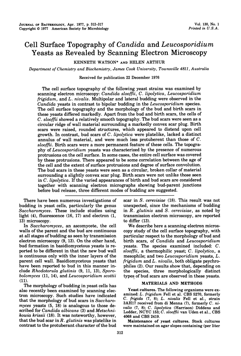
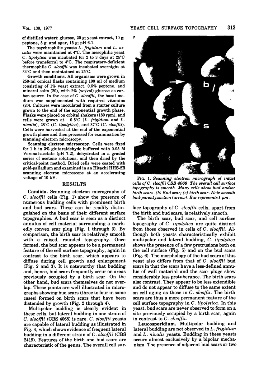
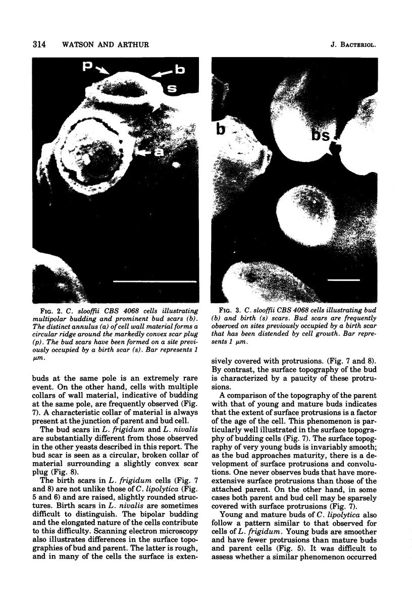
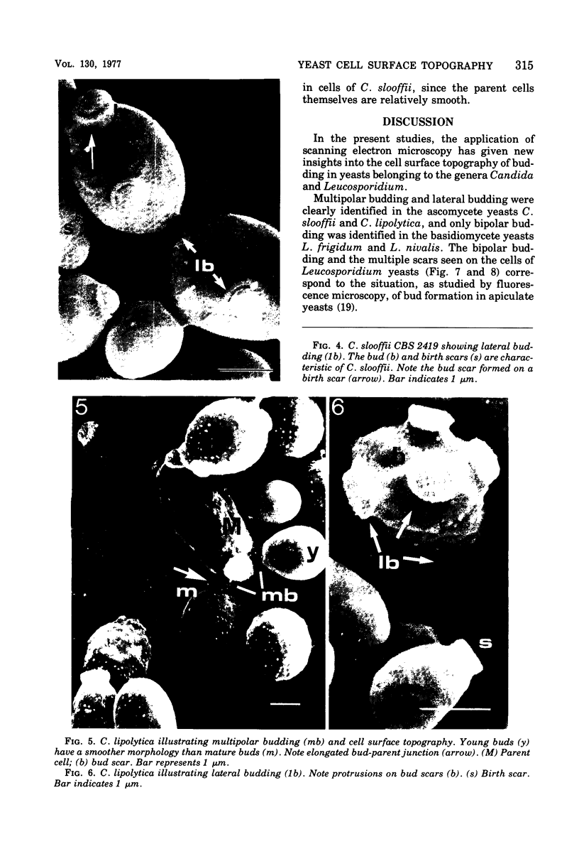
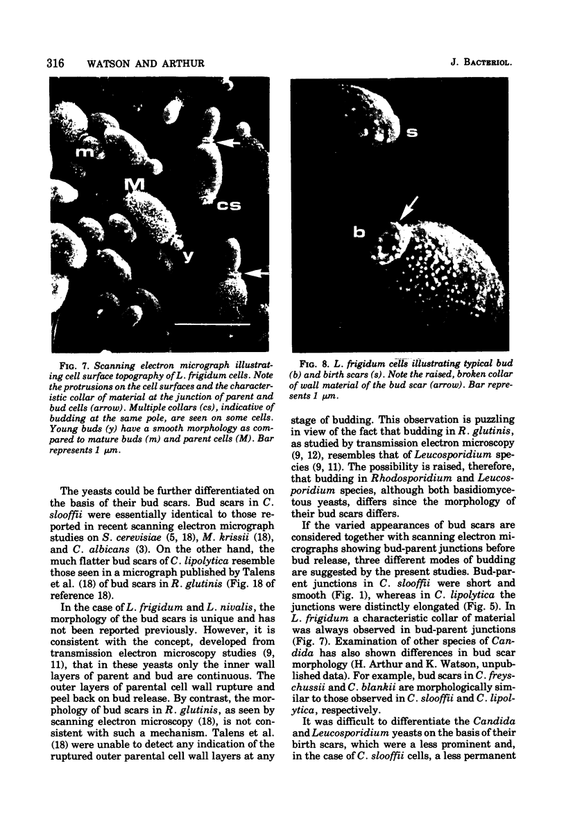
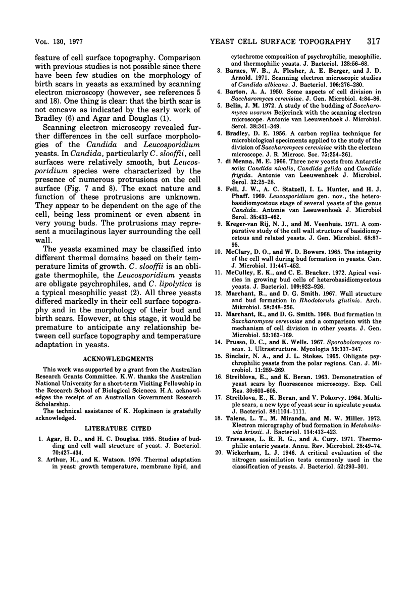
The cell surface topography of the following yeast strains was examined by scanning electron microscopy: Candida slooffii, C. lipolytica, Leucosporidium frigidum, and L. nivalis. Multipolar and lateral budding were observed in the Candida yeasts in contrast to bipolar budding in the Leucosporidium species. The cell surface topography and the morphology of the bud and birth scars in these yeasts differed markedly. Apart from the bud and birth scars, the cells of C. slooffii showed a relatively smooth topography. The bud scars were seen as a circular ridge of wall material surrounding a markedly convex scar plug. Birth scars were raised, rounded structures, which appeared to distend upon cell growth. In contrast, bud scars of C. lipolytica were platelike, lacked a distinct annulus of wall material, and were much less protuberent than those of C. slooffii. Birth scars were a more permanent feature of these cells. The topography of Leucosporidium yeasts was characterized by the presence of numerous protrusions on the cell surface. In some cases, the entire cell surface was covered by these protrusions. There appeared to be some correlations between the age of the cell and the extent of surface protrusions and degree of surface convolution...
Full text
PDF
Images in this article
Selected References
These references are in PubMed. This may not be the complete list of references from this article.
- AGAR H. D., DOUGLAS H. C. Studies of budding and cell wall structure of yeast; electron microscopy of the sections. J Bacteriol. 1955 Oct;70(4):427–434. doi: 10.1128/jb.70.4.427-434.1955. [DOI] [PMC free article] [PubMed] [Google Scholar]
- Arthur H., Watson K. Thermal adaptation in yeast: growth temperatures, membrane lipid, and cytochrome composition of psychrophilic, mesophilic, and thermophilic yeasts. J Bacteriol. 1976 Oct;128(1):56–68. doi: 10.1128/jb.128.1.56-68.1976. [DOI] [PMC free article] [PubMed] [Google Scholar]
- BARTON A. A. Some aspects of cell division in saccharomyces cerevisiae. J Gen Microbiol. 1950 Jan;4(1):84–86. doi: 10.1099/00221287-4-1-84. [DOI] [PubMed] [Google Scholar]
- BRADLEY D. E. A carbon replica technique for microbiological specimens applied to the study of the division of Saccharomyces cerevisiae with the electron microscope. J R Microsc Soc. 1956 Dec;75(4):254–261. doi: 10.1111/j.1365-2818.1955.tb00433.x. [DOI] [PubMed] [Google Scholar]
- Barnes W. G., Flesher A., Berger A. E., Arnold J. D. Scanning electron microscopic studies of Candida albicans. J Bacteriol. 1971 Apr;106(1):276–280. doi: 10.1128/jb.106.1.276-280.1971. [DOI] [PMC free article] [PubMed] [Google Scholar]
- Belin J. M. A study of the budding of Saccharomyces uvarum Beijerinck with the scanning electron microscope. Antonie Van Leeuwenhoek. 1972;38(3):341–349. doi: 10.1007/BF02328103. [DOI] [PubMed] [Google Scholar]
- Di Menna M. E. Three new yeasts from Antarctic soils: Candida nivalis, Candida gelida and Candida frigida spp.n. Antonie Van Leeuwenhoek. 1966;32(1):25–28. doi: 10.1007/BF02097442. [DOI] [PubMed] [Google Scholar]
- Fell J. W., Statzell A. C., Hunter I. L., Phaff H. J. Leucosporidium gen. n., the heterobasidiomycetous stage of several yeasts of the genus Candida. Antonie Van Leeuwenhoek. 1969;35(4):433–462. [PubMed] [Google Scholar]
- MCCLARY D. O., BOWERS W. D., Jr THE INTEGRITY OF THE CELL WALL DURING BUD FORMATION IN YEASTS. Can J Microbiol. 1965 Jun;11:447–452. doi: 10.1139/m65-059. [DOI] [PubMed] [Google Scholar]
- Marchant R., Smith D. G. Bud formation in Saccharomyces cerevisiae and a comparison with the mechanism of cell division in other yeasts. J Gen Microbiol. 1968 Sep;53(2):163–169. doi: 10.1099/00221287-53-2-163. [DOI] [PubMed] [Google Scholar]
- Marchant R., Smith D. G. Wall structure and bud formation in rhodotorula glutinis. Arch Mikrobiol. 1967;58(3):248–256. doi: 10.1007/BF00408807. [DOI] [PubMed] [Google Scholar]
- McCully E. K., Bracker C. E. Apical vesicles in growing bud cells of heterobasidiomycetous yeasts. J Bacteriol. 1972 Feb;109(2):922–926. doi: 10.1128/jb.109.2.922-926.1972. [DOI] [PMC free article] [PubMed] [Google Scholar]
- SINCLAIR N. A., STOKES J. L. OBLIGATELY PSYCHROPHILIC YEASTS FROM THE POLAR REGIONS. Can J Microbiol. 1965 Apr;11:259–269. doi: 10.1139/m65-032. [DOI] [PubMed] [Google Scholar]
- STREIBLOVA E., BERAN K., POKORNY V. MULTIPLE SCARS, A NEW TYPE OF YEAST SCAR IN APICULATE YEASTS. J Bacteriol. 1964 Oct;88:1104–1111. doi: 10.1128/jb.88.4.1104-1111.1964. [DOI] [PMC free article] [PubMed] [Google Scholar]
- Talens L. T., Miranda M., Miller M. W. Electron Micrography of Bud Formation in Metschnikowia krissii. J Bacteriol. 1973 Apr;114(1):413–423. doi: 10.1128/jb.114.1.413-423.1973. [DOI] [PMC free article] [PubMed] [Google Scholar]
- Travassos L. R., Cury R. Thermophilic enteric yeasts. Annu Rev Microbiol. 1971;25:49–74. doi: 10.1146/annurev.mi.25.100171.000405. [DOI] [PubMed] [Google Scholar]
- Wickerham L. J. A Critical Evaluation of the Nitrogen Assimilation Tests Commonly Used in the Classification of Yeasts. J Bacteriol. 1946 Sep;52(3):293–301. [PMC free article] [PubMed] [Google Scholar]