Abstract
Immune responses of the malaria vector mosquito Anopheles gambiae were monitored systematically by the induced expression of five RNA markers after infection challenge. One newly isolated marker encodes a homologue of the moth Gram-negative bacteria-binding protein (GNBP), and another corresponds to a serine protease-like molecule. Additional previously described markers that respond to immune challenge encode the antimicrobial peptide defensin, a putative galactose lectin, and a putative serine protease. Specificity of the immune responses was indicated by differing temporal patterns of induction of specific markers in bacteria-challenged larvae and adults, and by variations in the effectiveness of different microorganisms and their components for marker induction in an immune-responsive cell line. The markers exhibit spatially distinct patterns of expression in the adult female mosquito. Two of them are highly expressed in different regions of the midgut, one in the anterior and the other in the posterior midgut. Marker induction indicates a significant role of the midgut in insect innate immunity. Immune responses to the penetration of the midgut epithelium by a malaria parasite occur both within the midgut itself and elsewhere in the body, suggesting an immune-related signaling process.
Insects are known to mount potent cellular and humoral innate immune reactions in response to infection by bacteria, fungi, and macroparasites. The humoral response to bacteria and fungal pathogens is characterized by the transient synthesis of a battery of antibacterial/antifungal peptide factors (reviewed in ref. 1). In recent years much progress has been made in the analysis of humoral immune responses in model dipteran and lepidopteran insects (2). A number of antimicrobial peptides now have been identified, transcriptional activation mechanisms that control their production have been elucidated, and at least two distinct signaling pathways leading to transcriptional activation of “immune-genes” in Drosophila have been defined by molecular genetic analysis (3, 4). In contrast, relatively little is known concerning the early stages of insect immune induction. Recognition of nonself is believed to be mediated by various pattern recognition receptors able to bind specific microbial cell surface structures, possibly involving the activation of proteolytic cascades (1, 2). At present our knowledge of immune responses in medically important insects is still quite limited (5).
We are interested in the immune responses of Anopheles gambiae, the major African vector of human malaria, a disease of enormous social importance. The parasites of mammalian malaria (genus Plasmodium) infect relatively few mosquito species of the genus Anopheles, in which they undergo complex growth and differentiation events essential for disease transmission (6). In A. gambiae, susceptibility to malaria parasites can vary from strain to strain; genetically selected strains exist that block parasite development through lysis of the early ameboid ookinete stage, during penetration of the midgut epithelium (7), and others that melanotically encapsulate the ookinetes after penetration, as they transform into oocysts at the basal side of the epithelium (8). An understanding of the diverse processes of mosquito innate immunity at the molecular level can greatly facilitate the dissection of the mechanisms conferring Plasmodium refractory and susceptible phenotypes.
We previously have identified two immune responsive markers of A. gambiae: Gambif-1 (9) with homology to the Drosophila immune-responsive transcription factor Dorsal (10), and defensin (11), an antibacterial peptide. Moreover, a tissue-specific putative serine protease sequence, which was isolated by differential mRNA display, proved to be inducible by bacterial challenge of larvae (12). Here we characterize additional infection-responsive markers and report the use of a panel of five markers to explore the properties and specificity of the mosquito immune responses to microorganisms or bacterial cell-wall components in larvae, adults, and an established A. gambiae cell line. The markers show regional specificity of expression in different body parts. All five markers are expressed in the midgut, and two are highly enriched, one in the anterior and the other in the posterior midgut. We demonstrate that a Plasmodium species (P. berghei) can elicit specific induction of immune markers both within the midgut and elsewhere in the body.
MATERIALS AND METHODS
Mosquito Colonies and Bacterial Infections.
The A. gambiae strains G3, Suakoko, and L3–5 were maintained at 28°C, 75% humidity, with a 12-hr light/dark cycle. Adult mosquitoes were maintained on a 10% sucrose solution. Blood-feeding of female mosquitoes was performed on anesthetized BALB/c mice. Bacterial infection of third and fourth instar larvae and adult female mosquitoes were performed by pricking with a needle dipped in a concentrated solution of Escherichia coli (strain 1106) and Micrococcus luteus (strain A270). After pricking, the mosquitoes were allowed to recover over several different time periods. The results reported here were obtained with the G3 strain (8).
Plasmodium berghei Infections.
Four-day-old female mosquitoes were fed on anesthetized infected BALB/c mice that had been assayed for high levels of parasitemia and the presence of gametocyte-stage parasites (exflagellation) essentially as described (13). The mosquitoes thereafter were maintained at 19°C, 75% humidity, with a 12-hr light/dark cycle for 24 hr before dissections and RNA extraction.
Cell Line Maintenance and Infections.
The A. gambiae cell line Sua1B was established from triturated neonate larvae (H.-M.M., unpublished work) and cultured at 27°C in Schneider’s insect medium (Sigma) supplemented with 10% heat-inactivated fetal bovine serum, 100 units/ml penicillin, and 100 μg/ml streptomycin. Immune challenge of the cell line was performed by exposing a confluent culture for 8 hr to: heat-inactivated E. coli (1106) or M. luteus (A270) at amounts corresponding to 100 or 1,000 bacteria per SuaB1 cell; yeast (EGY40) at 100 yeast cells per SualB cell; lipopolysaccharide (LPS, E. coli 055:B5, Sigma), or lipoteichoic acid (LTA, Staphylococcus aureus, Sigma) at a final concentration of 20 μg/ml.
Dissections.
Tissues to be used for RNA extraction were dissected in PBS or Aedes saline solution (14) (0.6 mM MgCl2/4 mM KCl/1.8 mM NaHCO3/150 mM NaCl/25 mM Hepes/1.7 mM CaCl2, pH 7) and immediately frozen on dry ice.
RNA Extraction.
Total RNA was prepared from intact larvae or adult females, or from dissected body parts of adult female A. gambiae or cell lines using the RNaid PLUS kit (bio 101) according to the manufacturer’s instructions.
Differential Display.
This procedure was performed as described (12, 15) using the 10-mer primer L3 (5′-CCAGCAGCTT-3′; Operon Technologies, Alameda, CA), comparing cDNAs from naive and bacteria-challenged larvae at 12 hr after infection.
Cloning and Sequencing.
A degenerate primer AgP504 (5′-GCCGCTCGAGMGIGCIAARYTICCIMMIGGNGA-3′), based on a highly conserved peptide region shared between Bombyx mori Gram-negative bacteria-binding protein (GNBP) and the putative polysaccharide binding region of Bacillus circulans glucanase A1 (16), was used in combination with the T7 sequencing primer to amplify a 0.8-kb region from the 3′ end of the A. gambiae homologue, from a directionally cloned bacteria-challenged larval cDNA library (9). This PCR product and a 0.45-kb product from the differential display (corresponding to ISPL5) were gel-purified and cloned using the TA Cloning Kit (Invitrogen). The United States Biochemical Sequenase Kit was used for sequencing according to the manufacturer’s instructions. To obtain full-length GNBP and ISPL5 clones, the initial PCR fragments were used as probes to screen the above-mentioned cDNA library. Analysis of sequences was performed using the GCG software (17), and the databases were searched using the blast programs (18).
Expression Analysis by Reverse Transcription–PCR (RT-PCR).
This method was performed as described (12). For the expression analysis of the markers in parasite infected mosquitoes, radioactive RT-PCRs were performed by adding 0.05 μl radiolabeled α-[P32]dATP to each PCR reaction. The radioactive reactions were electrophoresed on 6% acrylamide gels and visualized after autoradiography on x-ray film (Kodak). The ribosomal protein S7 gene (19) sequence was used as a normalization standard. PCR cycle numbers were chosen empirically to attain comparable band intensities for the different markers in each experiment while avoiding saturation (except when the abundance of the sequence was very disparate between biological samples). The number of PCR cycles was constant for a particular sequence in the multiple samples analyzed in a given experiment and are recorded adjacent to the data panels in the figures. The primers used were as follows: S7A, 5′-GGCGATCATCATCTACGT-3′ and S7B, 5′-GTAGCTGCTGCAAACTTCGG-3′; ISP13A, 5′-GTCCTGGGGAGGTATTCC-3′ and ISP13B, 5′-AGCACTTCATTTGAAGCC-3′; ISPL5A, 5′-AAAGACCTTGTGATGGAGATG-3′ and ISPL5B, 5′-CTTCAATAAAAACGTACAACAT-3′; GNBPA, 5′-GCAACGAGAATCTGTACC-3′ and GNBPB, 5′-TAACCACCAGCAACGAGG-3′; DEFA, 5′-CTGTGCCTTCCTAGAGCAT-3′ and DEFB, 5′- CACACCCTCTTCCCAGGAT-3′; and IGALE20A, 5′-CCTGTCCAGAAGAAGTCC-3′ and IGALE20B, 5′-TAGATGTGAATGACATGG-3′.
RESULTS
Cloning of Two Infection-Responsive Markers.
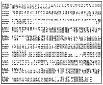
We have identified and characterized two additional mosquito transcripts that are induced upon bacterial challenge. One of these, AgGNBP, encodes a homologue of GNBP of the moth B. mori (16); it was first isolated by PCR (see Materials and Methods) based on a degenerate primer that corresponds to a peptide segment shared between B. mori GNBP and the β-1,3 glucanase A1 (glc1) of B. circulans (20). The encoded peptide sequence shows extensive similarities (Fig. 1) both to GNBP and to glucanase, including a putative polysaccharide-binding domain (see Discussion).
Figure 1.
Full-length A. gambiae GNBP compared with B. mori GNBP (accession no. L38591) and B. circulans glucanase A1 (accession no. P23903). Residues matching in at least two of the sequences are shaded, and the highly conserved region of the putative B. circulans polysaccharide binding domain is indicated by ∗. The first 24 residues of AgGNBP show features suggestive of a signal peptide. The potential glycosylphosphatidylinositol anchor sequences of AgGNBP are underlined. Numbers indicate position of residues from the N terminus.
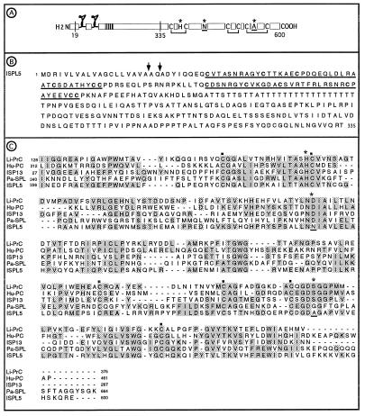
The other marker, ISPL5 (immune-related serine protease-like sequence 5), was first identified through differential display, comparing cDNAs of bacteria-challenged and naive mosquito larvae (see Materials and Methods). The encoded sequence (Fig. 2) shows features reminiscent of serine proteases involved in hemolymph clotting, innate immunity, and development, but it lacks two residues of the catalytic triad that are necessary for enzymatic activity (see Discussion).
Figure 2.
(A) Schematic sequence features of full-length ISPL5 showing the proposed signal peptide cleavage site (amino acid 19), the putative cysteine knots, the polythreonine stem region (vertical stripes), the putative activation site (amino acid 335), cysteines and disulfide bridges that are conserved in serine proteases, and the catalytic triad (∗) with two nonconserved residues underlined. (B) The amino terminal region of ISPL5 with the putative cysteine knots underlined. Two possible signal peptide cleavage sites are marked with arrows. (C) ISPL5 serine protease domain compared with ISP13 (accession no. Z69978), Limulus (Tachypleus tridentatus) clotting factor 2, which converts coagulen to insoluble coagulen gel (Li-PrC, accession no. P21902), human blood coagulation regulator protein C (Hu-PC, accession no. P04070), and Pacifastacus leniusculus (crayfish) hemocyte-specific serine protease-like protein (Pa-SPL, unpublished data; accession no. Y11145). The six conserved cysteines of serine proteases are marked with ▪; the residues of the serine protease catalytic triad are marked with ∗, with the nonconserved residues underlined as in A above. Aligned residues that match in two or more sequences are shaded. Numbers indicate position of residues from the N terminus.
Induction of Five Bacterial Infection-Responsive Markers.
The experiments shown in Fig. 3, using mosquito larvae, adults, and cultured cells, establish that AgGNBP and ISPL5 are induced by bacteria and bacterial cell wall components, as are three other previously described mosquito sequences. The latter encode the antibacterial peptide defensin (11), the infection-responsive putative serine protease ISP13 previously named G13 (12), and the putative infection-responsive galactose lectin IGALE20 previously designated G20 (12). Induction was demonstrated by RT-PCR, relative to the ribosomal protein S7 transcript that was not affected by bacterial challenge and served as a constitutive control. For each sequence, the appropriate number of PCR cycles was selected after preliminary tests to avoid saturation and was constant for that sequence within an experiment.
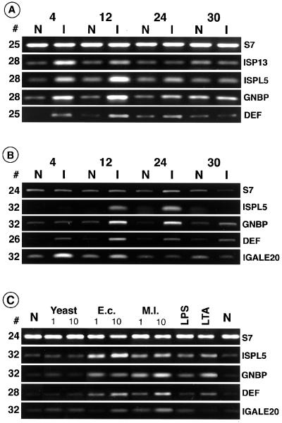
Figure 3.
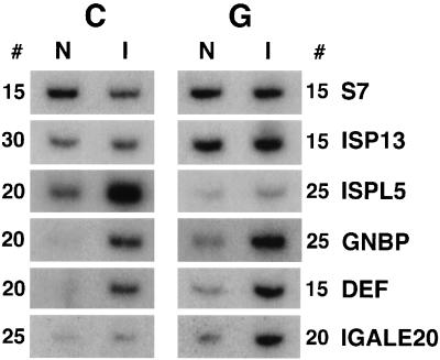
Expression profiles of immune markers in bacterially challenged (I) larvae (A) and adults (B) at 4, 12, 24, and 30 hr after infection compared with unchallenged naive animals (N). Changes in expression levels of specific sequences are detected relative to the control ribosomal protein S7 transcript. In this and subsequent figures the PCR cycle number for each sequence is indicated to the side of the data panel. The induction profiles of ISP13 and defensin in larvae were reported previously (11, 12). [Part of A reproduced from ref. 11 with permission (Copyright 1996, Royal Entomological Society) and part modified from figure 2 in ref. 12.] (C) Background levels of the markers in naive cells (N) and induced levels (I) after challenge with 10-fold differing concentrations of yeast, E. coli (E.c) and M. luteus (M.l.), or with LPS and LTA at concentrations as indicated in Materials and Methods.
Fig. 3 A and B records induction in bacteria-infected (I) versus naive (N) mosquito larvae and adult females, respectively, as a function of time after infection. The markers shown all were induced transiently and with varying kinetics. In larvae (Fig. 3A), for all four usable markers, induction was notable at 4 hr and essentially had subsided by 30 hr. It was earliest for ISP13, which showed a maximum at 4 hr and subsided by 24 hr. The other three markers were maximally induced at 12 hr and still were elevated at 24 hr. Bacterial induction of IGALE20 was not detectable in larvae against the background of a development-related increase in larval expression (ref. 12 and data not shown). In adult females (Fig. 3B) the kinetics of response were quite diverse and generally later than in larvae. Induction of IGALE20 was early and transient, being limited to 4 and 12 hr. Defensin was induced throughout from 4 to 30 hr, induction of GNBP was notable at 12 and 24 hr, and ISPL5 was detectably increased only at these two times. Adult ISP13 expression was not included in the analysis as no induction could be detected against its high constitutive expression level.
A number of A. gambiae-derived cell lines have been established in our laboratory, which display features characteristic of insect hemocytes, including phagocytosis of bacteria (H.M.M., unpublished data). As documented in Fig. 3C, both E. coli (Gram-negative) and M. luteus (Gram-positive) induce expression of four markers in the cell line Sua1B, in a dosage-dependent manner. Yeast (Saccharomyces cerevisiae) is not an effective elicitor, only very weakly inducing IGALE20 expression. Relative responsiveness to Gram-positive and Gram-negative bacteria varies: ISPL5 is induced most effectively by E. coli, in contrast to GNBP and IGALE20, which are clearly more responsive to M. luteus. Differential responses also are elicited by LPS (a cell-wall constituent of Gram-negative bacteria) and LTA (a cell-wall constituent of Gram-positive bacteria). The latter is a generally more effective inducer than LPS at the same concentration; however, it is ineffective in the case of IGALE20, despite the latter’s inducibility by Gram-positive bacteria. The ISP13 marker is not expressed in the Sua1B cell line and was not included in this analysis.
Regional Expression of the Markers in Adult Mosquitoes.
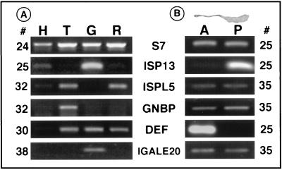
Localized expression of the infection-responsive markers in various body parts was determined in the experiments shown in Fig. 4 A and B, using naive adult female mosquitoes (which, however, normally harbor some microorganisms in the gut; ref. 21). Four body parts were analyzed in Fig. 4A: the head, the thorax, the midgut, and the remaining abdomen including ovaries and Malphigian tubules. Each of the markers is distinct in terms of preferential distribution. The midgut is the main site of expression of ISP13 and IGALE20, whereas AgGNBP is largely expressed in the thorax. Defensin is strongly expressed in the thorax, midgut, and the rest of the abdomen, whereas strong expression of ISPL5 is limited to the thorax and abdomen. The head expresses both ISP13 and ISPL5 at low levels.
Figure 4.
(A) Regional expression profiles, determined by RT-PCR, in naive adult head (H), thorax (T), midgut (G), and remaining abdominal tissues (R; which includes hindgut, Malphigian tubules and ovaries). ISP13 is largely amplified from the gut cDNA but also shows lower level expression in the head and abdomen. ISPL5 is mainly amplified in the thorax and abdomen. GNBP is mainly amplified from the thorax, defensin from the thorax, midgut and abdomen, and IGALE20 from the midgut. (B) Specific expression of the markers in anterior (A) versus posterior (P) regions of the unchallenged midgut (shown in a photograph at the top). ISP13 is posterior midgut specific and defensin is limited to the anterior midgut. ISPL5, GNBP and IGALE20 amplify weakly, approximately equally from both posterior and anterior midguts.
The mosquito midgut is subdivided (Fig. 4B) into a narrow anterior section and a larger, sac-like posterior region that expands substantially upon blood feeding and is the site of blood meal digestion and Plasmodium penetration after infection. Interestingly, constitutive high-level defensin expression is exclusively detected in the anterior and ISP13 in the posterior midgut region. ISPL5, AgGNBP, and IGALE20 are found in both regions at relatively low abundance.
Immune Response to Plasmodium in the Midgut and in Other Tissues.
In the experiment shown in Fig. 5, adult female mosquitoes were fed on naive mouse blood, or blood infected with the rodent malaria, P. berghei. After 24 hr the midgut was dissected from the remaining body, and both compartments were analyzed by RT-PCR. AgGNBP and defensin were strongly induced in both compartments; strong induction of ISPL5 was mostly concentrated in the carcass, and of IGALE20 mostly in the gut. Induction of ISP13 was only marginal in the midgut.
Figure 5.
Induction of immune markers after infection with P. berghei. Expression levels were assayed by radioactive RT-PCR in the midgut (G) and the remaining carcass (C) of mosquitoes fed 24 hr earlier on naive (N) or parasite-infected (I) mice. Note the prominent induction of IGALE20, defensin, and GNBP in the gut, as well as ISPL5, GNBP, and defensin in the carcass. Marginal levels of induction were observed for ISP13 in the infected gut and for IGALE20 in the carcass.
DISCUSSION
The Nature of the Immune-Related Markers.
Systematic examination of a diverse set of markers shows that they respond in a complex manner to bacterial challenge. The markers are immune-related, in that they are induced by infection (or bacterial constituents) relative to a constitutive ribosomal protein marker. With one exception, their physiological roles remain to be established. The exception is the previously reported antibacterial peptide, defensin (11), which is known to be active against Gram-positive bacteria (22). A second previously reported sequence, ISP13, has features indicative of an enzymatically competent serine protease, and resembles mammalian protease family members that are involved in immune response such as kallikreins, mast cell proteases, and plasminogens (12). Finally, the known IGALE20 sequence shows substantial similarity to mammalian galactose-specific lectins throughout its length, including some of the essential amino acids of the carbohydrate recognition domain (12).
Our set of markers includes two additional mosquito sequences. Within the aligned regions, AgGNBP shows an overall 32.6% amino acid sequence identity to a B. mori homologue (16), as well as 34.6% identity to the β-1,3 glucanase A1 from B. circulans, a bacterium that lyses fungi, including yeast (20). The three-way sequence similarity is highest in a region thought to constitute the polysaccharide binding domain of glucanases (16, 20). Bacterial and plant glucanases can participate in antifungal defense by recognizing and degrading structural polysaccharides of the cell wall (23). However, B. mori GNBP has been reported to lack glucanase activity but to bind Gram-negative bacteria (16). AgGNBP begins with a 24-residue region resembling a signal peptide (24), ends with a C-terminal hydrophobic region, and also contains sequence features of glycosylphosphatidylinositol anchor attachment sites (25). Follow-up studies at the protein level will be necessary to determine whether AgGNBP is membrane bound or secreted, whether it can function as a pattern recognition receptor, and if so what its binding specificity is.
The fifth marker, ISPL5, belongs to the serine protease family on the basis of sequence similarity, although it is unlikely to function as a protease. The C-terminal half is protease-like but lacks two of the three essential residues of the catalytic triad. It retains, however, six conserved cysteine residues thought to be important in stabilization of the serine protease fold (26). This 265-residue region shows greatest similarity to coagulation factors of the horseshoe crab, to a hemocyte-specific serine protease of the crayfish Pacifastacus, and to vertebrate mast cell proteases and complement activating factors. The N-terminal region contains two disulfide knot motifs, each including six conserved cysteine residues. Among other proteins that show this type of motif are Limulus pro-clotting enzyme (27) and the proteases encoded by the Drosophila genes easter, snake, stubble, and masquerade, which function in development (28). In the Limulus pro-clotting enzyme the disulfide knot is known to function as a receptor for the activating protein that cleaves at the zymogen activation site (27). The N-terminal region of ISPL5 also contains 18 contiguous threonine residues, a motif that may function as a flexible stem region, separating the disulfide knotted domain from the protease domain.
Another example of a serine protease-like protein that has the sequence features required for proper folding and substrate binding, although not for enzymatic catalysis, is the Masquerade protein of Drosophila (28). During development Masquerade is involved in muscle attachment by cell-matrix adhesion and is thought to act either as an adhesion molecule itself or as a competitive antagonist of serine proteases. More generally, serine proteases and protease-like molecules are implicated in developmental processes and in immune related mechanisms, frequently acting as positive and negative regulators of finely tuned enzymatic or signaling cascades. The biochemical and cellular function(s) of ISPL5 remain to be established by studies at the protein level.
Immune Reactions of A. gambiae.
Our parallel use of five infection-responsive markers has illuminated several distinct features of the mosquito response to bacteria.
First, the time-course studies on larval and adult immunity have revealed sequential stages in response to injected bacteria and differences in the induction kinetics between these two developmental stages (Fig. 3 A and B). In larvae responses initiate rapidly (especially for ISP13), with all four markers showing substantial induction by 4 hr and decreasing by 24 hr. The induction kinetics are more diverse and generally later in the adults. IGALE20 shows early induction, ISPL5 and GNBP are induced primarily at 12 and 24 hr, and defensin is broadly inducible. It is not known whether sequential induction reflects a cascade mechanism or differences in the mechanisms of induction in different tissues where these markers are expressed. For example, if induction is especially rapid in the fat body, the quicker response in larvae could be due to the greater presence of this tissue in larvae than in adults. However, the complex kinetics in the adult cannot readily dbe explained by the regional specificity of the markers; for example, injections of bacteria are made in the thorax, and yet GNBP induction is late relative to IGALE20.
Second, studies on a cell line revealed a degree of immune specificity, in that the markers are differentially induced by various elictors (Fig. 3C). Yeast is generally ineffective as an inducer. ISPL5 expression is enhanced preferentially by the Gram-negative bacterium E. coli rather than M. luteus, unlike the other three markers tested. Marker induction is also dose-dependent and can be elicited by purified bacterial cell wall components, with the Gram-positive component LTA being generally more effective than the Gram-negative LPS. Surprisingly, AgGNBP is more responsive to Gram-positive bacteria and LTA than to Gram-negative bacteria and LPS, even though the B. mori homologue binds exclusively Gram-negative bacteria (16). Provisionally we retain the name of this marker as a mnemonic of its homology with the B. mori sequence. We cannot exclude the possibility that AgGNBP has evolved a different recognition specificity, or that it is a related sequence (paralogue) rather than a true orthologue of the B. mori GNBP.
Third, we showed that the markers are distinct in terms of regional specificity (Fig. 4), potentially reflecting distinct roles in the mosquito immune response. The fat body is considered the main immune organ in insects, with the hemocytes and hypodermis also functioning in immunity (5). Fat body tissues are widely distributed and believed to be regionally specialized: in some insects, the visceral part surrounding the posterior midgut in the abdomen is preferentially active in protein synthesis, whereas the peripheral part in the thorax contains glycogen reserves (29). The fat body of the head is of unknown function. The possibility that the fat body may have region-specific immune functions has not been investigated. The pattern of distribution of ISPL5 in head, thorax, and abdomen (minus gut) may reflect generalized expression in the fat body or in the fat body and hypodermis. On the other hand, the predominant expression of AgGNBP in the thorax may reflect a regional specialization of the fat body or localization of hemocytes. The B. mori GNBP is concentrated in the hemolymph, and its mRNA is expressed in the fat body and (at a low level) in the epidermis of larvae (16).
Fourth, a potentially important observation was made that all five immune markers are expressed in the midgut. The two that are most abundant also are highly regionalized, with ISP13 represented only in the posterior and defensin only in the anterior section of the adult mosquito midgut. ISPL5 and AgGNBP are present at very low levels in the midgut and in greater abundance in other tissues. IGALE20 is expressed at low levels but almost exclusively in the midgut. Gut-specific lectins are believed to participate in host defense by binding to microorganisms (21), and lysozyme (30) as well as cecropin (31) have been observed in the Drosophila gut. However, the expression of antibacterial genes in the gut had not been documented previously in hematophagous insects (5). The midgut of insects is a major interface with the environment, and thus an immune-competence makes biological sense; although it has not been widely appreciated to date, it was clearly revealed in our malaria infection studies as well upon exposure of midgut tissues to heat killed bacteria in vitro (data not shown).
The Immune Reaction of A. gambiae to the Malaria Parasite.
Using our battery of markers that respond to bacterial infection challenge, we were able to show that significant immune reactions are elicited by the malaria parasite, both locally (in the midgut) and systemically (in the rest of the body). Defensin and AgGNBP participate strongly in both responses; IGALE20 is predominantly induced locally and ISPL5 at a distance. ISP13, which is constitutively expressed at high levels in the midgut, shows no more than marginal induction. These reactions are observed at a time when the parasites are physically constrained within the gut lumen, or within the midgut epithelial cell layer (ref. 32 and R. Cantera, personal communication), long before the sporozoite stage is released in the hemolymph. Thus, the systemic response of the carcass suggests some type of signaling, from the challenged midgut to other tissues presumably including the fat body. Indications are that cytokine-like molecules exist in invertebrates (33), but they have not been studied adequately at the molecular level.
In additional experiments (34), we have shown that formation of ookinetes leading to penetration of the midgut is required for the immune reaction; the mere presence of asexual parasites in the bloodmeal does not suffice. We also have shown that the response is not due to opportunistic infection of the hemolymph by gut bacteria during parasite passage through the epithelium: pretreatment with antibiotics suppresses the gut flora without affecting the immune response. It is unclear as yet whether the observed reaction is triggered by the parasite interacting with receptors that function in immune surveillance or is a result of injury that the parasite inflicts in traversing the epithelium. In this respect, it will be interesting to monitor the reaction in coadapted vector-parasite systems, such as in A. gambiae infected by the human malaria parasite P. falciparum.
The significance of the observed immune reaction to the parasite remains to be determined in conjunction with understanding the physiological roles of our immune markers, which are presently only broadly suggested by these sequences. Defensin has been shown to have activity against Plasmodium acting on late stages, rather than on the stage that traverses the gut (M. Shahabbudin, personal communication). Significantly, immune responsiveness is not limited to the parasite-refractory strain of the mosquito used in the experiments presented here. In this strain the parasite is melanotically encapsulated (presumably through a protease-regulated phenoloxidase cascade) and killed within the midgut epithelial tissue (8). However, defensin, AgGNBP and IGALE20 also are induced in a susceptible A. gambiae strain that fails to encapsulate the parasite (data not shown). At least these markers are unlikely to regulate the melanotic encapsulation process, although we will need to monitor their induction in the two strains quantitatively, at both the nucleic acid and protein levels. We also will need to consider the possibility of undetected sequence polymorphisms between the strains, which may affect activity or specificity of the marker gene products. In any case, the present systematic study has revealed that the innate immune system of the mosquito does respond to the presence of the parasite and must be considered in the context of parasite-vector interactions that can influence malaria transmission.
Acknowledgments
We thank P. Brey for making available the B. mori GNBP sequence before publication, D. Seeley for expert management of the mosquito colonies, C. Barillas-Mury for helpful advice, and other members of our laboratory for discussions. This work was supported by grants from the John D. and Catherine T. MacArthur Foundation, the Training and Mobility of Researchers (TMR) Programme of the European Union, and the United Nations Development Program/World Bank/World Health Organization Special Programme for Research and Training in Tropical Diseases. G.D. was supported by a TMR postdoctoral fellowship and H.-M.M. by a Deutsche Forschungsgemeinschaft fellowship.
ABBREVIATIONS
- GNBP
Gram-negative bacteria-binding protein
- LPS
lipopolysaccharide
- LTA
lipoteichoic acid
- RT-PCR
reverse transcription–PCR
Note Added in Proof
Expression of midgut-specific defensin sequences, exclusively localized in the anterior midgut, has been documented in the fly Stomoxys calcitrans by Lehane et al. (35).
Footnotes
References
- 1.Hoffman J A, Reichhard J-M, Hetru C. Curr Opin Immunol. 1996;8:8–13. doi: 10.1016/s0952-7915(96)80098-7. [DOI] [PubMed] [Google Scholar]
- 2.Hultmark D. Trends Genet. 1993;9:178–183. doi: 10.1016/0168-9525(93)90165-e. [DOI] [PubMed] [Google Scholar]
- 3.Cociancich S, Ghazi A, Hetru C, Hoffman J, A, Letellier L. J Biol Chem. 1993;268:19239–19245. [PubMed] [Google Scholar]
- 4.Lemaitre B, Kromer-Metzger E, Michaut L, Nicolas E, Meister M, Georgel P, Reichhart M, Hoffman J A. Proc Natl Acad Sci USA. 1995;92:9465–9469. doi: 10.1073/pnas.92.21.9465. [DOI] [PMC free article] [PubMed] [Google Scholar]
- 5.Paskewitz S M, Christensen B M. In: The Biology of Disease Vectors. Beaty B J, Marquardt W C, editors; Beaty B J, Marquardt W C, editors. Boulder: Univer. Press of Colorado; 1997. pp. 371–392. [Google Scholar]
- 6.Warburg A, Miller L H. Parasitol Today. 1991;7:179–181. doi: 10.1016/0169-4758(91)90127-a. [DOI] [PubMed] [Google Scholar]
- 7.Vernick K D, Fujioka H, Seeley D C, Tandler B, Aikawa M, Miller L H. Exp Parasitol. 1995;80:583–595. doi: 10.1006/expr.1995.1074. [DOI] [PubMed] [Google Scholar]
- 8.Collins F H, Sakai R K, Vernick K D, Paskewitz S, Seeley D C, Miller L H, Collins W E, Campbell C C, Gwadz R W. Science. 1986;234:607–610. doi: 10.1126/science.3532325. [DOI] [PubMed] [Google Scholar]
- 9.Barillas-Mury C, Charlesworth A, Gross I, Richman A, Hoffman J A, Kafatos F C. EMBO J. 1996;15:4961–4701. [PMC free article] [PubMed] [Google Scholar]
- 10.Steward R. Cell. 1989;55:487–495. doi: 10.1016/0092-8674(88)90035-9. [DOI] [PubMed] [Google Scholar]
- 11.Richman A M, Bulet P, Hetru C, Barillas-Mury C, Hoffman J A, Kafatos F C. Insect Mol Biol. 1996;5:203–210. doi: 10.1111/j.1365-2583.1996.tb00055.x. [DOI] [PubMed] [Google Scholar]
- 12.Dimopoulos G, Richman A, della Torre A, Kafatos F C, Louis C. Proc Natl Acad Sci USA. 1996;93:13066–13071. doi: 10.1073/pnas.93.23.13066. [DOI] [PMC free article] [PubMed] [Google Scholar]
- 13.Sinden W E. In: The Molecular Biology of Insect Disease Vectors. Crampton J M, Beard C B, Louis C, editors; Crampton J M, Beard C B, Louis C, editors. London: Chapman & Hall; 1997. pp. 67–91. [Google Scholar]
- 14.Hagedorn H H, Turner S, Hagedorn E A, Pontecorvo D, Greenbaum P, Pfeiffer D, Wheelock G, Flanagan T R. J Insect Physiol. 1997;23:203–206. doi: 10.1016/0022-1910(77)90030-0. [DOI] [PubMed] [Google Scholar]
- 15.Dimopoulos G, Louis C. In: The Molecular Biology of Insect Disease Vectors. Crampton J M, Beard C B, Louis C, editors; Crampton J M, Beard C B, Louis C, editors. London: Chapman & Hall; 1997. pp. 261–267. [Google Scholar]
- 16.Lee W-J, Lee J-D, Kravchenko V V, Ulevitch R, Brey P. Proc Natl Acad Sci USA. 1996;93:7888–7893. doi: 10.1073/pnas.93.15.7888. [DOI] [PMC free article] [PubMed] [Google Scholar]
- 17.Devereux J, Haeberli P, Smithies O. Nucleic Acids Res. 1984;12:387–395. doi: 10.1093/nar/12.1part1.387. [DOI] [PMC free article] [PubMed] [Google Scholar]
- 18.Altschul S F, Gish W, Miller W, Myers E W, Lipman D J. J Mol Biol. 1990;215:403–410. doi: 10.1016/S0022-2836(05)80360-2. [DOI] [PubMed] [Google Scholar]
- 19.Salazar C E, Mills-Hamm D, Kumar V, Collins F H. Nucleic Acids Res. 1993;21:4147. doi: 10.1093/nar/21.17.4147. [DOI] [PMC free article] [PubMed] [Google Scholar]
- 20.Yahata N, Watanabe T, Nakamura Y, Yamamoto Y, Kamimiya S, Tanaka H. Gene. 1990;86:113–117. doi: 10.1016/0378-1119(90)90122-8. [DOI] [PubMed] [Google Scholar]
- 21.Ham P J. In: Advances in Disease Vector Research. Harris K, editor; Harris K, editor. New York: Springer; 1992. pp. 101–149. [Google Scholar]
- 22.Cociancich S, Bulet P, Hetru C, Hoffman J A. Parasitol Today. 1994;10:132–139. doi: 10.1016/0169-4758(94)90260-7. [DOI] [PubMed] [Google Scholar]
- 23.Jach G, Gornhardt B, Mundy J, Logemann J, Pinsdorf E, Leah R, Schell J, Maas C. Plant J. 1995;8:97–109. doi: 10.1046/j.1365-313x.1995.08010097.x. [DOI] [PubMed] [Google Scholar]
- 24.von Heine G. Nucleic Acids Res. 1986;14:4683–4690. doi: 10.1093/nar/14.11.4683. [DOI] [PMC free article] [PubMed] [Google Scholar]
- 25.Coyne K E, Crisci A, Lublin D M. J Biol Chem. 1993;268:6689–6693. [PubMed] [Google Scholar]
- 26.Furie B, Bing D H, Feldmann R J, Robinson D J, Burnier J P, Furie B C. J Biol Chem. 1982;257:3875–3882. [PubMed] [Google Scholar]
- 27.Muta T, Hashimoto R, Miyata T, Nishimura H, Yoshihiro T, Iwanaga S. J Biol Chem. 1990;265:22426–22433. [PubMed] [Google Scholar]
- 28.Murugasu-Oei B, Rodrigues V, Yang X, Chia W. Genes Dev. 1995;9:139–154. doi: 10.1101/gad.9.2.139. [DOI] [PubMed] [Google Scholar]
- 29.van Heudsen M C. In: The Biology of Disease Vectors. Beaty B J, Marquardt W C, editors; Beaty B J, Marquardt W C, editors. Boulder: Univ. Press of Colorado; 1996. pp. 349–355. [Google Scholar]
- 30.Kylsten P, Kimbrell D A, Daffre S, Samakovlis C, Hultmark D. Mol Gen Genet. 1992;232:335–343. doi: 10.1007/BF00266235. [DOI] [PubMed] [Google Scholar]
- 31.Tryselius Y, Samakovlis C, Kimbrell D A, Hultmark D. Eur J Biochem. 1992;204:395–399. doi: 10.1111/j.1432-1033.1992.tb16648.x. [DOI] [PubMed] [Google Scholar]
- 32.Paskewitz S M, Brown M R, Lea A O, Collins F H. J Parasitol. 1988;74:432–439. [PubMed] [Google Scholar]
- 33.Beck G, Habicht G S. Mol Immunol. 1991;28:577–584. doi: 10.1016/0161-5890(91)90126-5. [DOI] [PubMed] [Google Scholar]
- 34.Richman, A., Dimopoulos, G., Seeley, D. & Kafatos, F. (1997) EMBO J., in press. [DOI] [PMC free article] [PubMed]
- 35.Lehane M J, Wu D, Lehane S M. Proc Natl Acad Sci USA. 1997;94:11502–11507. doi: 10.1073/pnas.94.21.11502. [DOI] [PMC free article] [PubMed] [Google Scholar]


