

Abstract
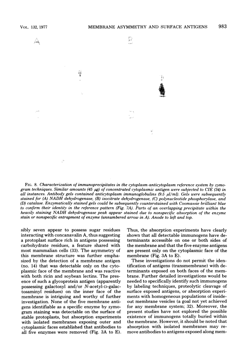
Crossed immunoelectrophoresis of Triton X-100-solubilized plasma membranes of Micrococcus lysodeikticus established the presence of 27 discrete antigens. Individual antigens were identified as membrane components possessing enzyme activity by zymogram staining procedures and by reactivity of certain antigens with a selection of four lectins in the crossed-immunoelectrophoresis (immunoaffinoelectrophoresis) system. Absorption experiments with intact, stable protoplasts and isolated membranes established the asymmetric nature of the M. lysodeikticus plasma membranes. Of the 14 antigens with determinants accessible solely on the cytoplasmic face of the membrane, four possessed individual dehydrogenase activities, and a fifth was identifiable as a component possessing adenosine triphosphatase (EC 3.6.1.3) activity. Evidence from absorption studies with isolated membranes suggested that antigens such as the adenosine triphosphatase complex were more readily accessible to reaction with antibodies than was succinate dehydrogenase (EC 1.3.99.1), for example. Twelve antigens were located on the protoplast surface as determined by antibody absorption, and the succinylated lipomannan was identified as a major antigen. At least five other antigens possessed sugar residues that interacted with concanavalin A. With the antisera generated to isolated membranes, there was no evidence suggesting that any of these antigens was not detectable on either surface of the plasma membrane. From absorption experiments with washed, whole cells of M. lysodeikticus, it was concluded that the immunogens on the protoplast surface were also detectable on the surface of the intact cell. However, some of the components such as the succinylated lipomannan appeared to be exposed to a greater extent than others. The cytoplasmic fraction from M. lysodeikticus was used as an antigen source to generate antibodies, and 97 immunoprecipitates were resolvable by crossed immunoelectrophoresis. In the cytoplasm-anticytoplasm reference immunoelectrophoresis pattern of precipitates, three of the immunoprecipitates unique to the cytoplasmic fraction were identifiable by zymogram staining procedures as catalase (EC 1.11.1.6), isocitrate dehydrogenase (EC 1.1.1.42), and polynucleotide phosphorylase (EC 2.3.7.8). The identification of membrane and cytoplasmic antigens (including the above-mentioned enzymes) provides a sensitive analytical system for monitoring cross-contamination and antigen distribution in cellular fractions.
Full text
PDF


Images in this article


Selected References
These references are in PubMed. This may not be the complete list of references from this article.
- Axelsen N. H. Antigen-antibody crossed electrophoresis (Laurell) applied to the study of the antigenic structure of Candida albicans. Infect Immun. 1971 Nov;4(5):525–527. doi: 10.1128/iai.4.5.525-527.1971. [DOI] [PMC free article] [PubMed] [Google Scholar]
- Barsukov L. I., Kulikov V. I., Bergelson L. D. Lipid transfer proteins as a tool in the study of membrane structure. Inside-outside distribution of the phospholipids in the protoplasmic membrane of Micrococcus lysodeikticus. Biochem Biophys Res Commun. 1976 Aug 9;71(3):704–711. doi: 10.1016/0006-291x(76)90888-3. [DOI] [PubMed] [Google Scholar]
- Bjerrum O. J., Bhakdi S., Knüfermann H., Bog-Hansen T. C. Immunoelectrophoretic heterogeneity and cross-reactions of individual "spectrin" components isolated by preparative sodium dodecylsulfate-polyacrylamide-gel electrophoresis. Biochim Biophys Acta. 1974 Nov 27;373(1):44–50. doi: 10.1016/0005-2736(74)90103-5. [DOI] [PubMed] [Google Scholar]
- Bjerrum O. J., Bog-Hansen T. C. The immunochemical approach to the characterization of membrane proteins. Human erythrocyte membrane proteins analysed as a model system. Biochim Biophys Acta. 1976 Nov 11;455(1):66–89. doi: 10.1016/0005-2736(76)90154-1. [DOI] [PubMed] [Google Scholar]
- Bjerrum O. J., Lundahl P., Brogren C. H., Hjertén S. Immunoabsorption of membrane-specific antibodies for determination of exposed and hidden proteins in human erythrocyte membranes. Biochim Biophys Acta. 1975 Jun 25;394(2):173–181. doi: 10.1016/0005-2736(75)90255-2. [DOI] [PubMed] [Google Scholar]
- Bog-Hansen T. C. Crossed immuno-affinoelectrophoresis. An analytical method to predict the result of affinity chromatography. Anal Biochem. 1973 Dec;56(2):480–488. doi: 10.1016/0003-2697(73)90215-7. [DOI] [PubMed] [Google Scholar]
- Gurd J. W., Evans W. H., Perkins H. R. Immunochemical characterization of proteins from mouse liver plasma membranes. Biochem J. 1973 Dec;135(4):827–832. doi: 10.1042/bj1350827. [DOI] [PMC free article] [PubMed] [Google Scholar]
- Harboe N., Ingild A. Immunization, isolation of immunoglobulins, estimation of antibody titre. Scand J Immunol Suppl. 1973;1:161–164. doi: 10.1111/j.1365-3083.1973.tb03798.x. [DOI] [PubMed] [Google Scholar]
- Johansson K. E., Hjertén S. Localization of the Tween 20-soluble membrane proteins of Acholeplasma laidlawii by crossed immunoelectrophoresis. J Mol Biol. 1974 Jun 25;86(2):341–348. doi: 10.1016/0022-2836(74)90023-0. [DOI] [PubMed] [Google Scholar]
- Joseph R., Shockman G. D. Synthesis and excretion of glycerol teichoic acid during growth of two streptococcal species. Infect Immun. 1975 Aug;12(2):333–338. doi: 10.1128/iai.12.2.333-338.1975. [DOI] [PMC free article] [PubMed] [Google Scholar]
- Kahane I., Furthmayr H., Marchesi V. T. Isolation of membrane glycoproteins by affinity chromatography in the presence of detergents. Biochim Biophys Acta. 1976 Mar 19;426(3):464–476. doi: 10.1016/0005-2736(76)90391-6. [DOI] [PubMed] [Google Scholar]
- LAURELL C. B. ANTIGEN-ANTIBODY CROSSED ELECTROPHORESIS. Anal Biochem. 1965 Feb;10:358–361. doi: 10.1016/0003-2697(65)90278-2. [DOI] [PubMed] [Google Scholar]
- LOWRY O. H., ROSEBROUGH N. J., FARR A. L., RANDALL R. J. Protein measurement with the Folin phenol reagent. J Biol Chem. 1951 Nov;193(1):265–275. [PubMed] [Google Scholar]
- Matsumoto I., Osawa T. Purification and characterization of an anti-H(O) phytohemagglutinin of Ulex europeus. Biochim Biophys Acta. 1969 Nov 11;194(1):180–189. doi: 10.1016/0005-2795(69)90193-7. [DOI] [PubMed] [Google Scholar]
- Möller G., Coutinho A., Persson U. Mechanism of b-lymphocyte activation: failure to obtain evidence of a direct role of the Ig receptors in the triggering process. Scand J Immunol. 1975;4(1):37–52. doi: 10.1111/j.1365-3083.1975.tb02598.x. [DOI] [PubMed] [Google Scholar]
- Nachbar M. S., Oppenheim J. D. The production and purification of specific anti-soybean agglutinin antibody by affinity chromatography. Biochim Biophys Acta. 1973 Sep 14;320(2):494–502. doi: 10.1016/0304-4165(73)90330-9. [DOI] [PubMed] [Google Scholar]
- Nicolson G. L., Blaustein J. The interaction of Ricinus communis agglutinin with normal and tumor cell surfaces. Biochim Biophys Acta. 1972 May 9;266(2):543–547. doi: 10.1016/0005-2736(72)90109-5. [DOI] [PubMed] [Google Scholar]
- Oppenheim J. D., Salton M. R. Localization and distribution of Micrococcus lysodeikticus membrane ATPase determined by ferritin labeling. Biochim Biophys Acta. 1973 Mar 16;298(2):297–322. doi: 10.1016/0005-2736(73)90360-x. [DOI] [PubMed] [Google Scholar]
- Owen P., Freer J. H. Isolation and properties of mesosomal membrane fractions from Micrococcus lysodeikticus. Biochem J. 1972 Oct;129(4):907–917. doi: 10.1042/bj1290907. [DOI] [PMC free article] [PubMed] [Google Scholar]
- Owen P., Salton M. R. A succinylated mannan in the membrane system of Micrococcus lysodeikticus. Biochem Biophys Res Commun. 1975 Apr 21;63(4):875–880. doi: 10.1016/0006-291x(75)90649-x. [DOI] [PubMed] [Google Scholar]
- Owen P., Salton M. R. Antigenic and enzymatic architecture of Micrococcus lysodeikticus membranes established by crossed immunoelectrophoresis. Proc Natl Acad Sci U S A. 1975 Sep;72(9):3711–3715. doi: 10.1073/pnas.72.9.3711. [DOI] [PMC free article] [PubMed] [Google Scholar]
- Owen P., Salton M. R. Isolation and characterization of a mannan from mesosomal membrane vesicles of Micrococcus lysodeikticus. Biochim Biophys Acta. 1975 Oct 6;406(2):214–234. doi: 10.1016/0005-2736(75)90006-1. [DOI] [PubMed] [Google Scholar]
- Pless D. D., Schmit A. S., Lennarz W. J. The characterization of mannan of Micrococcus lysodeikticus as an acidic lipopolysaccharide. J Biol Chem. 1975 Feb 25;250(4):1319–1327. [PubMed] [Google Scholar]
- Salton M. R., Freer J. H., Ellar D. J. Electron transport components localized in a lipid-depleted sheet isolated from Micrococcus lysodeikticus membranes by deoxycholate extraction. Biochem Biophys Res Commun. 1968 Dec 30;33(6):909–915. doi: 10.1016/0006-291x(68)90398-7. [DOI] [PubMed] [Google Scholar]
- Salton M. R., Owen P. Bacterial membrane structure. Annu Rev Microbiol. 1976;30:451–482. doi: 10.1146/annurev.mi.30.100176.002315. [DOI] [PubMed] [Google Scholar]
- Sharon N., Lis H. Lectins: cell-agglutinating and sugar-specific proteins. Science. 1972 Sep 15;177(4053):949–959. doi: 10.1126/science.177.4053.949. [DOI] [PubMed] [Google Scholar]
- Smyth C. J., Friedman-Kien A. E., Salton M. R. Antigenic analysis of Neisseria gonorrhoeae by crossed immunoelectrophoresis. Infect Immun. 1976 Apr;13(4):1273–1288. doi: 10.1128/iai.13.4.1273-1288.1976. [DOI] [PMC free article] [PubMed] [Google Scholar]