

Abstract
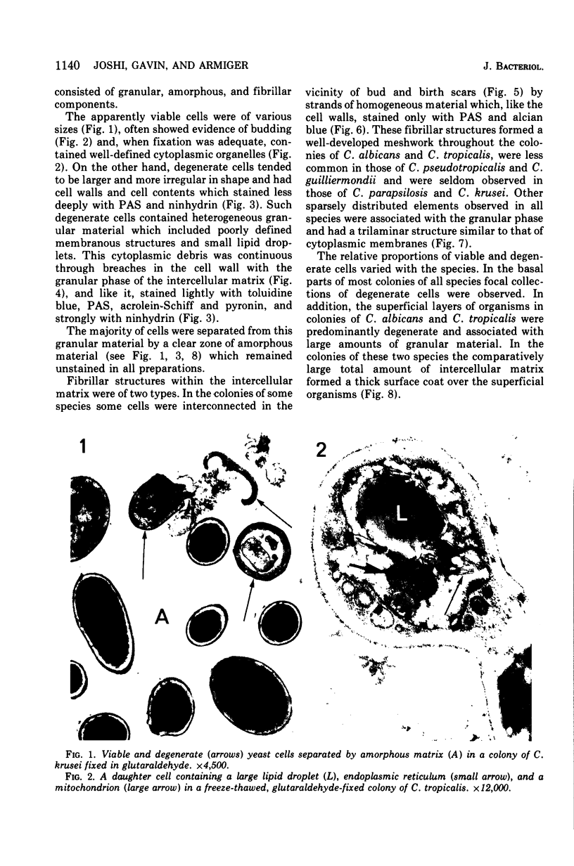
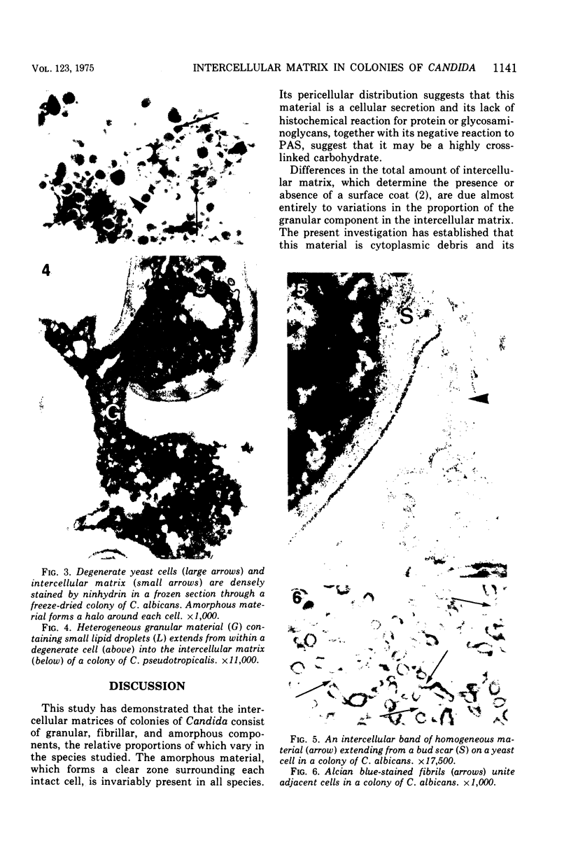
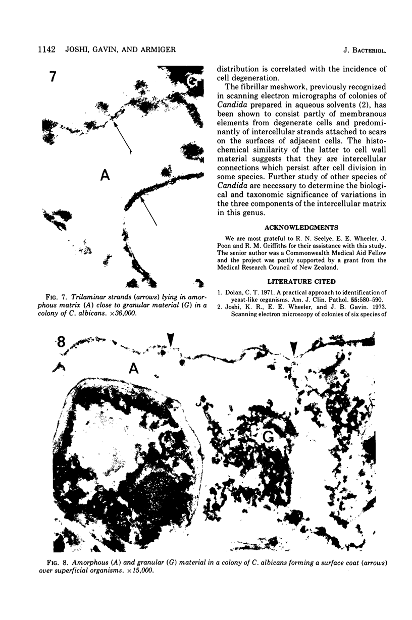
The histochemistry and fine structure of typical colonies of six species of Candida were studied, using a total of 31 clinical isolates. The colonies consisted of viable and degenerate cells which lay in an intercellular matrix. This matrix was made up of amorphous, granular, and fibrillar components, the relative proportions and total amount of which varied from species to species. The cells of all species were surrounded by a zone of homogeneous amorphus material, which may be a highly cross-linked carbohydrate. This separated intact cells from irregularly distributed granular debris derived from the cytoplasm of degenerate cells. Focal cellular degeneration and associated granular debris were present within the colonies of all species and were most common in the surface layers of cells of colonies of C. albicans and C. tropicalis. The large amounts of intercellular matrix in this region formed a surface coat on colonies of these two species. Intercellular strands of cell wall material, and to a lesser extent other membranous elements from degenerate cells, formed a prominent fibrillar meshwork in the colonies of C. albicans and C. tropicalis, but were less common in those of C. pseudotropicalis and C. guilliermondii and seldom seen in those of C. parapsilosis and C. krusei.
Full text
PDF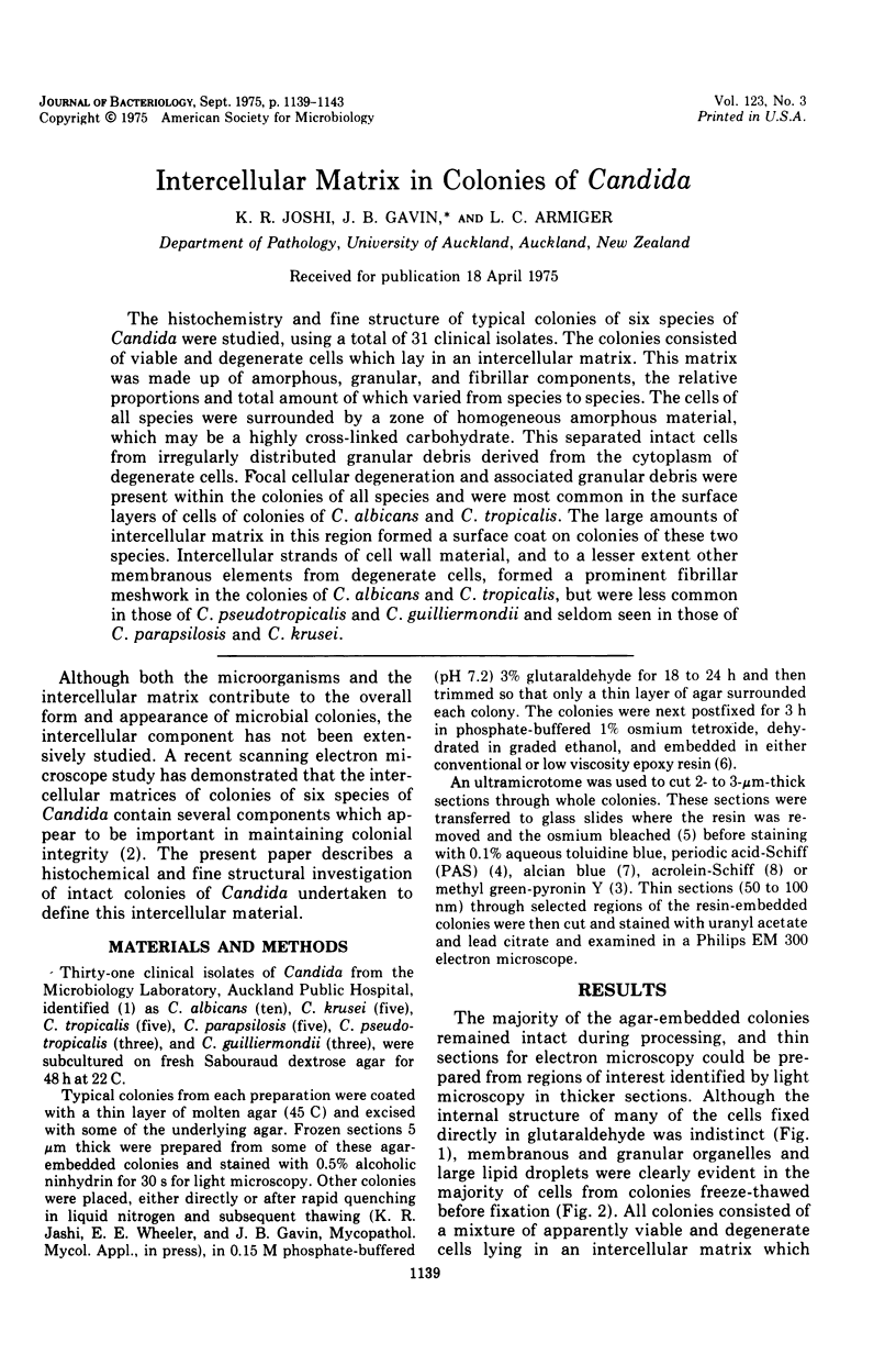
Images in this article
Selected References
These references are in PubMed. This may not be the complete list of references from this article.
- Dolan C. T. A practical approach to identification of yeast-like organisms. Am J Clin Pathol. 1971 May;55(5):580–590. doi: 10.1093/ajcp/55.5.580. [DOI] [PubMed] [Google Scholar]
- Joshi K. R., Wheeler E. E., Gavin J. B. Scanning electron microscopy of colonies of six species of Candida. J Bacteriol. 1973 Jul;115(1):341–348. doi: 10.1128/jb.115.1.341-348.1973. [DOI] [PMC free article] [PubMed] [Google Scholar]
- KURNICK N. B. Pyronin Y in the methyl-green-pyronin histological stain. Stain Technol. 1955 Sep;30(5):213–230. doi: 10.3109/10520295509114469. [DOI] [PubMed] [Google Scholar]
- Snodgress A. B., Dorsey C. H., Bailey G. W., Dickson L. G. Conventional histopathologic staining methods compatible with epon-embedded, osmicated tissue. Lab Invest. 1972 Mar;26(3):329–337. [PubMed] [Google Scholar]
- Spurr A. R. A low-viscosity epoxy resin embedding medium for electron microscopy. J Ultrastruct Res. 1969 Jan;26(1):31–43. doi: 10.1016/s0022-5320(69)90033-1. [DOI] [PubMed] [Google Scholar]
- van DUIJN Acrolein-Schiff, a new staining method for proteins. J Histochem Cytochem. 1961 May;9:234–241. doi: 10.1177/9.3.234. [DOI] [PubMed] [Google Scholar]