Abstract
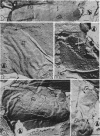
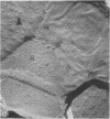
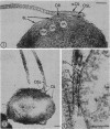
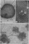
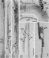
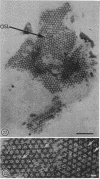
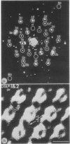
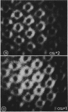
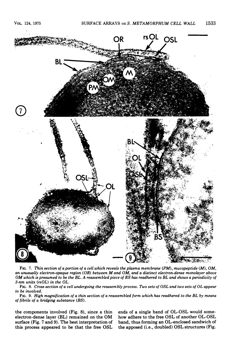
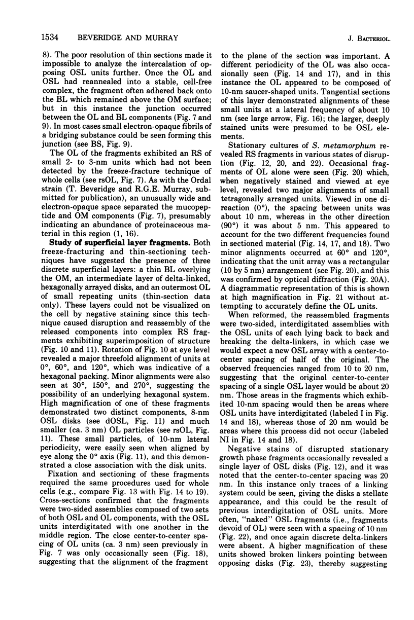
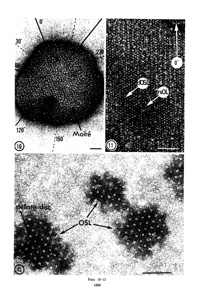
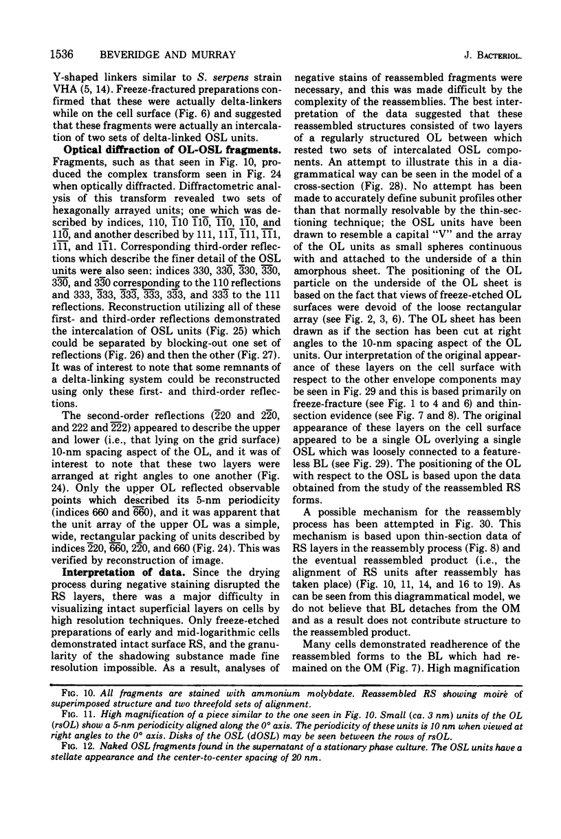
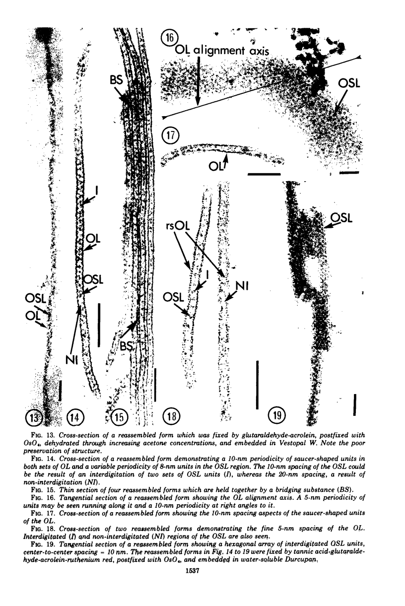
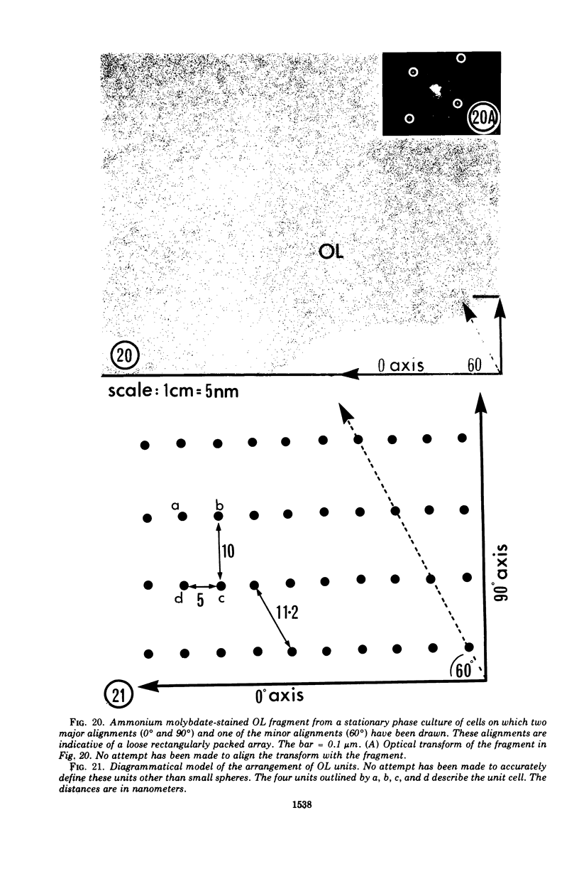
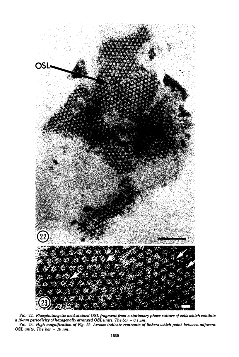
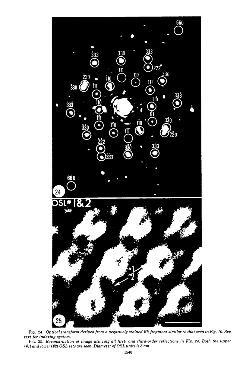
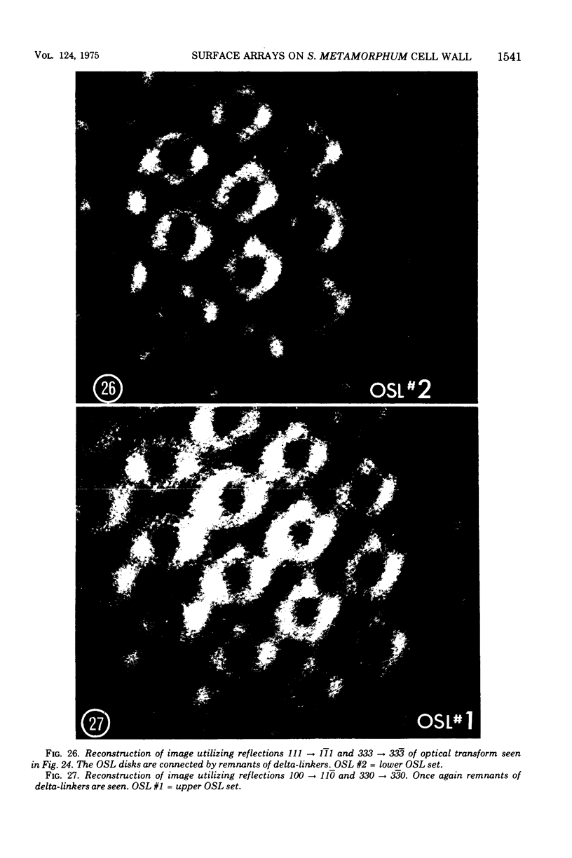
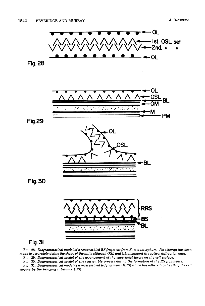
A complex and easily disrupted arrangement of macromolecules was present on the outer (lipopolysaccharide) membrane of the cell wall of Spirillum metamorphum. Separation of the arrays from the cell and spontaneous reassembly into regularly structured complexes usually occurred during preparation for electron microscopy. Freeze etchings, thin sections, and optical diffraction analysis of negatively stained fragments indicated that they consisted of two sets of a thin layer which was studied with 3-nm particles arranged in a loose (OL). The OSL consisted of a hexagonal arrangement of 8-nm disks and the OL of a thin layer which was studied with 3-nm particles arranged in a loose rectangular manner. The OSL of reassembled fragments displayed numerous broken delta-linkers between units and a center-to-center spacing of half the expected distance, which suggests that an interdigitation of two OSL arrays had occurred. The observations combined with freeze etchings and thin sections of whole cells suggested a possible reassembly mechanism. The normal surface arrangement of these layers on cells was thought to consist of the OL overlying one set of OSL which was loosely adherent to a thin amorphous backing layer.
Full text
PDF
Images in this article
Selected References
These references are in PubMed. This may not be the complete list of references from this article.
- BEER M. SELECTIVE STAINING FOR ELECTRON MICROSCOPY. Lab Invest. 1965 Jun;14:1020–1025. [PubMed] [Google Scholar]
- Beveridge T. J., Murray R. G. Superficial macromolecular arrays on the cell wall of Spirillum putridiconchylium. J Bacteriol. 1974 Sep;119(3):1019–1038. doi: 10.1128/jb.119.3.1019-1038.1974. [DOI] [PMC free article] [PubMed] [Google Scholar]
- Buckmire F. L., Murray R. G. Studies on the cell wall of Spirillum serpens. 1. Isolation and partial purification of the outermost cell wall layer. Can J Microbiol. 1970 Oct;16(10):1011–1022. doi: 10.1139/m70-171. [DOI] [PubMed] [Google Scholar]
- Buckmire F. L., Murray R. G. Studies on the cell wall of Spirillum serpens. II. Chemical characterization of the outer structured layer. Can J Microbiol. 1973 Jan;19(1):59–66. doi: 10.1139/m73-009. [DOI] [PubMed] [Google Scholar]
- DeVoe I. W., Costerton J. W., MacLeod R. A. Demonstration by freeze-etching of a single cleavage plane in the cell wall of a gram-negative bacterium. J Bacteriol. 1971 May;106(2):659–671. doi: 10.1128/jb.106.2.659-671.1971. [DOI] [PMC free article] [PubMed] [Google Scholar]
- Korn A. H., Feairheller S. H., Filachione E. M. Glutaraldehyde: nature of the reagent. J Mol Biol. 1972 Apr 14;65(3):525–529. doi: 10.1016/0022-2836(72)90206-9. [DOI] [PubMed] [Google Scholar]
- POLLOCK M. R., RICHMOND M. H. Low cyst(e)ine content of bacterial extracellular proteins: its possible physiological significance. Nature. 1962 May 5;194:446–449. doi: 10.1038/194446a0. [DOI] [PubMed] [Google Scholar]
- REYNOLDS E. S. The use of lead citrate at high pH as an electron-opaque stain in electron microscopy. J Cell Biol. 1963 Apr;17:208–212. doi: 10.1083/jcb.17.1.208. [DOI] [PMC free article] [PubMed] [Google Scholar]


