

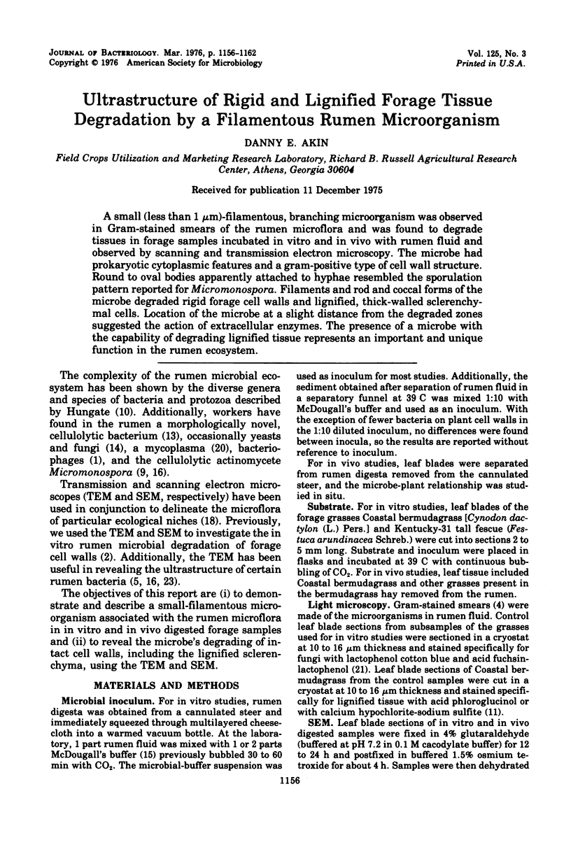
Abstract
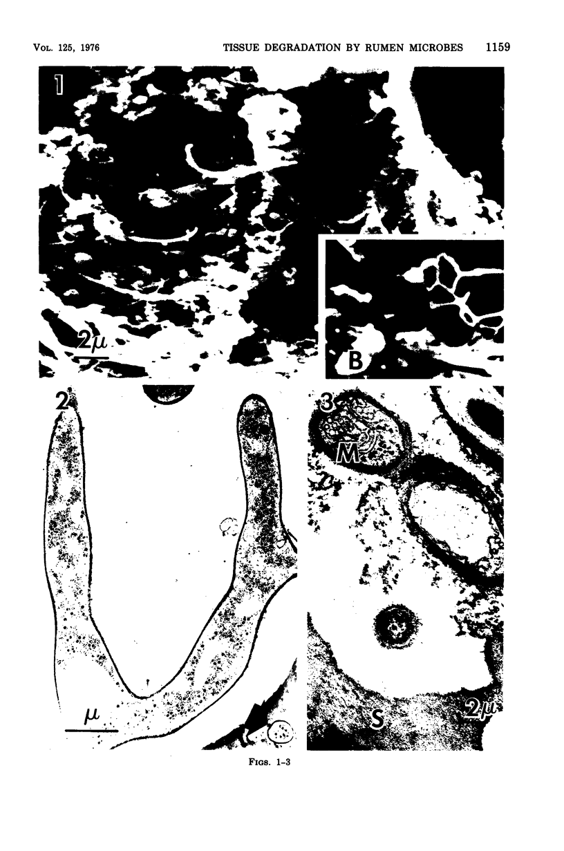
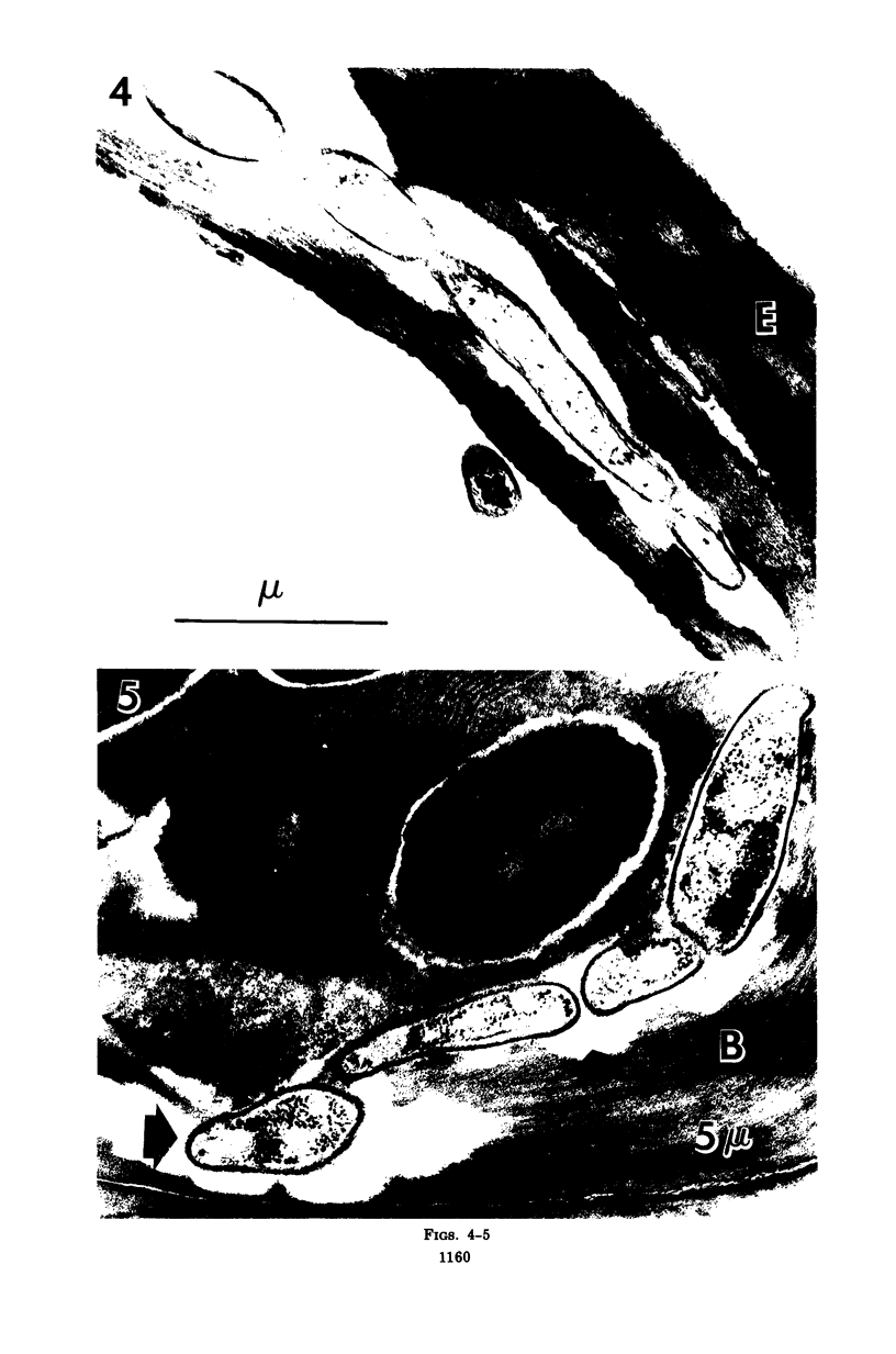
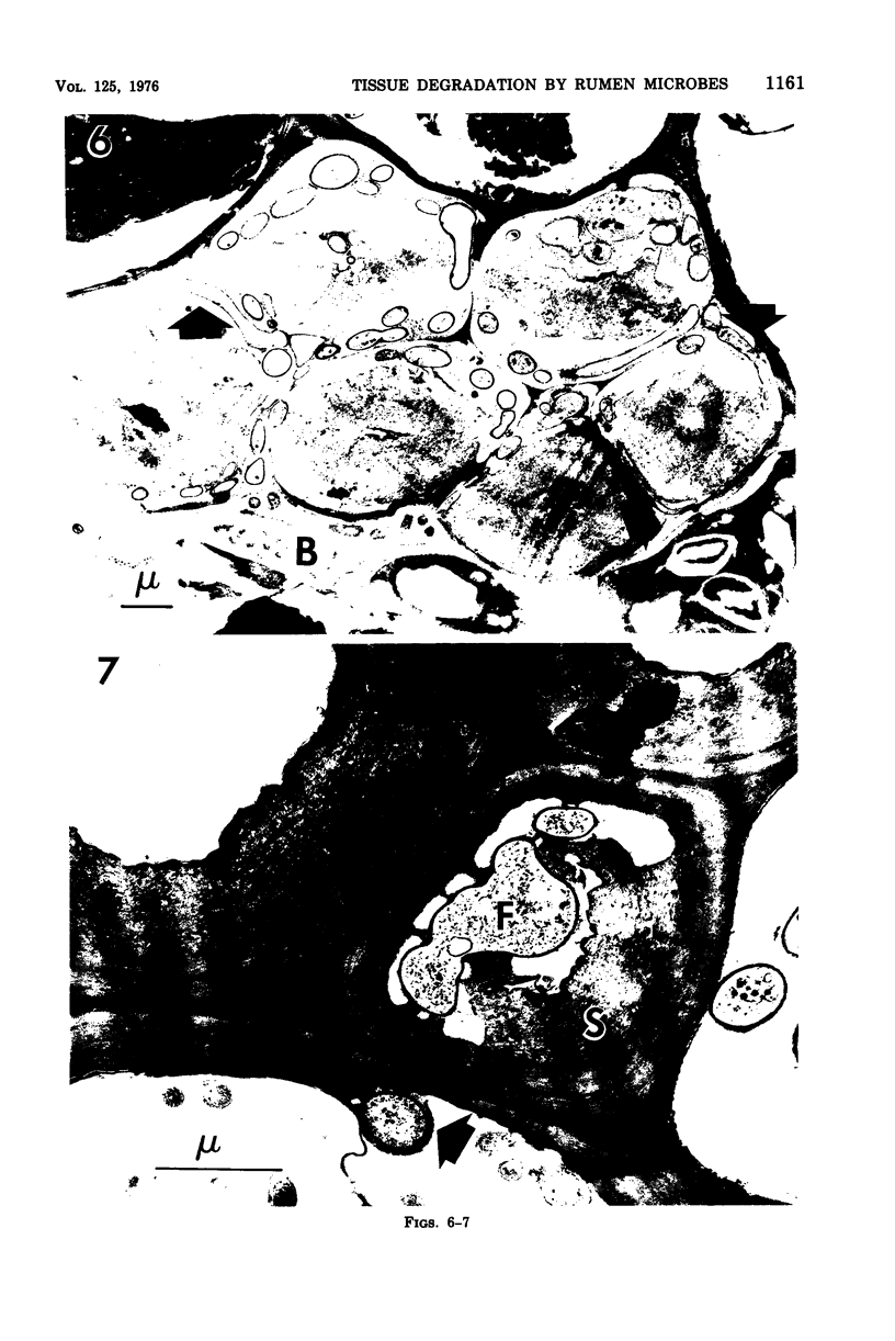
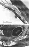
A small (less than 1 mum)-filamentous, branching microorganism was observed in Gram-stained smears of the rumen microflora and was found to degrade tissues in forage samples incubated in vitro and in vivo with rumen fluid and observed by scanning and transmission electron microscopy. The microbe had prokaryotic cytoplasmic features and a gram-positive type of cell wall structure. Round to oval bodies apparently attached to hyphae resembled the sporulation pattern reported for Micromonospora. Filaments and rod and coccal forms of the microbe degraded rigid forage cell walls and lignified, thick-walled sclerenchymal cells. Location of the microbe at a slight distance from the degraded zones suggested the action of extracellular enzymes. The presence of a microbe with the capability of degrading lignified tissue represents an important and unique function in the rumen ecosystem.
Full text
PDF
Images in this article

Selected References
These references are in PubMed. This may not be the complete list of references from this article.
- Akin D. E., Amos H. E. Rumen bacterial degradation of forage cell walls investigated by electron microscopy. Appl Microbiol. 1975 May;29(5):692–701. doi: 10.1128/am.29.5.692-701.1975. [DOI] [PMC free article] [PubMed] [Google Scholar]
- Akin D. E., Burdick D., Michaels G. E. Rumen bacterial interrelationships with plant tissue during degradation revealed by transmission electron microscopy. Appl Microbiol. 1974 Jun;27(6):1149–1156. doi: 10.1128/am.27.6.1149-1156.1974. [DOI] [PMC free article] [PubMed] [Google Scholar]
- Costerton J. W., Damgaard H. N., Cheng K. J. Cell envelope morphology of rumen bacteria. J Bacteriol. 1974 Jun;118(3):1132–1143. doi: 10.1128/jb.118.3.1132-1143.1974. [DOI] [PMC free article] [PubMed] [Google Scholar]
- Hungate R. E. Studies on Cellulose Fermentation: II. An Anaerobic Cellulose-decomposing Actinomycete, Micromonospora propionici, N. Sp. J Bacteriol. 1946 Jan;51(1):51–56. [PMC free article] [PubMed] [Google Scholar]
- Lacey J. Actinomycetes in soils, composts and fodders. Soc Appl Bacteriol Symp Ser. 1973 Jan;2:231–251. [PubMed] [Google Scholar]
- Leatherwood J. M., Sharma M. P. Novel anaerobic cellulolytic bacterium. J Bacteriol. 1972 May;110(2):751–753. doi: 10.1128/jb.110.2.751-753.1972. [DOI] [PMC free article] [PubMed] [Google Scholar]
- Lund A. Yeasts and moulds in the bovine rumen. J Gen Microbiol. 1974 Apr;81(2):453–462. doi: 10.1099/00221287-81-2-453. [DOI] [PubMed] [Google Scholar]
- Maluszyńska G. M., Janota-Bassalik L. A cellulolytic rumen bacterium, Micromonospora ruminantium sp.nov. J Gen Microbiol. 1974 May;82(1):57–65. doi: 10.1099/00221287-82-1-57. [DOI] [PubMed] [Google Scholar]
- McDougall E. I. Studies on ruminant saliva. 1. The composition and output of sheep's saliva. Biochem J. 1948;43(1):99–109. [PMC free article] [PubMed] [Google Scholar]
- Morrison I. M. Structural invesiigations on the lignin-carbohydrate complexes of Lolium perenne. Biochem J. 1974 Apr;139(1):197–204. doi: 10.1042/bj1390197. [DOI] [PMC free article] [PubMed] [Google Scholar]
- OVERMAN J. R., PINE L. ELECTRON MICROSCOPY OF CYTOPLASMIC STRUCTURES IN FACULTATIVE AND ANAEROBIC ACTINOMYCES. J Bacteriol. 1963 Oct;86:656–665. doi: 10.1128/jb.86.4.656-665.1963. [DOI] [PMC free article] [PubMed] [Google Scholar]
- Stafford H. A. Histochemical & Biochemical Differences Between Lignin-Like Materials in Phleum pratense L. Plant Physiol. 1962 Sep;37(5):643–649. doi: 10.1104/pp.37.5.643. [DOI] [PMC free article] [PubMed] [Google Scholar]
- Van Soest P. J. The uniformity and nutritive availability of cellulose. Fed Proc. 1973 Jul;32(7):1804–1808. [PubMed] [Google Scholar]
- Williams S. T., Davies F. L. Use of scanning electron microscope for the examination of actinomycetes. J Gen Microbiol. 1967 Aug;48(2):171–177. doi: 10.1099/00221287-48-2-171. [DOI] [PubMed] [Google Scholar]
- Williams S. T., Sharples G. P., Bradshaw R. M. The fine structure of the Actinomycetales. Soc Appl Bacteriol Symp Ser. 1973 Jan;2:113–130. [PubMed] [Google Scholar]