Abstract
Infection with the human gastric pathogen Helicobacter pylori is associated with a spectrum of diseases including gastritis, peptic ulcers, gastric adenocarcinoma, and gastric mucosa–associated lymphoid tissue lymphoma. The cytotoxin-associated gene A (CagA) protein of H. pylori, which is translocated into host cells via a type IV secretion system, is a major risk factor for disease development. Experiments in gastric tissue culture cells have shown that once translocated, CagA activates the phosphatase SHP-2, which is a component of receptor tyrosine kinase (RTK) pathways whose over-activation is associated with cancer formation. Based on CagA's ability to activate SHP-2, it has been proposed that CagA functions as a prokaryotic mimic of the eukaryotic Grb2-associated binder (Gab) adaptor protein, which normally activates SHP-2. We have developed a transgenic Drosophila model to test this hypothesis by investigating whether CagA can function in a well-characterized Gab-dependent process: the specification of photoreceptors cells in the Drosophila eye. We demonstrate that CagA expression is sufficient to rescue photoreceptor development in the absence of the Drosophila Gab homologue, Daughter of Sevenless (DOS). Furthermore, CagA's ability to promote photoreceptor development requires the SHP-2 phosphatase Corkscrew (CSW). These results provide the first demonstration that CagA functions as a Gab protein within the tissue of an organism and provide insight into CagA's oncogenic potential. Since many translocated bacterial proteins target highly conserved eukaryotic cellular processes, such as the RTK signaling pathway, the transgenic Drosophila model should be of general use for testing the in vivo function of bacterial effector proteins and for identifying the host genes through which they function.
Author Summary
Like many pathogens, the human gastric bacterium Helicobacter pylori orchestrates infection through the activity of proteins that it translocates into host cells. The H. pylori translocated protein, CagA, which shares no homology to any other proteins, is a significant risk factor for H. pylori–associated diseases including gastric cancer. Experiments in tissue culture cells have shown that CagA can activate SHP-2 phosphatase, a component of the receptor tyrosine kinase signaling pathway. Based on this activity, CagA has been proposed to function as a mimic of Gab proteins that serve as adaptors in this signaling pathway. We have developed a transgenic Drosophila model to test this hypothesis in the tissues of an organism. We demonstrate that CagA can substitute for Gab and restore developmental defects caused by the loss of the Drosophila Gab, including promoting photoreceptor specification in the developing eye. Furthermore, we show that CagA functions similarly to Gab because it requires the Drosophila SHP-2 to exert its effect on photoreceptor development. Our transgenic Drosophila model provides new insight into CagA's activity in tissues and will allow us to identify host factors through which CagA functions to manipulate cellular signaling pathways and promote disease.
Introduction
The human pathogen, Helicobacter pylori, infects the stomachs of at least half the world's population and chronic infection is associated with the development of diseases such as gastritis, peptic ulcers and gastric cancer [1]. A major virulence determinant of H. pylori is the cytotoxin associated gene A (CagA) which is translocated into host cells via a type four secretion system (reviewed in [2]). Inside host cells, CagA is phosphorylated by Src family kinases on tyrosines contained in repeated five-amino acid motifs (EPIYA) in CagA's carboxyl terminus. Phosphorylated CagA disrupts receptor tyrosine kinase (RTK) signaling pathways by directly activating Src homology 2 (SH2) domain containing tyrosine phosphatase (SHP-2) (reviewed in [3]). Normally SHP-2 is activated by the scaffolding adaptor Grb2-associated binder (Gab) proteins, thereby amplifying RTK signaling pathways to control cell growth, differentiation and survival (reviewed in [4]). The Gab proteins occupy a pivotal position in RTK signaling pathways by interacting directly with RTKs such as the c-Met receptor of the Hepatocyte growth factor/Scatter factor (HGF/SF) as well as downstream cytoplasmic proteins including SHP-2, v-crk sarcoma virus CT10 oncogene homolog (avian)-like (Crk(L)), and Growth factor receptor-bound protein 2 (Grb2) (reviewed in [5],[6],[7]). Although CagA shares no sequence similarity with Gab proteins, CagA has been shown to activate SHP-2 in tissue culture cells, resulting in cell elongation [8],[9]. Similarly, in tissue culture cells CagA has been found to associate with c-Met, Crk(L) and Grb2 [10],[11],[12]. Based on these interactions, CagA has been hypothesized to mimic Gab proteins and to function as an oncogene by over-activating RTK signaling [13]. The significance of CagA's interactions with RTK signaling pathway proteins, however, has only been explored in tissue culture cells.
We have developed transgenic Drosophila with inducible CagA expression as a model to understand CagA's mechanisms of action in complex epithelial tissues. In order to test the hypothesis that CagA can function as a Gab substitute, we investigated CagA activity in a well-characterized Gab-dependent process, the specification of photoreceptors in the Drosophila eye [14],[15],[16]. The Drosophila compound eye, whose crystalline array of facets or ommatidia are exquisitely sensitive to perturbations in cell specification, has been used as a powerful system for the discovery and genetic analysis of RTK signaling components [17],[18]. Drosophila RTK signaling proteins are highly conserved with their mammalian orthologues and oncogenic mutations in these proteins, such as those that constitutively activate RTK receptors or their downstream effectors, function similarly in both Drosophila and mammalian cells [19]. The Drosophila model also offers elegant tools for genetic manipulations including the UAS/GAL4 system [20] for expression of transgenes in a tissue specific manner, the FLP/FRT system for the generation of somatic mutant clones [21], and null mutations in most RTK signaling pathway members, which allow us to probe the in vivo requirements for CagA's activation of RTK signaling pathways. Finally, Drosophila are amenable to forward genetic approaches that will facilitate the discovery of host factors required for CagA function in eukaryotic cells [22].
RTK signaling is required for multiple steps of Drosophila photoreceptor development. The Drosophila epidermal growth factor receptor (EGFR) is necessary for cell proliferation in the early eye imaginal disc, cell survival in the differentiating region of the disc behind the morphogenetic furrow, and recruitment of all photoreceptors except R8 [23]. A second RTK, Sevenless (SEV) is required exclusively for the R7 photoreceptor to adopt the appropriate fate, as opposed to becoming a nonneuronal cone cell [24] (reviewed in [25]). The Drosophila Gab adaptor, Daughter of Sevenless (DOS) is required for full signaling through both the EGFR and SEV pathways [16]. Clones of eye imaginal cells lacking DOS activity fail to proliferate and produce few photoreceptors, similar to clones lacking EGFR [16],[26],[27]. The EGFR pathway is required additionally for multiple aspects of Drosophila development [28].
Here we show that CagA can substitute for the Drosophila Gab adaptor, DOS, and rescue phenotypes associated with loss of dos, including larval lethality and photoreceptor differentiation. We further demonstrate that CagA functions through the Drosophila SHP-2 homologue, Corkscrew (CSW) similar to Gab. Our work demonstrates the power of using a genetically tractable system like Drosophila to dissect the mechanism of action of a prokaryotic protein that modulates a conserved eukaryotic signaling pathway.
Results
CagA is phosphorylated, cortically-localized in Drosophila cells and disrupts eye development
To determine if the Drosophila system would be useful for dissecting the molecular mechanism of CagA-induced activation of RTK signaling, we examined whether CagA exhibited similar properties when expressed in Drosophila tissue to those previously observed in mammalian tissue culture cells. We used P-element mediated transgenesis to generate Drosophila with a transgene encoding an N-terminal hemagglutinin (HA) tagged CagA under control of the yeast GAL4 upstream activating sequence (UAS-CagA). Additionally, we generated transgenic flies with a mutated version of CagA lacking the EPIYA tyrosine phosphorylation motifs (UAS-CagAEPISA). These transgenic flies were crossed to flies that expressed the GAL4 transcription factor under tissue-specific or inducible promoters to express CagA in specific cells and at specific times during development. In the experiments described here, the GMR-GAL4 line was used to express CagA in all cells of the developing imaginal eye disc after the morphogenetic furrow.
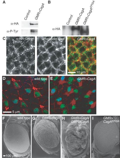
Western analysis of anti-HA affinity purified proteins from heads of adult UAS-CagA/GMR-GAL4 flies showed that CagA was expressed (α-HA) and phosphorylated (α-P-Tyr, Figure 1A). Similar to CagA's distribution in tissue culture cells [8],[29], we showed in the Drosophila eye disc CagA was localized predominantly to the cell cortex (Figure 1C). Examination of the cellular morphology of the pupal retina revealed that CagA expression caused disorganization of the epithelium. The wild type retinal epithelium is organized into regular cell clusters, each containing a single R7 and R8 photoreceptor (Figure 1D). In retina expressing CagA, the normal cell shapes and neighbor relationships were perturbed (Figure 1E), similar to CagA-dependent epithelial disorganization observed in mammalian tissue culture monolayers [29],[30]. When we examined the eyes of adult flies expressing a single copy of CagA with GMR-GAL4, we observed a perturbation of the normal crystalline array of the ommatidia (compare wild type, Figure 1F, with CagA expression, Figure 1G). Expression of two copies of the UAS-CagA transgene dramatically enhanced the eye phenotype, indicating that the developmental pathways disrupted were sensitive to the amount of CagA expressed (Figure 1H). Expressing one copy of the CagA mutant lacking the tyrosine phosphorylation sites (CagAEPISA) did not perturb the crystalline array of the adult eye to the extent caused by wild type CagA (Figure 1I) even though the CagAEPISA protein was expressed at similar levels as CagA (Figure 1B). Dose dependent perturbations of Drosophila eye patterning, as observed with CagA expression, have been used as the basis for genetic screens for modifiers of the rough eye phenotype to elucidate several signaling pathways, including RTK pathways. [17],[31]
Figure 1. CagA is phosphorylated, associates with the cortex in Drosophila cells and disrupts eye development.
(A) UAS-CagA was expressed in the Drosophila eye with GMR-GAL4. CagA protein (α-HA) was tyrosine phosphorylated (α-P-Tyr). Controls expressed only GMR-GAL4. (B) The CagA and CagAEPISA proteins were expressed in the eye to similar levels. (C) CagA (α-HA, green) localized to the cortex (phalloidin, red) of cells in the larval eye disc. Wild type (D) and UAS-CagA/GMR-GAL4 (E) pupal retinas were stained with MAb 24B10 (red) to outline the photoreceptors, and antibodies to visualize the R7 (green) and R8 (cyan) photoreceptors. Scanning electron microscope micrographs of adult eyes from flies with (F) one copy of GMR-GAL4 and no UAS transgene, (G) one copy of UAS-CagA, (H) two copies of UAS-CagA and (I) one copy of UAS-CagAEPISA.
CagA can substitute for the Drosophila Gab
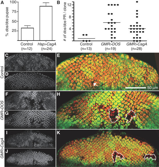
To test the hypothesis that CagA functions as a prokaryotic mimic of eukaryotic Gab proteins, we asked whether CagA expression could rescue phenotypes caused by the loss of the Drosophila Gab, DOS. DOS functions downstream of multiple RTKs during development, and homozygous dos loss-of-function mutants rarely develop into pupae and never survive to adulthood [16]. Rescue of dos mutants' lethality has been used as an in vivo assay to determine the function of specific domains of DOS [26]. We therefore determined the percentage of dos homozygous mutants that survived to the pupal stage of development with or without CagA expressed ubiquitously with temporal precision using the heat shock inducible Hsp-GAL4. The frequency of dos homozygous mutants was scored as a percentage of expected pupae that should develop if the dos mutants showed no lethality defect. As expected, a low percentage (33%) of homozygous dos mutant pupae expressing only Hsp-GAL4 were observed (Figure 2A). When CagA was expressed, we observed a significant increase to 89% of the pupae developing that lacked dos (Figure 2A). These results indicate that CagA can substitute for essential functions of DOS during Drosophila development.
Figure 2. CagA can substitute for the Drosophila Gab.
(A) Few homozygous Hsp-GAL4/+; dos/dos individuals survived to the pupal stage (control) measured as dos/dos pupae that developed as a percentage (%) of expected pupae that should develop if dos mutants were fully viable. Significantly more UAS-CagA/Hsp-GAL4; dos/dos pupae were recovered (Chi squared, p value<0.02). n = number experiments completed, with between 150-450 pupae examined in each experiment. Error bars indicate standard error. The dos/dos mitotic clones (marked by the absence of GFP (C, F, I, and green staining in E, H, K)), were induced using the FLP/FRT system in different genetic backgrounds, and photoreceptors were visualized with ELAV staining (D, G, J, and red staining in E, H, K). (B) The number of photoreceptors (PR) per dos/dos clones is shown for a representative experiment (n = number of eye discs examined). An area lacking GFP was counted as a clone if it was large enough to normally contain at least one ommatidium. (C–E) In control flies expressing GMR-GAL4, few dos/dos clones were observed. Any clones were miniscule (arrow in E) and contained few photoreceptors. Photoreceptors development was rescued by expression of either GMR-GAL4; UAS-DOS (F–H) or GMR-GAL4; UAS-CagA (I–K) as indicated by the formation of larger dos/dos clones containing several photoreceptors (clones are outlined in H and K).
To specifically test whether CagA could substitute for Gab in photoreceptor development, we generated mitotic dos/dos clones within the eye using the FLP/FRT recombinase system [27],[32]. In these experiments the dos mutation was recombined onto a chromosome arm containing a centromere proximal FRT recombination site and maintained in trans to a chromosome containing the same FRT site as well as a GFP transgene. By expressing FLP recombinase in the developing eye we induced mitotic recombination between FRT sites, which generated clones of homozygous cells (+/+ and dos/dos) in an otherwise heterozygous background (dos/+). The dos/dos mutant cells were distinguished by their lack of GFP, and the photoreceptors were visualized by staining for the photoreceptor-specific protein ELAV. As previously reported [16],[26] the dos/dos clones rarely contained photoreceptors and were composed of very few cells (Figure 2B–E), due to the dual requirements for EGFR signaling in cell survival and photoreceptor specification [23]. As expected, expression of DOS with GMR-GAL4 in dos/dos cells resulted in much larger clones with increased numbers of photoreceptors (Figure 2B, F–H). Expression of CagA in dos/dos cells was able to rescue clone size and photoreceptor development similarly to expression of DOS with the same driver (Figure 2B, I–K). Two independent dos mutants gave similar results (Figure 2 and data not shown). These data demonstrate that CagA can substitute for DOS during the development of photoreceptors.
CagA's specification of photoreceptors requires SHP-2/CSW
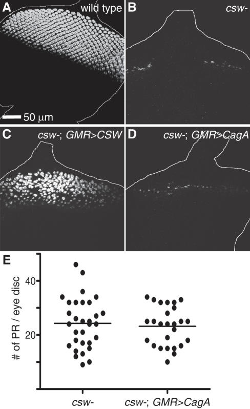
We predicted that if CagA functions similarly to Gab, then CagA would require the downstream signaling molecule SHP-2/CSW to promote photoreceptor development. As a downstream component of RTK pathways, CSW is required for photoreceptor development [17]. In contrast to wild type larval eye discs, in which thousands of photoreceptors are specified (Figure 3A), in larval eye discs of csw null mutants only a few photoreceptors develop along the morphogenetic furrow (Figure 3B, E) as described previously [33]. The residual photoreceptors in the csw eye discs were mostly R8 cells (data not shown), the only photoreceptor class that does not require RTK signaling for its specification [23]. A significant increase in photoreceptor number could be achieved in the csw mutant eye discs by expression of UAS-CSW with GMR-GAL4 (Figure 3C) or Hsp-GAL4 (data not shown). However, expression of CagA from multiple different transgenic lines using either GMR-GAL4 or Hsp-GAL4 failed to increase the number of photoreceptors in two different csw null mutants (Figure 3D, E, data not shown). These results argue that CagA, like DOS, requires SHP-2/CSW to promote photoreceptor development.
Figure 3. CagA's specification of photoreceptors requires SHP-2/CSW.
(A) Wild type larval eye discs contain thousands of photoreceptors, which were visualized by anti-ELAV staining. (B) Few photoreceptors develop in csw mutant larval eye discs. (C) Expression of UAS-CSW using GMR-GAL4 in the csw mutant partially rescued the lack of photoreceptor development but expression of UAS-CagA did not (D). (E) There was no significant difference (T-test, p value>0.2) in the number of photoreceptors (PR) that developed in csw mutants with or without CagA expression.
Discussion
We used a transgenic Drosophila system to test the hypothesis that H. pylori's virulence factor CagA can substitute for the Gab adaptor in RTK signaling pathways. This system is ideal for these studies because RTK signaling pathway components can be genetically manipulated, resulting in interpretable phenotypic consequences for tissue development. First, we have demonstrated that CagA in Drosophila tissue is phosphorylated, that it associates with the cell cortex, and that its expression causes epithelial disorganization as in mammalian tissue culture cells. Second, we have provided genetic evidence that CagA can substitute for Gab by demonstrating that CagA expression restores larval viability and photoreceptor development in mutants lacking the Drosophila Gab, DOS. Our inability to rescue dos mutants to adulthood with CagA expression may be due to differences in RTK activation or to non-overlapping functions of Gab and CagA. Indeed too much CagA expression (using an actin-GAL4 driver) is lethal to flies (unpublished results), which is not the case for ubiquitous expression of DOS [26]. Third, our genetic epistasis analysis with mutants lacking csw has shown that CagA functions through the Drosophila SHP-2 homologue, similar to results from tissue culture experiments [8],[9].
RTK signaling is essential for several fundamental biological processes and erroneous signaling can promote tumor formation [19]. Gain-of-function mutations of SHP-2 have been established as oncogenic in numerous leukemia types as well as other diseases like Noonan's Syndrome [4],[34],[35]. Over-expression of the Gab scaffolding adaptor proteins is associated with the development of several types of cancers, including breast cancer [6],[7] and gastric cancer [36]. The specific cancers that develop as a result of these mutations reflect tissue sensitivities to increased Gab and SHP-2. In the case of H. pylori infection, CagA provides a tissue specific activation of RTK signaling that can precipitate events leading to gastric carcinogenesis [37], as suggested by a recent report of CagA-expressing transgenic mice [38].
Our approach of examining the cellular effects of CagA expression in Drosophila tissue takes advantage of the fact that bacterial proteins frequently target essential, highly conserved cell-signaling pathways. Drosophila has been employed traditionally as a model organism for dissecting signaling pathways in development, but in recent years it has also proven useful in understanding host-pathogen interactions (reviewed in [39],[40]), and in one instance has been used as a heterologous system for expression of the bacterial toxins, anthrax lethal and edema factors [41]. Here we have exploited Drosophila eye development to demonstrate CagA's capacity to function as a RTK adaptor. Future studies using this transgenic Drosophila model will allow us to better understand the cellular and tissue-wide consequences of CagA's disruption of eukaryotic signaling pathways and to identify candidate host factors through which CagA functions.
Materials and Methods
Construction of UAS-CagA and UAS-CagAEPISA
CagA cDNA was amplified from genomic DNA from H. pylori G27. The CagAEPISA (lacking EPIYA tyrosine phosphorylation motifs) cDNA was amplified from a plasmid provided by Manuel Amieva (originally from Markus Stein [42]). CagAEPISA lacks the tyrosines in the four 5-amino acid motifs, EPIYA, which are phosphorylated by host kinases (point mutations at nucleotide 2684 [A→C] and 2740 [A→C] and a deletion at nucleotide 2878 to 3082). CagA and CagAEPISA were cloned into a modified pUAST vector with an N-terminal hemagglutinin (HA) tag (provided by Chris Q. Doe). Transgenic lines were generated by injecting Qiagen-purified plasmid DNA into y,w1118 embryos. Several independent transformant lines were established for each construct.
Drosophila Strains
Genetic null alleles of csw (cswC114 and csw13-87) and dos (dos1.46 and dos2.46) were obtained from Michael Simon. The UAS-DOS strain was from Thomas Raabe and the UAS-CSW strain (UAS-flgcsw[WTCIM]) from Lizabeth Perkins. UAS-CagA and UAS-CagAEPISA (lacking EPIYA tyrosine phosphorylation motifs) transgenes were expressed in the eye using P{w[+mC] = GAL4-ninaE.GMR}12 (GMR-GAL4, Bloomington Stock Center (BSC) # 1104). P{GAL4-Hsp70.PB}2 (Hsp-GAL4, BSC # 2077) was used for heat-shock inducible expression of transgenes.
Scanning Electron Microscopy
Fly heads were fixed overnight at 4°C in 2% gluteraldehyde in 0.1 M sodium cacodylate buffer (pH 7.2) and dehydrated through an ethanol series (30%, 50%, 70%, 80%, 90% 95%, three times in absolute ethanol) at room temperature for 10 minutes in each solution. Samples were critically point dried, sputter coated with gold and viewed using a JEOL 6400 SEM.
Larval Eye Discs
Eye imaginal discs were dissected from third instar wandering larvae, fixed for 30 minutes (4% formaldehyde, 0.1 M PIPES (pH 6.9), 0.3% Triton X-100, 2 mM EGTA, 1 mM MgSO4). Discs were washed (0.3% Triton X-100 in phosphate buffered saline, PBS) and blocked for one hour (1% BSA, 0.3% Triton X-100 in PBS). Primary antibodies included rat anti-ELAV 1∶10 (05HB 7E8A10, from Chris Q. Doe), rat anti-HA 1∶100 (Roche) and chicken anti-GFP 1∶2,000 (Chemicon). Secondary antibodies included anti- rat conjugated Rhodamine Red 1∶200 (Jackson ImmunoResearch), anti-rat conjugated AlexaFluor 488 1∶200 (Molecular Probes), anti-mouse conjugated Cy3 1∶200 (Jackson ImmunoResearch) and anti-chicken conjugated Cy2 1∶100 (Jackson ImmunoResearch). Phalloidin conjugated to Tetramethyl Rhodamine Iso-Thiocyanate (TRITC, Sigma Aldrich, 1∶500) was used to stain F-actin. Imaginal discs were visualized using a Nikon TE2000 U with C1 Digital Eclipse confocal microscope.
Pupal Retinas
Wandering third instar larvae were placed at 25°C and approximately 50 hours later the pupal retinas were dissected (50% pupal stage). Retinas were dissected in PBS, fixed for 20 minutes (4% paraformaldehyde in PBS) and washed three times in PBT (0.5% Triton X-100 in PBS). Retinas were blocked at least 15 minutes in 10% normal goat serum in PBT. Antibodies were diluted in the blocking solution. Primary antibodies included mouse MAb 24B10 which stains all photoreceptors and their axons [43] (Developmental Studies Hybridoma Bank, 1∶200), rabbit anti-SAL, which stains R7 and R8 nuclei (also called SPALT, provide by Reinhard Schuh [44], 1∶100), guinea pig anti-SENSELESS, which stains R8 nuclei (proved by Hugo Bellen [45], 1∶1000). Secondary antibodies from Molecular Probes included AlexaFluor 555 conjugated anti-mouse, AlexaFluor 488 conjugated anti-rabbit and AlexaFluor 633 conjugated anti-guinea pig, which were all used at 1∶250. Pupal retinas were visualized using a Leica TCS SP5 confocal microscope.
Western Analysis
Fly heads were collected by flash freezing adult flies in liquid nitrogen, shaking the flies in a conical tube, and then separating the heads from the bodies using a mesh sieve. Heads (∼1.5 mL) were homogenized in ice cold lysis buffer (50 mM Hepes, 150 mM NaCl, 1 mM EDTA, 1 mM Na3VO4, 0.5% Triton X-100 and Complete protease inhibitors [Roche]) and then centrifuged at 16,000 G for 5 minutes. Supernatant from the lysate solution (1.5 mL) was added to 50 µL anti-HA Affinity Matrix (Roche) which was incubated overnight at 4°C with gentle agitation. The anti-HA affinity matrix was washed 4 times with ice-cold lysis buffer. CagA was eluted from the matrix by boiling in 100 uL sample loading buffer and separated using manufactures protocols for 7% NuPAGE® Novex Tris-Acetate gels, transferred to polyvinylidene difluoride membranes, blocked overnight at 4°C (200 mM Tris pH 7.5, 100 mM NaCl, 0.1% Tween-20 and 3% BSA (Fisher)), probed using appropriate antibodies and detected using enhanced chemiluminescene (ECL plus, Amersham Biosciences). Mouse anti-HA was used at 1∶1,000 (Babco). Mouse anti-phospho tyrosine was used at 1∶2,000 (Cell Signal Technologies). Horseradish peroxidase-conjugated sheep anti-mouse (Amersham Biosciences) was used at 1∶5,000.
dos Assays
Hsp70-GAL4 balanced over CyO, P{Ubi-GFP} with dos2.42 over TM3, P{Act-GFP}, Ser were crossed to dos1.46/TM3, P{Act-GFP}, Ser (negative control) or UAS-CagA; dos1.46/TM3,P{Act-GFP}, Ser. Progeny were raised at 30°C and pupae were examined for GFP florescence using a Stemi SV 11 Apo Zeiss microscope. The number of non-GFP expressing progeny was scored as a percentage of the total number of pupae that developed per bottle and averaged across bottles of the same genotype. At least 12 bottles were scored per cross with between 150–450 pupae examined per bottle.
The FLP/FRT recombinase system was used to induce somatic clones in the eye [27]. Males y w,ey-FLP 3.5/Y; GMR-GAL4; FRT2, dos1.46/CyO-TM6B were crossed to P{ey-FLP.N}6, ry506 (BSC #5577); P{Ubi-GFP.nls}3L1 P{Ubi-GFP.nls}3L2 P{FRT(whs)}2A (BSC #5825) (negative control) or UAS-DOS; P{Ubi-GFP.nls}3L1 P{Ubi-GFP.nls}3L2 P{FRT(whs)}2A (positive control). Male GMR-GAL4, UAS-CagA; FRT2, dos1.46/CyO-TM6B were crossed to P{ey-FLP.N}6, ry506; P{Ubi-GFP.nls}3L1 P{Ubi-GFP.nls}3L2 P{FRT(whs)}2A. Imaginal eye discs were stained with anti-ELAV and anti-GFP antibodies.
csw Assay
Two genetic null alleles of csw were used to examine if CagA could rescue loss of csw. The cswC114 or csw13-87 alleles were balanced over FM7, P{Act-GFP} with GMR-GAL4 balanced over CyO, P{Ubi-GFP} on the second chromosome. These females were then crossed to males y1w1118, P{Ubi-GFP.nls}X1 P{FRT(whs)}9-2 (BSC # 5832)/Y (negative control), y1w1118, P{Ubi-GFP.nls}X1 P{FRT(whs)}9-2/Y; UAS-CSW (positive control) or y1w1118, P{Ubi-GFP.nls}X1 P{FRT(whs)}9-2/Y; UAS-CagA. Eye imaginal discs were dissected from male larvae.
Acknowledgments
We thank Michael Simon, Thomas Raabe, Lizabeth Perkins, and members of the Herman, Doe, and Johnson labs for reagents and technical assistance; David Reid, Lucy Cho, Mischa McGibney, and Chloe Slichter for help with fly husbandry; and Tory Herman, Adam Miller, Manuel Amieva, and members of the Guillemin lab for helpful discussions and critically reading the manuscript. The 24B10 antibody developed by S. Benzer was obtained from the Developmental Studies Hybridoma Bank developed under the auspices of the NICHD and maintained by The University of Iowa, Department of Biological Sciences, Iowa City, IA 52242.
Footnotes
The authors have declared that no competing interests exist.
This work was supported by NIH grant R01 DK075667 to KG and an American Heart Association pre-doctoral fellowship to CMB.
References
- 1.Rothenbacher D, Brenner H. Burden of Helicobacter pylori and H. pylori-related diseases in developed countries: recent developments and future implications. Microbes Infect. 2003;5:693–703. doi: 10.1016/s1286-4579(03)00111-4. [DOI] [PubMed] [Google Scholar]
- 2.Bourzac KM, Guillemin K. Helicobacter pylori-host cell interactions mediated by type IV secretion. Cell Microbiol. 2005;7:911–919. doi: 10.1111/j.1462-5822.2005.00541.x. [DOI] [PubMed] [Google Scholar]
- 3.Hatakeyama M. The role of Helicobacter pylori CagA in gastric carcinogenesis. Int J Hematol. 2006;84:301–308. doi: 10.1532/IJH97.06166. [DOI] [PubMed] [Google Scholar]
- 4.Neel BG, Gu H, Pao L. The ‘Shp’ing news: SH2 domain-containing tyrosine phosphatases in cell signaling. Trends Biochem Sci. 2003;28:284–293. doi: 10.1016/S0968-0004(03)00091-4. [DOI] [PubMed] [Google Scholar]
- 5.Liu Y, Rohrschneider LR. The gift of Gab. FEBS Lett. 2002;515:1–7. doi: 10.1016/s0014-5793(02)02425-0. [DOI] [PubMed] [Google Scholar]
- 6.Gu H, Neel BG. The “Gab” in signal transduction. Trends Cell Biol. 2003;13:122–130. doi: 10.1016/s0962-8924(03)00002-3. [DOI] [PubMed] [Google Scholar]
- 7.Nishida K, Hirano T. The role of Gab family scaffolding adapter proteins in the signal transduction of cytokine and growth factor receptors. Cancer Sci. 2003;94:1029–1033. doi: 10.1111/j.1349-7006.2003.tb01396.x. [DOI] [PMC free article] [PubMed] [Google Scholar]
- 8.Higashi H, Tsutsumi R, Muto S, Sugiyama T, Azuma T, et al. SHP-2 tyrosine phosphatase as an intracellular target of Helicobacter pylori CagA protein. Science. 2002;295:683–686. doi: 10.1126/science.1067147. [DOI] [PubMed] [Google Scholar]
- 9.Higuchi M, Tsutsumi R, Higashi H, Hatakeyama M. Conditional gene silencing utilizing the lac repressor reveals a role of SHP-2 in cagA-positive Helicobacter pylori pathogenicity. Cancer Sci. 2004;95:442–447. doi: 10.1111/j.1349-7006.2004.tb03229.x. [DOI] [PMC free article] [PubMed] [Google Scholar]
- 10.Mimuro H, Suzuki T, Tanaka J, Asahi M, Haas R, et al. Grb2 is a key mediator of Helicobacter pylori CagA protein activities. Mol Cell. 2002;10:745–755. doi: 10.1016/s1097-2765(02)00681-0. [DOI] [PubMed] [Google Scholar]
- 11.Churin Y, Al-Ghoul L, Kepp O, Meyer TF, Birchmeier W, et al. Helicobacter pylori CagA protein targets the c-Met receptor and enhances the motogenic response. J Cell Biol. 2003;161:249–255. doi: 10.1083/jcb.200208039. [DOI] [PMC free article] [PubMed] [Google Scholar]
- 12.Suzuki M, Mimuro H, Suzuki T, Park M, Yamamoto T, et al. Interaction of CagA with Crk plays an important role in Helicobacter pylori-induced loss of gastric epithelial cell adhesion. J Exp Med. 2005;202:1235–1247. doi: 10.1084/jem.20051027. [DOI] [PMC free article] [PubMed] [Google Scholar]
- 13.Hatakeyama M. Helicobacter pylori CagA--a potential bacterial oncoprotein that functionally mimics the mammalian Gab family of adaptor proteins. Microbes Infect. 2003;5:143–150. doi: 10.1016/s1286-4579(02)00085-0. [DOI] [PubMed] [Google Scholar]
- 14.Herbst R, Zhang X, Qin J, Simon MA. Recruitment of the protein tyrosine phosphatase CSW by DOS is an essential step during signaling by the sevenless receptor tyrosine kinase. EMBO J. 1999;18:6950–6961. doi: 10.1093/emboj/18.24.6950. [DOI] [PMC free article] [PubMed] [Google Scholar]
- 15.Herbst R, Carroll PM, Allard JD, Schilling J, Raabe T, et al. Daughter of sevenless is a substrate of the phosphotyrosine phosphatase Corkscrew and functions during sevenless signaling. Cell. 1996;85:899–909. doi: 10.1016/s0092-8674(00)81273-8. [DOI] [PubMed] [Google Scholar]
- 16.Raabe T, Riesgo-Escovar J, Liu X, Bausenwein BS, Deak P, et al. DOS, a novel pleckstrin homology domain-containing protein required for signal transduction between sevenless and Ras1 in Drosophila. Cell. 1996;85:911–920. doi: 10.1016/s0092-8674(00)81274-x. [DOI] [PubMed] [Google Scholar]
- 17.Simon MA, Bowtell DD, Dodson GS, Laverty TR, Rubin GM. Ras1 and a putative guanine nucleotide exchange factor perform crucial steps in signaling by the sevenless protein tyrosine kinase. Cell. 1991;67:701–716. doi: 10.1016/0092-8674(91)90065-7. [DOI] [PubMed] [Google Scholar]
- 18.Freeman RM, Jr, Plutzky J, Neel BG. Identification of a human src homology 2-containing protein-tyrosine-phosphatase: a putative homolog of Drosophila corkscrew. Proc Natl Acad Sci USA. 1992;89:11239–11243. doi: 10.1073/pnas.89.23.11239. [DOI] [PMC free article] [PubMed] [Google Scholar]
- 19.Schlessinger J. Cell signaling by receptor tyrosine kinases. Cell. 2000;103:211–225. doi: 10.1016/s0092-8674(00)00114-8. [DOI] [PubMed] [Google Scholar]
- 20.Duffy JB. GAL4 system in Drosophila: a fly geneticist's Swiss army knife. Genesis. 2002;34:1–15. doi: 10.1002/gene.10150. [DOI] [PubMed] [Google Scholar]
- 21.Theodosiou NA, Xu T. Use of FLP/FRT system to study Drosophila development. Methods. 1998;14:355–365. doi: 10.1006/meth.1998.0591. [DOI] [PubMed] [Google Scholar]
- 22.St Johnston D. The art and design of genetic screens: Drosophila melanogaster. Nat Rev Genet. 2002;3:176–188. doi: 10.1038/nrg751. [DOI] [PubMed] [Google Scholar]
- 23.Dominguez M, Wasserman JD, Freeman M. Multiple functions of the EGF receptor in Drosophila eye development. Curr Biol. 1998;8:1039–1048. doi: 10.1016/s0960-9822(98)70441-5. [DOI] [PubMed] [Google Scholar]
- 24.Tomlinson A, Ready DF. Sevenless: A Cell-Specific Homeotic Mutation of the Drosophila Eye. Science. 1986;231:400–402. doi: 10.1126/science.231.4736.400. [DOI] [PubMed] [Google Scholar]
- 25.Voas MG, Rebay I. Signal integration during development: insights from the Drosophila eye. Dev Dyn. 2004;229:162–175. doi: 10.1002/dvdy.10449. [DOI] [PubMed] [Google Scholar]
- 26.Bausenwein BS, Schmidt M, Mielke B, Raabe T. In vivo functional analysis of the daughter of sevenless protein in receptor tyrosine kinase signaling. Mech Dev. 2000;90:205–215. doi: 10.1016/s0925-4773(99)00252-x. [DOI] [PubMed] [Google Scholar]
- 27.Xu T, Rubin GM. Analysis of genetic mosaics in developing and adult Drosophila tissues. Development. 1993;117:1223–1237. doi: 10.1242/dev.117.4.1223. [DOI] [PubMed] [Google Scholar]
- 28.Bier E. Localized activation of RTK/MAPK pathways during Drosophila development. Bioessays. 1998;20:189–194. doi: 10.1002/(SICI)1521-1878(199803)20:3<189::AID-BIES1>3.0.CO;2-N. [DOI] [PubMed] [Google Scholar]
- 29.Bagnoli F, Buti L, Tompkins L, Covacci A, Amieva MR. Helicobacter pylori CagA induces a transition from polarized to invasive phenotypes in MDCK cells. Proc Natl Acad Sci U S A. 2005;102:16339–16344. doi: 10.1073/pnas.0502598102. [DOI] [PMC free article] [PubMed] [Google Scholar]
- 30.Amieva MR, Vogelmann R, Covacci A, Tompkins LS, Nelson WJ, et al. Disruption of the epithelial apical-junctional complex by Helicobacter pylori CagA. Science. 2003;300:1430–1434. doi: 10.1126/science.1081919. [DOI] [PMC free article] [PubMed] [Google Scholar]
- 31.Firth L, Manchester J, Lorenzen JA, Baron M, Perkins LA. Identification of genomic regions that interact with a viable allele of the Drosophila protein tyrosine phosphatase corkscrew. Genetics. 2000;156:733–748. doi: 10.1093/genetics/156.2.733. [DOI] [PMC free article] [PubMed] [Google Scholar]
- 32.Perrimon N. Creating mosaics in Drosophila. Int J Dev Biol. 1998;42:243–247. [PubMed] [Google Scholar]
- 33.Allard JD, Chang HC, Herbst R, McNeill H, Simon MA. The SH2-containing tyrosine phosphatase corkscrew is required during signaling by sevenless, Ras1 and Raf. Development. 1996;122:1137–1146. doi: 10.1242/dev.122.4.1137. [DOI] [PubMed] [Google Scholar]
- 34.Mohi MG, Neel BG. The role of Shp2 (PTPN11) in cancer. Curr Opin Genet Dev. 2007;17:23–30. doi: 10.1016/j.gde.2006.12.011. [DOI] [PubMed] [Google Scholar]
- 35.Ostman A, Hellberg C, Bohmer FD. Protein-tyrosine phosphatases and cancer. Nat Rev Cancer. 2006;6:307–320. doi: 10.1038/nrc1837. [DOI] [PubMed] [Google Scholar]
- 36.Lee SH, Jeong EG, Nam SW, Lee JY, Yoo NJ, et al. Increased expression of Gab2, a scaffolding adaptor of the tyrosine kinase signalling, in gastric carcinomas. Pathology. 2007;39:326–329. doi: 10.1080/00313020701329773. [DOI] [PubMed] [Google Scholar]
- 37.Mueller A, Falkow S, Amieva MR. Helicobacter pylori and gastric cancer: what can be learned by studying the response of gastric epithelial cells to the infection? Cancer Epidemiol Biomarkers Prev. 2005;14:1859–1864. doi: 10.1158/1055-9965.EPI-04-0820. [DOI] [PubMed] [Google Scholar]
- 38.Ohnishi N, Yuasa H, Tanaka S, Sawa H, Miura M, et al. Transgenic expression of Helicobacter pylori CagA induces gastrointestinal and hematopoietic neoplasms in mouse. Proc Natl Acad Sci U S A. 2008 doi: 10.1073/pnas.0711183105. [DOI] [PMC free article] [PubMed] [Google Scholar]
- 39.Vodovar N, Acosta C, Lemaitre B, Boccard F. Drosophila: a polyvalent model to decipher host-pathogen interactions. Trends Microbiol. 2004;12:235–242. doi: 10.1016/j.tim.2004.03.007. [DOI] [PubMed] [Google Scholar]
- 40.Dorer MS, Isberg RR. Non-vertebrate hosts in the analysis of host-pathogen interactions. Microbes Infect. 2006;8:1637–1646. doi: 10.1016/j.micinf.2005.11.020. [DOI] [PubMed] [Google Scholar]
- 41.Guichard A, Park JM, Cruz-Moreno B, Karin M, Bier E. Anthrax lethal factor and edema factor act on conserved targets in Drosophila. Proc Natl Acad Sci USA. 2006;103:3244–3249. doi: 10.1073/pnas.0510748103. [DOI] [PMC free article] [PubMed] [Google Scholar]
- 42.Stein M, Bagnoli F, Halenbeck R, Rappuoli R, Fantl WJ, et al. c-Src/Lyn kinases activate Helicobacter pylori CagA through tyrosine phosphorylation of the EPIYA motifs. Mol Microbiol. 2002;43:971–980. doi: 10.1046/j.1365-2958.2002.02781.x. [DOI] [PubMed] [Google Scholar]
- 43.Fujita SC, Zipursky SL, Benzer S, Ferrus A, Shotwell SL. Monoclonal antibodies against the Drosophila nervous system. Proc Natl Acad Sci U S A. 1982;79:7929–7933. doi: 10.1073/pnas.79.24.7929. [DOI] [PMC free article] [PubMed] [Google Scholar]
- 44.Kuhnlein RP, Frommer G, Friedrich M, Gonzalez-Gaitan M, Weber A, et al. spalt encodes an evolutionarily conserved zinc finger protein of novel structure which provides homeotic gene function in the head and tail region of the Drosophila embryo. Embo J. 1994;13:168–179. doi: 10.1002/j.1460-2075.1994.tb06246.x. [DOI] [PMC free article] [PubMed] [Google Scholar]
- 45.Nolo R, Abbott LA, Bellen HJ. Senseless, a Zn finger transcription factor, is necessary and sufficient for sensory organ development in Drosophila. Cell. 2000;102:349–362. doi: 10.1016/s0092-8674(00)00040-4. [DOI] [PubMed] [Google Scholar]


