Abstract
Kinematic properties of trunk extension are considered sensitive differentiators of movement between asymptomatic and low back pain subjects. The aim of this study was to quantify the continuous interaction of the hip and lumbar spine kinematics and temporal characteristics as a function of direction during the task of trunk bending backwards and returning to the upright position in healthy young subjects. The sagittal hip and lumbar spine kinematics during the extension task were examined in 18 healthy male subjects. Five trials of trunk extension were recorded for each subject and paired t-tests were then used to determine significant differences (P < 0.05) between the mean lumbar and the hip time-normalized kinematic and temporal variables. The data from the full cycle of trunk extension was analyzed with respect to movement initiation, time to reach peak velocity and peak angular displacement during the full cycle of trunk extension. Three distinct phases of movements were identified based on the continuous movement trajectories of velocity and angular displacement in the lumbar spine and hip; that of extension, return and, a terminal overcorrection phase. There were significant differences identified in the respective mean peak angular velocities of the lumbar spine (21.7 ± 8.6, 37.0 ± 14.7, 8.3 ± 5.0 deg/s) when compared with those of hip (14.6 ± 6.1, 21.7 ± 8.5, 5.4 ± 3.5 deg/s) in each of these three phases. The lumbar spine initiated the movement of trunk extension when bending backwards and returning to the upright position significantly early than that of the hip. These results highlight that in normal healthy adults there is the tendency for the lumbar spine to dominate over the hip during the task of backward trunk bending in terms of the amount and velocity of movement. At the end of extension the kinematics of the lumbar spine and hip kinematic are characterized by a terminal overcorrection phase marking the completion of the movement.
Keywords: Lumbar spine, Hip, Trunk extension, Kinematics, Velocity
Introduction
The movement of trunk extension is achieved through the coordinated rotation of the hip and the lumbar spine in the sagittal plane [5]. Trunk extension is clearly an important component of many functional activities and is considered essential in the clinical assessment of low back pain LBP [16]. Treatment protocols that incorporate extension of lumbar spine by moving the trunk backwards in the lying or standing position are also advocated by some to aid the resolution of LBP symptoms [16, 17].
Kinematic properties of trunk extension are considered sensitive differentiators of movement between asymptomatic and low back pain subjects. The motion of the lumbar spine and hip has been described using the kinematic parameters of angular displacement, velocity and acceleration [2, 8, 13, 18, 27]. Alterations in these variables are considered to be important indicators of spinal disorders [8–13]. For example, the results of studies on normal and LBP patients indicate that velocity during trunk extension is significantly reduced in LBP patients and that this loss is more marked than that seen in flexion [10, 12]. Further, the kinematic parameters of extension such as velocity and acceleration yield stronger potential to discriminate LBP populations than those of trunk flexion in certain conditions such as spondylolisthesis [10]. However, while extension velocity promises to be a useful measure to serve as an indicator of the trunk’s musculoskeletal status [12] there are still some confounding results on the most sensitive variable to use when discriminating LBP populations on the basis of time-indexed kinematic characteristics. For example, researchers report significant losses in the magnitude of lumbar spine movement in all directions, loss of hip flexion and altered hip and lumbar spine kinematics in LBP subjects while performing trunk movements [27] and between LBP subgroups, some of which involve the movement of lumbar spine extension [4, 25].
Even within the normative data, there is a lack of information regarding the lumbar spine and hip characteristics during the full cycle of trunk extension that is, from the fully upright position to extension and then back to the upright position. Previous kinematic studies investigating extension have either been directed to examination of thoracic spine relative to the hip [15] or, to that of lumbar spine and pelvic movement, from the fully flexed to the upright position [13, 19].
A further issue in kinematic studies of spinal motion is that while mean values of magnitude and velocity may reveal significant differences between healthy and LBP population groups, such measures may not be sensitive enough to detect individuals at their initial presentation [15]. However, Pal et al. [18] have shown that analysis of continuous motion profiles of lumbar spine and hip movement during trunk flexion effectively discriminates different movement patterns between individuals, even within a healthy population. These investigators found that by incorporating additional kinematic and temporal parameters such as the point of initiation in the movement cycle and peak velocity it was also possible to detect differences in the lumbar spine and hip characteristics during flexion and the corresponding return movement cycle [18]. Surprisingly, a thorough examination of the kinematic characteristics of the full cycle of trunk extension has not been thoroughly examined and it remains to be seen whether a more complete set of temporal and kinematic variables is able to discriminate between the lumbar spine and hip within this movement. More objective functional information regarding dynamic trunk performance is required to support the on-going work in the classification of symptom-based low back disorders [10]. Therefore, the aim of this study is to quantify the time-dependent hip and lumbar spine kinematics as a function of direction during the task of trunk bending backwards and returning to the upright position in healthy young subjects.
Materials and methods
Participants
Twenty young asymptomatic male participants were recruited from a university student population for the study. However, the kinematic and temporal data used for analysis was from 18 subjects as two participants had moved beyond 10° at their knees during the recording session and hence their data were removed from the analysis.
The anthropometric details of the 18 participants are: mean age, 20.7 ± 1.2 years (mean ± SD), mean height 1.80 ± 0.10 m, mean weight of 77.0 ± 12.10 kg and a mean body mass index (BMI) of 23.80 ± 3.8 kg/m2. The exclusion criteria for the study participants were no previous history of LBP requiring clinical intervention, orthopaedic disorders of the lower limb, neurological disorder or systemic connective tissue disease. Approval for the study was granted by the University Ethics Committee with all participants providing written informed consent prior to testing.
Measures
A three-dimensional (3-D) Motion Analysis System1 (MAS) with a set of 12 opto-electric cameras supported by EVaRT™ 4.0 software was used to record the sagittal movement pathways at a sampling rate of 60 Hz. In this current study, the calibration estimate of each camera within the MAS to detect a single point in the x-axis, in keeping the agreed acceptable error for this system, was 0.45 mm (±0.14 mm). Assessment of the validity of the method using skin surface measurement when applied to spinal motion analysis indicates that there is good agreement of the rotation angle of the lumbar spine and X-ray findings [22]. The trunk motion characteristics to be examined in this study of angular displacement and velocity have been shown to be highly repeatable especially when performed in the sagittal plane [10].
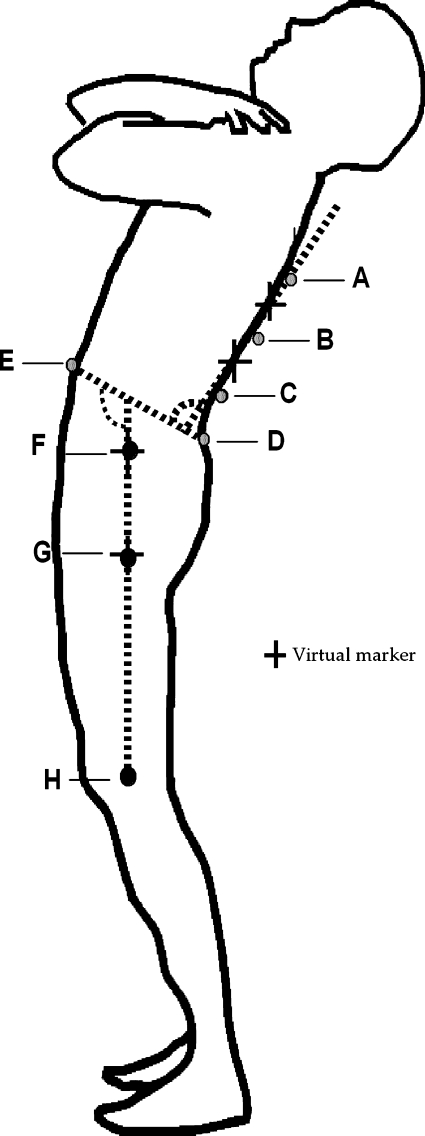
Thirteen retro-reflective surface markers (diameter of marker = 13 mm) were attached with double-sided adhesive tape to the lower limb, pelvic and spinal landmarks by an experienced musculoskeletal physiotherapist. The landmarks served to define a right-handed local co-ordinate system comprising the T9, T12 and L3 spinous processes, and bilaterally, the anterior and posterior superior iliac spines (ASIS, PSIS), greater trochanters (GT), lateral femoral epicondyles (LFE). Another surface marker was located at one-third of the distance between the GT and LFE (1/3 thigh) (Fig. 1). A full description of the measurement process and calculations of angular displacement and velocity variables are detailed in Pal et al. [18].
Fig. 1.
Schematic diagram of landmarks and virtual markers used to define the lumbar spine and hip angles. A T9, B T12, C L3, D posterior superior iliac spine, E anterior superior iliac spine, F greater trochanter, G 1/3 thigh and H lateral femoral epicondyle
Procedure
Following the collection of anthropometric measurements, each participant went through a standardised warm-up preparatory session of walking on a treadmill for 5 min at their own pace. The movements of extension (and return) and flexion (and return) were then demonstrated to the participant followed by a brief practice session. The movement sequence was randomised between extension (and return) and flexion (and return) and five trials of each movement were recorded for each participant. In order to standardise foot placement of the participants for the recording session, both feet were placed at a distance of 1/10 of the participant’s body height [6]. The participants were then instructed to extend backwards and return to the upright position without bending their knees. The knee angle for each trial was observed during the post-processing stage and if any subjects had an angle greater than 10° then that subject was eliminated from the study. It was emphasized in the instructions to the participants that they should move to the self-determined maximum end range of motion rather than pushing to the extreme possible limits of spinal motion. The participants moved at their own comfortable pace within a 10 s time frame at the sound of an audible signal. They were instructed to hold the extended position to the count of two before returning to the upright position. This latter instruction was included so that the movement patterns of extension and return could be distinguished from each other in the post-processing part of the analysis.
In the post-processing phase of the data the individual reflective marker paths of the recorded motion sequences were observed for potential error before smoothing with a 6 Hz Butterworth filter. The kinematic data describing the extension and return cycles for each of the five trials for 18 participants were then time normalized to 100 data points and transferred to a Microsoft Excel 2003 programme for analysis.
Analysis
The normalized data from the five trials from each participant (n = 18) were averaged in order to determine angular displacement (degrees), angular velocity (deg/s), time taken for the hip and lumbar spine during extension and return (expressed as a percentage of the total movement task time). The onset of lumbar spine and hip movement in each trial with respect to time was taken to be the first point in the cycle beyond 0.05% of angular displacement. The values were then pooled to provide absolute mean values (±95% confidence intervals). The absolute mean angular displacement and velocity patterns were presented graphically and descriptively. Paired t-tests were used to determine significant differences (P < 0.05) between the lumbar and hip variables during extension and return with respect to movement initiation, time to reach peak velocity and peak angular displacement.
Results
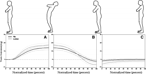
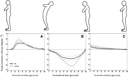
Analysis of the lumbar spine and hip kinematic data revealed three different phases of movement within the full cycle of trunk extension based on the continuous movement trajectories of velocity and displacement in the lumbar spine and hip. For the purposes of description these three phases are referred to as extension, return from extension and terminal overcorrection. The graphs of lumbar and hip absolute mean angular displacement and velocity during these three phases are plotted in Figs. 2a–c, 3a–c). The cycle duration in these graphs is depicted as percentage normalized to mean task time of each phase. The mean time taken to complete the extension, return from extension and terminal correction phases were calculated to be 2.8 ± 0.5, 1.6 ± 0.5 and 1.7 ± 0.8 s, respectively (Fig. 2).
Fig. 2.
A–C Mean sagittal angular displacement (deg) (±95% confidence intervals) about the lumbar spine and hip (n = 18) during the three phases of A Trunk extension B Return to the upright position and C Terminal overcorrection. The mean (standard deviation) time (s) taken to complete extension, return to the upright position and the terminal overcorrection phase were 2.80(0.50), 1.60(0.50) and 1.70(0.80) s, respectively
Fig. 3.
A–C Mean sagittal angular velocity (deg/s) (±95% confidence intervals) about the lumbar spine and hip (n = 18) during the three phases of A Extension from B Return to the upright position and C Terminal overcorrection
Lumbar spine and hip movement velocity
The lumbar spine and hip mean peak velocity values obtained during extension, return from extension and terminal overcorrection are given in Table 1. The mean peak angular velocities of the lumbar spine were statistically significantly greater than the hip in all three phases of movement (Table 1). However, it was only in the extension phase that a statistically significant difference (P < 0.011) was identified in the time taken to reach lumbar spine and hip mean peak angular velocity (32.3 ± 9.1 vs. 38.1 ± 10.0%, respectively (Table 2).
Table 1.
Summary of the kinematic interactions of the lumbar spine and hip during the three phases of trunk extension (n = 18)
| Kinematic variable | Lumbar spine mean (SD) | Hip mean (SD) | Mean differences (95% confidence interval) | P value |
|---|---|---|---|---|
| Trunk extension | ||||
| Peak angular displacement (deg) | 17.5 (5.6) | 13.9 (4.7) | 3.6 (0.6 to 6.6) | 0.022* |
| Peak velocity (deg/s) | 21.7 (8.6) | 14.6 (6.1) | 7.1 (3.3 to 10.9) | 0.001** |
| Return from extension | ||||
| Peak velocity (deg/s) | 37.0 (14.7) | 21.7 (8.5) | 15.3 (10.8 to 19.8) | 0.001** |
| Terminal overcorrection phase | ||||
| Peak angular displacement (deg) | −5.5 (3.5) | −1.7 (1.9) | −3.7 (−5.2 to −2.2) | 0.001** |
| Peak velocity (deg/s) | 8.3 (5.0) | 5.4 (3.5) | 2.9 (1.2 to 4.6) | 0.003* |
*P < 0.05, **P < 0.001
Table 2.
Summary of the temporal interactions of the lumbar spine and hip during the three phases of trunk extension (n = 18)
| Percent of movement cycle | Lumbar spine mean (SD) | Hip mean (SD) | Mean difference (95% confidence interval) | P value |
|---|---|---|---|---|
| Trunk extension | ||||
| Time to initiate extension (%) | 19.2 (4.2) | 24.3 (7.7) | 5.1 (8.1 to 2.1) | 0.002* |
| Time to reach peak extension velocity (%) | 32.3 (9.1) | 38.1 (10.0) | 5.8 (10.1 to 1.5) | 0.011* |
| Return trunk movement | ||||
| Time to initiate return (%) | 15.0 (5.2) | 20.7 (9.9) | 5.7 (10.8 to 19.8) | 0.001** |
| Terminal overcorrection phase | ||||
| Peak terminal overcorrection velocity (deg/s) | 8.3 (5.0) | 5.4 (3.5) | 2.9 (1.2 to 4.6) | 0.003* |
*P < 0.05, **P < 0.001
Movement profiles
The mean peak values of angular displacement of the lumbar spine and hip during extension and terminal correction are given in Fig. 2a–c and Table 1. Lumbar spine and hip angular displacement values were found to be significantly different in the extension phase (17.5 ± 5.6° vs. 13.9 ± 4.7°, respectively). At completion of the return from extension phase both the lumbar spine (5.5 ± 3.5°) and the hip values moved consistently into flexion (1.7 ± 1.9°) although there was a statistically significant difference in effect magnitude between these two structures. Fig. 2a–c illustrates the interaction between angular displacement of the lumbar spine and hip throughout the extension, return from extension and terminal correction phases. Within the temporal characteristics it can be seen that the lumbar spine initiated movement significantly earlier than the hip in both the upright to extension and return to upright phases (Table 2 ; Figs. 2, 3a–c).
Discussion
This study provides the first description of three phases of sagittal plane lumbar spine and hip movement, which takes place when the trunk is bent backwards and returns to the neutral posture position; that of extension, return to the upright position and a terminal overcorrection phase. The results also showed that there was a tendency for more movement to occur in the lumbar spine rather than the hip in all of these three phases. The impression that the lumbar spine dominated the extension and return movement task is supported further by the finding that the mean peak velocity of the lumbar spine was significantly different than that of the hip (Table 1) and, that the lumbar spine, not the hip, initiated the first two phases of movement.
The identification of a distinct overcorrection phase at the end of the movement whereby the lumbar spine and hip moved into ∼2°–6° flexion resulted from careful observation of the continuous motion profile data in the post-processing phase. The overcorrection phase is not an artifact as this feature was a consistent finding in each movement profile for all participants. Further, a similar pattern of overcorrection can be seen in the results of previous investigators where complete record tracings of trunk extension and return to the upright position are provided [20]. It also appears that the overcorrection phase is specific to the extension task as, to date, no previous investigators examining the kinematic properties of lumbar spine and hip flexion have reported any such phenomenon.
Whether or not the overcorrection phase is a local or centrally driven motor phenomenon is not clear but it is recognized that the spinal muscles operate with a complex strategy to control stability and motion [23]. This is particularly the case as the spine moves through the Neutral Zone, or the range of low passive stiffness [21] and where, the spinal muscles alone control the spine during dynamic flexion/extension movements [3, 23]. The overcorrection phase observed in this study may hypothetically be a response to the bracing effect of the abdominal and extensor muscles as the trunk moves beyond a certain biomechanical boundary.
An alternative proposal is that the overcorrection phase may be the manifestation of intersegmental motion phase lags, which take place in a stepwise fashion within the lumbar spine vertebrae during dynamic trunk flexion [7]. Our data suggest that flexion of the lumbar spine commenced in the terminal 30% of the return to the upright position, at which stage the motion lag within the spine would have commenced. Further, neither of these proposals excludes the possibility that the vestibular and associated motor control system may be driving the overcorrection phase and that it represents a novel postural strategy to maintain upright posture.
During both extension and return, movement initiation was observed primarily at the lumbar spine followed by the hip. We are not aware of this result being reported previously in the literature. The lumbar spine was also the predominant contributor to angular displacement although the overlap of confidence intervals suggests similar contributions of both structures to these movements, a result consistent with the research of Lee and Wong [8] and Wong and Lee [27]. The values for peak angular displacement for the lumbar spine and hip were also consistent with the values obtained from other studies [8, 27]. Although the lumbar spine demonstrated a significantly greater peak velocity during both extension and return it should also be noted that the kinematic patterns for both displacement and velocity for the lumbar spine and hip are closely matched.
The clinical assessment of patients with lumbar spine dysfunction includes testing for aberrant gross range of motion about the three axes of motion. The knowledge that the lumbar spine tends to undergo a greater magnitude of movement during the extension and return movement task than the hip is not adequate to assist with any differential diagnoses between the hip or lumbar region. However, coupled with the knowledge that the flexion and return movement involves different relative contributions from the hip and lumbar spine regions [8, 18, 27] than in the extension cycle, may be clinically relevant in understanding symptom behavior. This information may also be of value when designing therapeutic exercise programmes.
There are several limitations to the present study. Only one form of free-standing extension was examined and possible diurnal variations in data gathering were not taken into account. Some researchers are critical of evaluating motion within defined time limits [26] but the 10 s time frame used to complete the extension task in this study was very generous and allowed participants to complete the extension task in an unhurried manner. Further, in terms of test repeatability testing at a subject’s preferred speed during flexion and extension movements will produce more consistent readings of range of motion and velocity characteristics [14]. The present study was limited to male participants but here again, research indicates gender does not influence 3-D lumbar spine kinematics [24]. Finally, this study has used a non-invasive approach to kinematic measurement of free standing extension involving the use of surface markers to define bony landmarks. There is still debate about the error associated with the skin markers relative to the underlying skeleton. However, it is generally agreed that large motions such as flexion and extension have acceptable error limits with skin-base marker systems [1]. Furthermore the consistent pattern of inter-subject movement observations made in this study infers stable kinematic and temporal characteristics between the lumbar spine and hip during trunk extension at least in healthy subjects.
Conclusions
The results suggest that healthy subjects have an invariant pattern of lumbar spine and hip kinematic characteristics when extending the trunk and returning to the upright position. The healthy lumbar spine appears to dominate over the hip in a wide range of pelvic kinematic characteristics as the trunk extends and then reverses back to the upright position. The terminal overcorrection phase identified at the completion of the movement trunk extension cycle is a phenomenon, which may prove useful in further discrimination of the lumbar spine and hip kinematics of between and within different LBP population groups.
Footnotes
Motion Analysis™ Motion Analysis Corporation, California, USA.
References
- 1.Alexander EJ, Andriacchi TP. Correction for deformation in skin-based marker systems. J Biomech. 2001;34:355–361. doi: 10.1016/S0021-9290(00)00192-5. [DOI] [PubMed] [Google Scholar]
- 2.Esola MA, McClure PW, Fitzgerald GK, Siegler S. Analysis of lumbar spine and hip motion during forward bending in subjects with and without a history of low back pain. Spine. 1996;21:71–78. doi: 10.1097/00007632-199601010-00017. [DOI] [PubMed] [Google Scholar]
- 3.Gay RE, Ilharreborde B, Zhao K, Zhao C, An KN. Sagittal plane motion in the human lumbar spine: comparison of the in vitro quasistatic neutral zone and dynamic motion parameters. Clin Biomech. 2006;21:914–919. doi: 10.1016/j.clinbiomech.2006.04.009. [DOI] [PubMed] [Google Scholar]
- 4.Gombatto SP, Collins DR, Sahrmann SA, Engsberg JR, Dillen LR. Patterns of lumbar region movement during trunk lateral bending in 2 subgroups of people with low back pain. Phys Ther. 2007;87:441–454. doi: 10.2522/ptj.20050370. [DOI] [PubMed] [Google Scholar]
- 5.Granata KP, England SA. Stability of dynamic trunk movement. Spine. 2006;31:E271–276. doi: 10.1097/01.brs.0000216445.28943.d1. [DOI] [PMC free article] [PubMed] [Google Scholar]
- 6.Jayaraman G, Nazre AA, McCann V, Redford JB. A computerized technique for analysing lateral bending behaviour of subjects with normal and impaired lumbar spine. A pilot study. Spine. 1994;19:824–832. doi: 10.1097/00007632-199404000-00017. [DOI] [PubMed] [Google Scholar]
- 7.Kanayama M, Abumi K, Kaneda K, Tadano S, et al. Phase lag of the intersegmental motion in flexion-extension of the lumbar and lumbosacral spine. An in vivo study. Spine. 1996;21:1416–1422. doi: 10.1097/00007632-199606150-00004. [DOI] [PubMed] [Google Scholar]
- 8.Lee RYW, Wong TKT. Relationship between the movements of the lumbar spine and hip. Hum Mov Sci. 2002;21:481–494. doi: 10.1016/S0167-9457(02)00117-3. [DOI] [PubMed] [Google Scholar]
- 9.Marras WS, Lewis KE, Ferguson SA, Parnianpour M. Impairment magnification during dynamic trunk motions. Spine. 2000;25:587–595. doi: 10.1097/00007632-200003010-00009. [DOI] [PubMed] [Google Scholar]
- 10.Marras WS, Parnianpour M, Ferguson SA, Kim JY, et al. The classification of anatomic- and symptom-based low back disorders using motion measure models. Spine. 1995;20:2531–2546. doi: 10.1097/00007632-199512000-00013. [DOI] [PubMed] [Google Scholar]
- 11.Marras WS, Parnianpour M, Ferugson SA, Kim JY, et al. Quantification and classification of low back disorders based on trunk motion. Eur J Phys Med Rehabil. 1993;3:218–235. [Google Scholar]
- 12.Marras WS, Wongsam PE. Flexibility and velocity of the normal and impaired lumbar spine. Arch Phys Med Rehabil. 1986;67:213–217. [PubMed] [Google Scholar]
- 13.McClure PW, Esola M, Schreier R, Siegler S. Kinematic analysis of lumbar and hip motion while rising from a forward, flexed position in patients with and without a history of low back pain. Spine. 1997;22:552–558. doi: 10.1097/00007632-199703010-00019. [DOI] [PubMed] [Google Scholar]
- 14.McGregor AH, Hughes SPF. The effect of test speed on the motion characteristics of the lumbar spine during an A–P flexion–extension test. J Back Musculoskeletal Rehabil. 2000;14:99–104. [Google Scholar]
- 15.McGregor AH, McCarthy ID, Hughes SP. Motion characteristics of the lumbar spine in the normal population. Spine. 1995;20:2421–2428. doi: 10.1097/00007632-199511001-00009. [DOI] [PubMed] [Google Scholar]
- 16.McKenzie RA. The lumbar spine: mechanical diagnosis and therapy. Waikanae: Spinal Publications; 1981. [Google Scholar]
- 17.McKenzie RA, May S. The lumbar spine: mechanical diagnosis and therapy. Waikanae: Spinal Publications; 2003. [Google Scholar]
- 18.Pal P, Milosavljevic S, Sole G, Johnson GJ. Hip and lumbar continuous motion characteristics during flexion and return in young healthy males. Eur Spine J. 2006;16:741–747. doi: 10.1007/s00586-006-0200-2. [DOI] [PMC free article] [PubMed] [Google Scholar]
- 19.Paquet N, Malouin F, Richards CL. Hip-spine movement interaction and muscle activation patterns during sagittal trunk movements in low back pain patients. Spine. 1994;19:596–603. doi: 10.1097/00007632-199403000-00016. [DOI] [PubMed] [Google Scholar]
- 20.Pearcy MJ, Gill JM, Whittle MW, Johnson GR. Dynamic back movement measured using a three-dimensional television system. J Biomech. 1987;20:943–949. doi: 10.1016/0021-9290(87)90323-X. [DOI] [PubMed] [Google Scholar]
- 21.Scanell JP, McGill SM. Lumbar posture–should it, and can it, be modified? A study of passive tissue stiffness and lumbar position during activities of daily living. Phys Ther. 2003;83:907–17. [PubMed] [Google Scholar]
- 22.Schuit D, Petersen C, Johnson R, Levine P, Knecht H, Goldberg D. Validity and reliability of measures obtained from the OSI CA-6000 spine motion analyzer for lumbar spinal motion. Man Ther. 1997;2:206–215. doi: 10.1054/math.1997.0301. [DOI] [PubMed] [Google Scholar]
- 23.Thompson RE, Barker TM, Pearcy MJ. Defining the neutral zone of sheep intervertebral joints during dynamic motions: an in vitro study. Clin Biomech. 2003;18:89–98. doi: 10.1016/S0268-0033(02)00180-8. [DOI] [PubMed] [Google Scholar]
- 24.Vachalathiti R, Crosbie J, Smith R. Effects of age, gender and speed on three dimensional lumbar spine kinematics. Aust J Physiother. 1995;41:245–253. doi: 10.1016/S0004-9514(14)60433-5. [DOI] [PubMed] [Google Scholar]
- 25.Dillen LR, Gombatto SP, Collins DR, Engsberg JR, Sahrmann SA. Symmetry of timing of hip and lumbopelvic rotation motion in 2 different subgroups of people with low back pain. Arch Phys Med Rehabil. 2007;88:351–60. doi: 10.1016/j.apmr.2006.12.021. [DOI] [PubMed] [Google Scholar]
- 26.Wong KW, Luk KD, Leong JC, Wong SF, et al. Continuous dynamic spinal motion analysis. Spine. 2006;31:414–419. doi: 10.1097/01.brs.0000199955.87517.82. [DOI] [PubMed] [Google Scholar]
- 27.Wong TKT, Lee RYW. Effects of low back pain on the relationship between the movements of the lumbar spine and hip. Hum Mov Sci. 2004;23:21–34. doi: 10.1016/j.humov.2004.03.004. [DOI] [PubMed] [Google Scholar]


