Abstract
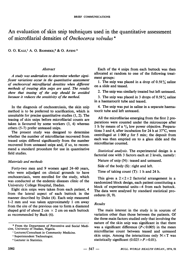
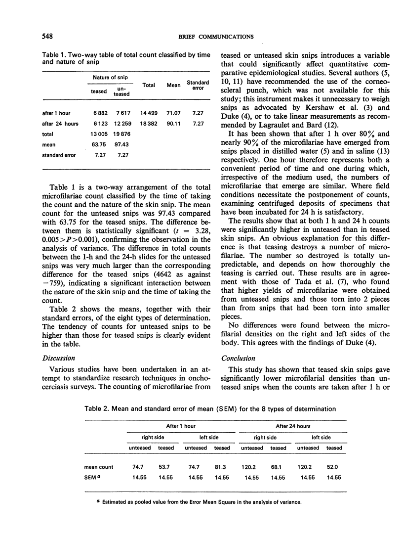
A study was undertaken to determine whether significant variations occur in the quantitative assessment of onchocercal microfilarial densities when different methods of treating skin snips are used. The results show that teasing of the snip should be avoided because it reduces the sensitivity of the method.
Full text
PDF


Selected References
These references are in PubMed. This may not be the complete list of references from this article.
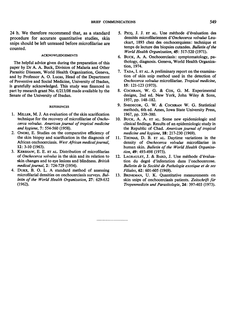
- KERSHAW W. E., DUKE B. O., BUDDEN F. H. Distribution of microfilariae of O. volvulus in the skin; its relation to the skin changes and to eye lesions and blindness. Br Med J. 1954 Sep 25;2(4890):724–729. doi: 10.1136/bmj.2.4890.724. [DOI] [PMC free article] [PubMed] [Google Scholar]
- Lagraulet J., Bard J. Une méthode d'évaluation du degré d'infestation dans l'onchocercose. Bull Soc Pathol Exot Filiales. 1969 May-Jun;62(3):601–605. [PubMed] [Google Scholar]
- MILLER M. J. An evaluation of the skin scarification technic for the recovery of microfilariae of Onchocerca volvulus. Am J Trop Med Hyg. 1958 Sep;7(5):554–557. doi: 10.4269/ajtmh.1958.7.554. [DOI] [PubMed] [Google Scholar]
- Picq J. J., Coz J., Jardel J. P. Une methode d'evaluation des densites microfilariennes d'Onchocerca volvulus Leuckart, 1893 chez des onchocerquiens: technique et temps de lecture des biopsies cutanees. Bull World Health Organ. 1971;45(4):517–520. [PMC free article] [PubMed] [Google Scholar]
- Thomas D. B., Anderson R. I., MacRae A. A. Daytime variation in the density of Onchocerca volvulus microfilariae in human skin. Bull World Health Organ. 1973;49(5):493–498. [PMC free article] [PubMed] [Google Scholar]


