
Fig. 1.
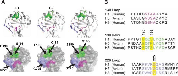
Structural and genetic basis for hemagglutinin mutations. (A) The RBDs of alternative viral hemagglutinins are shown. (B) Comparison of amino acid sequences in the major 130 and 220 loops and the 190 helix, color-coded in purple, lavender, and green, respectively.