Abstract
The tail of the earliest known articulated fully skeletonized vertebrate, the arandaspid Sacabambaspis from the Ordovician of Bolivia, is redescribed on the basis of further preparation of the only specimen in which it is most extensively preserved. The first, but soon discarded, reconstruction, which assumed the presence of a long horizontal notochordal lobe separating equal sized dorsal and ventral fin webs, appears to have considerable merit. Although the ventral web is significantly smaller than the dorsal one, the presence of a very long notochordal lobe bearing a small terminal web is confirmed. The discrepancy in the size of the ventral and dorsal webs rather suggests that the tail was hypocercal, a condition that would better accord with the caudal morphology of the living agnathans and the other jawless stem gnathostomes.
Keywords: Vertebrata, Arandaspida, Ordovician, caudal fin
1. Introduction
The anatomy of the earliest known articulated vertebrate possessing an extensive dermal skeleton, Sacabambaspis janvieri (Gagnier, Blieck & Rodrigo, 1986), from the Ordovician (Llanvirn and Caradoc) of South America, has been described in detail by Gagnier (1993a), on the basis of several specimens from the locality of Sacabambilla, Cochabamba area, Bolivia. Although the head armour, body scales and histology of this ‘ostracoderm’ (armoured jawless vertebrate) are now relatively well known (Gagnier 1993a,b; Sansom et al. 2005), the morphology of its caudal fin remains a puzzle and has been interpreted in a number of different ways. Further preparation of the only specimen that displays the caudal fin web now allows its reconstruction, which lends support to Gagnier's (1989) long debated reconstruction although with some modification, and provides clear evidence for the structure of the oldest recorded ostracoderm tail fin.
2. Material and methods
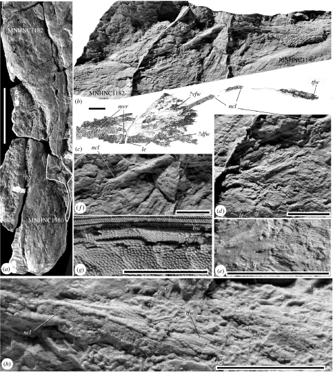
The material considered comes from the Ordovician (Caradoc) Anzaldo Formation of Bolivia. The articulated Sacabambaspis material from Sacabambilla consists of a number of three-dimensional specimens preserved in a very large concretion and, at least, six dorsoventrally flattened specimens preserved in a large sandstone slab. The specimens are housed in the Museo de Historia Natural Alcide d'Orbigny (MHNC), Cochabamba and (as a temporary deposit) in the Museum National d'Histoire Naturelle, Paris. The specimen MHNC 1182 (figure 1a), which displays the caudal fin, comes from the sandstone slab and has been further prepared by removing a small part of the overlying head shield of another, neighbouring, articulated specimen (MHNC 1180). The dermoskeleton of the caudal region has been removed with dilute hydrochloric acid, an elastomer cast of the resulting external mould made, whitened with magnesium and photographed.
Figure 1.
Sacabambaspis janvieri Gagnier et al. 1986, Ordovician (Caradoc), Anzaldo Formation, Sacabambilla Bolivia. (a) Specimens MHNC 1182 and 1180 in ventral view, caudal region outlined (thick white line) and median ventral ridge indicated (thin white line). (b–f, h) Elastomer cast of a portion of the counterpart of MHNC 1182 and 1180. (b) General view of the caudal region of MHNC 1182 and part of the ventral shield of MHNC 1180 (partly removed to expose the tip of the notochordal lobe). (c) Explanatory drawing of the part of the caudal region in (b). (d) Ventral and dorsal fin webs. (e) Detail view of the parallel scale series in the presumed dorsal web. (f) Detail view of the proximal part of the notochordal lobe; compare with the epibranchial plate of MHNC 1011 illustrated in (g). (h) Posterior end of the notochordal lobe and adjacent fin ray-like scale rows of the terminal fin web. Scale bars, (a) 100 mm, (b–h) 10 mm. Abbreviations: brpl, branchial plates; bsc, body scales; ?dfw, probable dorsal fin web or epichordal lobe; epl, epibranchial plate; le, leading edge of the fin web; mvr, median ventral ridge; ncl, notochordal lobe; tfw, terminal fin web; ?vfw, probable ventral fin web or anal fin.
3. Previous interpretations
The overall morphology of Sacabambaspis has previously been reconstructed on the basis of a dozen of more or less complete articulated specimens. These show elongate, dorsally flattened and ventrally inflated head shields, and a trunk covered with elongated flank scales arranged in chevrons. Based upon a single specimen (MHNC 1182, which forms the basis for the current study), Gagnier reconstructed the tail region as having a symmetrical caudal fin web with an elongate cylindrical process emerging from the rear, originally interpreted as a horizontal notochordal lobe, analogous to that of the living coelacanth. This early reconstruction of the tail by Gagnier (1989, fig. 2) (Blieck et al. 1991, fig. 10a; Gagnier & Blieck 1992, fig. 3) still appears in some popular illustrations. Gagnier (1993a) mentioned a second specimen (MHNC 1186) that may display part of the tail, but the latter only shows a poorly informative patch of fin web.
Shortly after Gagnier's first descriptions, this interpretation of the tail was questioned (Soehn & Wilson 1990; Sansom et al. 2001), because no other fossil or living jawless vertebrate possesses a caudal fin with a long, axial, notochordal lobe, and owing to issues over the preservation of the tail region in this single specimen. The posterior extremity of the presumed notochordal lobe of the specimen MHNC 1182 was partly covered by the head shield of another Sacabambaspis specimen (MHNC 1180; figure 1a). Therefore, it was assumed that the minute square-shaped scales of the presumed notochordal lobe were in fact not part of the tail, but merely the impression of either an isolated epibranchial plate (figure 1g) or the shield margin of another, underlying, specimen. Subsequent reconstructions of Sacabambaspis thus show (as dashed lines) a leaf shaped, isocercal caudal fin, ending with an incomplete axial lobe (Gagnier 1992, fig. 4, 1993a, fig. 4; Janvier 1996, figs 1.1, 4.2b(i)).
4. Description
Considering the importance of this unique source of information about the structure of the tail in Ordovician vertebrates, since no other caudal fin is known to date in Ordovician fully skeletonized vertebrates, we decided to further prepare this specimen at the cost of the destruction of a small part of the overlying head shield MHNC 1180 that hid it. As described by Gagnier (1993a,b) and reiterated here, the body scales of MHNC 1182 (figure 1a) are exposed in ventral view and pass progressively to large patches of minute, elongated scales arranged in rows, which clearly indicate the presence of caudal fin webs (figure 1b,c–e). The posterior extension of the body axis (originally interpreted as the notochordal lobe) is definitely not part of an underlying epibranchial plate or shield margin (figure 1b–f, h). It continues posteriorly over about 7 cm, in the form of a roughly cylindrical squamation composed of slightly disjunct, square-shaped scales (ncl, figure 1f,h), and ends posteriorly with a small web covered with elongated scales that are similar to those of the larger two webs situated more anteriorly (tfw, figure 1h).
5. Results and discussion
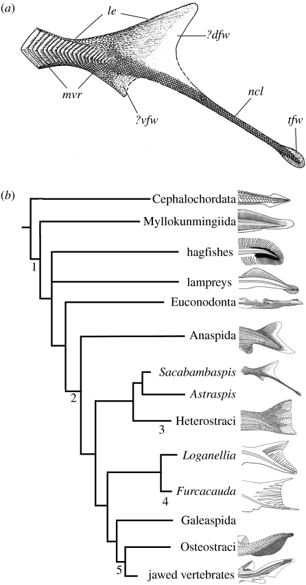
Gagnier's (1989) first reconstruction of the tail of Sacabambaspis, though subsequently discarded, is largely confirmed here, although with some significant modifications. The tail consists of relatively large dorsal and ventral webs and an elongated notochordal lobe, the posterior end of which is bordered by a small fin web (figure 2a). This tail structure clearly differs from that of heterostracans, which are currently grouped with arandaspids and astraspids in the clade Pteraspidomorphi (Gagnier 1993b, 1995; Donoghue & Smith 2001; Sansom et al. 2005), in which the caudal fin looks diphycercal (i.e. symmetrical) and strengthened by a few large radials (figure 2b; Janvier 1996).
Figure 2.
(a) Reconstruction of the caudal region in Sacabambaspis janvieri, assuming a moderately hypocercal condition, and the presence of a small ventral web. Same abbreviations as for figure 1. (b) Distribution of the hypo- and epicercal conditions of the tail in one of the current phylogenies of the major living and fossil vertebrate taxa. The position of the notochord (grey) is entirely hypothetical in the anaspids, heterostracans, osteostracans and the thelodonts Furcacauda and Loganellia. (Acrania as sister group to vertebrates; tree topology after Sansom et al. 2005.) See text for the characters at nodes (after Wilson & Caldwell 1993; Janvier 1996; Donoghue et al. 2000; Zhang & Hou 2004).
Despite these new data about the tail structure in Sacabambaspis, there remain some questions regarding its morphology, because the specimen MHNC 1182 is exposed in ventral aspect (figure 1a–c), but the caudal fin seems essentially exposed in lateral aspect. The tail must have been twisted at the level of the tail pedicle, and part of the fin web obscures the proximal part of the notochordal lobe (figure 1b,d). The scales of the median ventral ridge (mvr, figure 1c) seem to be in continuity with the smaller mass of fin web scales (?vfw, figure 1c) and, further back, the notochordal lobe, suggesting a considerable discrepancy in the size of the two fin webs. The actual outline of the larger, and presumably dorsal, mass of the fin web scale (?dfw, figure 1c) is unclear, except for the anterior part of its leading edge (le, figure 1c,d), which has been collapsed laterally.
A possibility is that the tail of Sacabambaspis may have been isocercal, with almost equal-sized webs extending dorsal and ventral to a horizontal notochordal lobe, as initially reconstructed by Gagnier (1989), and much like the tail of coelacanths, certain onychodonts, the primitive actinopterygian Dialipina, and certain fossil chondrichthyans, among crown-group gnathostomes (Janvier 1996; Schultze & Cumbaa 2001). This condition could be regarded as primitive for vertebrates, as it vaguely resembles the isocercal tail of cephalochordates, whose median fins are nevertheless not supported by cartilaginous radials (figure 2b). However, a more plausible interpretation that accounts for the discrepancy in fin web sizes and the relative position of the notochordal lobe is that it is moderately hypocercal (i.e. the caudal part of the notochord tapers posteroventrally), with an epichordal fin web extending dorsally to a smaller ventral web, possibly homologous to the anal fin (figure 2a). One may even wonder whether there are two webs, or a single, large, dorsal one, collapsed over the notochordal lobe during decay. The best proxy for the caudal fin of Sacabambaspis could be that of thelodonts, such as Loganellia, which possess an elongated notochordal lobe (figure 2b; Turner 1991). Indeed, this proposition seems to be supported by the collapse of the specimen itself, the fine scales within what we describe as the dorsal fin web covers a considerably greater area than the ventral fin web (figure 1c), this orientation being supported by the positioning of the scale masses with respect to the presumed median ventral ridge scales (mvr, figures 1c and 2a).
A hypocercal interpretation would make more sense in the light of the similar tail structure of most fossil and living jawless vertebrates (myllokunmingiids, hagfishes, lampreys, euconodonts, anaspids, most thelodonts; figure 2b); in all of these taxa, the notochord shows a slight ventral flexure and the notochordal lobe of the tail bends posteroventrally (Janvier 1996; Donoghue et al. 2000, Zhang & Hou 2004). In hagfishes, the hypocercal condition is not visible externally, but the tip of the notochord clearly bends posteroventrally (figure 2b). Osteostracans are the only jawless vertebrates that share with gnathostomes an epicercal tail; that is, the caudal part of the notochord tapers posterodorsally (figure 2b). The apparently diphycercal, hand-shaped, aspect of the tail in heterostracans and furcacaudiform thelodonts (e.g. Furcacauda; figure 2b; Wilson & Caldwell 1993; Janvier 1996) may merely be a particular case of the hypocercal condition, where the epichordal web and the notochordal lobe have become equal in length. However, the position of the notochord in these taxa remains unknown.
This interpretation of the Sacabambaspis tail would also better agree with the distribution of tail characters in current vertebrate phylogenies (e.g. Donoghue & Smith 2001), where a hypocercal condition seems to be the general condition for vertebrates (1, figure 2b), subsequently modified into superficially diphycercal (heterostracans, furcacaudiforms; 3, 4, figure 2b) and actually epicercal (osteostracans, jawed gnathostomes; 5, figure 2b) conditions. This, however, implies that the anal fin (2, figure 2b), possibly represented by the ventral web (if present) in Sacabambaspis, has been lost in the other pteraspidomorphs, galeaspids and osteostracans.
References
- Blieck A, Elliott D.K, Gagnier P.-Y. Some questions concerning the phylogenetic relationships of heterostracans, Ordovician to Devonian jawless vertebrates. In: Chang M.M, Liu Y.H, Zhang G.R, editors. Early vertebrates and related problems of evolutionary biology. Science Press; Beijing, China: 1991. pp. 1–17. [Google Scholar]
- Donoghue P.C.J, Smith M.P. The anatomy of Turinia pagei (Powrie) and the phylogenetic status of the Thelodonti. Trans. R. Soc. Edin. (Earth Sci.) 2001;92:15–37. [Google Scholar]
- Donoghue P.C.J, Forey P.L, Aldridge R.J. Conodont affinity and chordate phylogeny. Biol. Rev. 2000;75:191–251. doi: 10.1017/s0006323199005472. doi:10.1017/S0006323199005472 [DOI] [PubMed] [Google Scholar]
- Gagnier P.-Y. The oldest vertebrate: a 470 million year-old fish, Sacabambaspis janvieri, from the Ordovician of Bolivia. Natl Geogr. Res. 1989;5:250–253. [Google Scholar]
- Gagnier P.-Y. Ordovician vertebrates from Bolivia. In: Suarez-Soruco R, editor. Fosiles y facies de Bolivia. I. Vertebrados. Revista Técnica de YPFB. Vol. 12. 1992. 371–379. [Google Scholar]
- Gagnier P.-Y. Sacabambaspis janvieri, Vertébré ordovicien de Bolivie. 1, Analyse morphologique. Ann. Paléontol. 1993a;79:19–69. [Google Scholar]
- Gagnier P.-Y. Sacabambaspis janvieri, Vertébré ordovicien de Bolivie. 2, Analyse phylogénétique. Ann. Paléontol. 1993b;79:119–166. [Google Scholar]
- Gagnier P.-Y. Ordovician vertebrates and agnathan phylogeny. Bull. Mus. Natl Hist. Nat. 1995;17:1–37. Paris. [Google Scholar]
- Gagnier P.-Y, Blieck A. On Sacabambaspis janvieri and the vertebrate diversity in Ordovician seas. In: Mark-Kurik E, editor. Fossil fishes as living animals. Academia. Vol. 1. 1992. 9–20. [Google Scholar]
- Gagnier P.-Y, Blieck A, Rodrigo G. First Ordovician vertebrate from South America. Geobios. 1986;19:629–634. [Google Scholar]
- Janvier P. Oxford University Press; Oxford, UK: 1996. Early vertebrates. [Google Scholar]
- Sansom I.J, Smith M.M, Smith M.M. The Ordovician radiation of vertebrates. In: Ahlberg P.E, editor. Major events in vertebrate evolution. Taylor & Francis; London, UK: 2001. pp. 156–171. [Google Scholar]
- Sansom I.J, Donoghue P.C.J, Albanesi G. Histology and affinity of the earliest armoured vertebrate. Biol. Lett. 2005;1:446–449. doi: 10.1098/rsbl.2005.0349. doi:10.1098/rsbl.2005.0349 [DOI] [PMC free article] [PubMed] [Google Scholar]
- Schultze H.-P, Cumbaa S.L. Dialipina and the characters of basal actinopterygians. In: Ahlberg P.E, editor. Major events in vertebrate evolution. Taylor & Francis; London, UK: 2001. pp. 315–332. [Google Scholar]
- Soehn K.L, Wilson M.V.H. A complete, articulated heterostracan from Wenlockian (Silurian) beds of the Delorme Group, Mackenzie Mountains, Northwest Territories, Canada. J. Vert. Paleontol. 1990;10:405–419. [Google Scholar]
- Turner S. Monophyly and interrelationships of the Thelodonti. In: Chang M.M, Liu Y.H, Zhang G.R, editors. Early vertebrates and related problems of evolutionary biology. Science Press; Beijing, China: 1991. pp. 87–119. [Google Scholar]
- Wilson M.V.H, Caldwell M.W. New Silurian and Devonian fork-tailed “thelodonts” and jawless vertebrates with stomachs and deep bodies. Nature. 1993;361:442–444. doi:10.1038/361442a0 [Google Scholar]
- Zhang Y.-G, Hou X.-G. Evidence for a single median fin-fold in tail in the Lower Cambrian vertebrate, Haikouichthys ercaicunensis. J. Evol. Biol. 2004;17:1162–1166. doi: 10.1111/j.1420-9101.2004.00741.x. doi:10.1111/j.1420-9101.2004.00741.x [DOI] [PubMed] [Google Scholar]


