


Abstract
The free β-subunit of human chorionic gonadotropin β is expressed in several nontrophoblastic tumours and this is usually associated with aggressive disease. Little is known about human chorionic gonadotropin β expression in renal cancer. We determined the pretreatment levels of human chorionic gonadotropin β in serum of patients with renal cell carcinoma, and studied whether elevated levels predicted the clinical outcome. Serum samples were collected before surgery from 177 patients with renal cell carcinoma and from 84 apparently healthy controls. Human chorionic gonadotropin β in serum was measured by a highly sensitive time-resolved immunofluorometric assay. The prognostic value of human chorionic gonadotropin β, and of usual clinical and pathological variables was analyzed by the Kaplan-Meier method, the log rank test and Cox multiple hazard regression. The serum concentrations of human chorionic gonadotropin β were increased in 23% of the renal cell carcinoma patients and they were significantly higher in patients with renal cell carcinoma than in controls (P<0.0001). The concentrations did not correlate with clinical stage and histopathological grade, but patients with increased human chorionic gonadotropin β levels had significantly shorter survival time than those with levels below the median (cut-off 1.2 pmol l−1, P=0.0029). In multivariate analysis human chorionic gonadotropin β, tumour stage and grade were independent prognostic variables. The serum concentration of human chorionic gonadotropin β is an independent prognostic variable in renal cell carcinoma. The preoperative value of human chorionic gonadotropin β in serum may be used to identify patents with increased risk of progressive disease.
British Journal of Cancer (2002) 86, 185–189. DOI: 10.1038/sj/bjc/6600050 www.bjcancer.com
© 2002 The Cancer Research Campaign
Keywords: renal cell carcinoma, stage, grade, hCGβ, prognosis
The incidence of urological malignancies such as prostate cancer, bladder cancer and kidney cancer has been steadily increasing in the Western world during the past decades (Parkin et al, 1999). Renal cell carcinoma (RCC) arising in the renal parenchyma is the most common malignancy of the kidney. Conventional renal cell carcinoma (CRCC) and papillary renal cell carcinoma account for 85–90% of the five distinct types of RCCs. The fairly indolent chromophobe renal cell carcinoma and the unclassified category account for approximately 5% each, while the rarest and most aggressive type is collecting duct carcinoma (Kovacs et al, 1997). Clinical stage and nuclear grade are the most important prognostic factors in RCC (Fuhrman et al, 1982), but within a given disease stage the outcome is highly variable, and other prognostic indicators are needed to identify patients with high risk of progressive disease (Golimbu et al, 1986). Cell proliferation markers, p53 mutations, growth factor expression, and intratumoural microvessel density have been investigated as prognostic indicators, with partly contradicting results (Gelb et al, 1997; Grignon et al, 1995). Among the serum markers studied, VEGF, interleukin-10, CA-125, and tumour-associated trypsin inhibitor (TATI) have been shown a prognostic value, but no specific markers are available (Grankvist et al, 1997; Jacobsen et al, 2000; Paju et al, 2001; Wittke et al, 1999).
Human chorionic gonadotropin (hCG) is a 38 kDa glycoprotein hormone consisting of two dissimilar subunits, α and β (hCGα and hCGβ) (Bahl et al, 1972). HCG is normally produced by trophoblasts during pregnancy and it is a very sensitive marker for trophoblastic tumours (Vaitukaitis and Ebersole, 1976). Expression of hCGβ is a fairly common phenomenon in several non-trophoblastic tumours (Alfthan et al, 1992a; Marcillac et al, 1992), and in transitional cell carcinoma of the bladder (Iles et al, 1989) and several other tumours this is associated with aggressive and therapy resistant disease. Tissue expression of hCGβ seems to have prognostic significance in carcinomas of the prostate (Sheaff et al, 1996), and hCGβ in serum has been found to be of prognostic value in ovarian cancer (Ind et al, 1997). The aim of this study was to evaluate the prognostic value of hCGβ in sera of patients with RCC in relation to established prognostic factors.
MATERIALS AND METHODS
Patients
Data for this retrospective analysis was collected from 177 patients with RCC from whom pretreatment serum samples were available. The patients underwent radical nephrectomy at the University Hospital of Umeå, Sweden between 1983 and 1995. The study included 111 men and 66 women with a mean age of 65 years (range 25–85) (Table 1). Informed consent was obtained from all patients included in the study. Serum samples were collected before surgery and stored at −80°C until analysis. Staging procedures included physical examination, chest radiography, ultrasonography and computer tomography. Patients with skeletal symptoms or elevated serum alkaline phosphatase were assessed by bone scintigraphy and skeletal radiography. After nephrectomy, all patients were followed up with clinical and radiological examinations. At the last follow-up, 47 of 177 patients were alive with a median follow-up time of 124 months, (range 34–191 months). Among 130 patients that had died, 95 had died of RCC and 35 of other causes (Table 1). Sera from 84 apparently healthy volunteers was used as a reference group.
Table 1. Patient and tumour characteristics.

Tumour staging
Tumour staging was performed according to 1997 TNM Stage classification (Sobin and Wittekind, 1997). Among the 177 patients, 70 patients had TNM stage I and II (pT1–pT2, N0, M0), 54 stage III (pT1–T3a–c, N0–N1, M0) and 53 were of stage IV (pT1–T4, N0–N3, M1). Nuclear grading was performed according to Skinner et al (1971) and DNA ploidy according to Ljungberg et al (1996).
Serum determinations
HCGβ in serum was determined by a time-resolved immunofluorometric assay as earlier described (Alfthan et al, 1988). With a sample volume of 25 μl in a total assay volume of 225 μl the detection limit was 0.45 pmol l−1. The upper reference limit of the assay for hCGβ in serum was 2 pmol l−1. The reference range is identical in women and men and it is not dependent on age. Serum creatinine was measured by a routine method in the Laboratory of Clinical Chemistry, University Hospital, Umeå, Sweden. The upper reference limit was 125 μmol l−1.
Statistical analysis
Differences in serum hCGβ concentrations in patients with various stages and grades and controls were analyzed by the Mann-Whitney U-test. Survival curves were plotted using the Kaplan-Meier method, and comparison of survival times was performed with the log-rank test. Serum hCGβ was also studied as a categorical variable using quartiles (hCGβ <0.8 pmol l−1, 0.8–1.2 pmol l−1, 1.2–1.95 pmol l−1 and hCGβ >1.95 pmol l−1). The independence of hCGβ as a predictor of survival was analyzed by multivariate analysis using the Cox proportional hazard model with serum hCGβ, nuclear grade and stage as input variables. The statistical endpoint was survival measured from the date of nephrectomy to date of death or date of last follow-up. Cases of death from unrelated causes were censored. All tests were two-sided and P-values below 0.05 were considered significant.
RESULTS
The median concentration of creatinine in serum of RCC patients was 86 μmol l−1 (range 45–353 μmol l−1), and elevated levels occurred in 13%. The serum concentrations of hCGβ were not related to serum creatinine.
The concentration of hCGβ in serum was elevated (>2 pmol l−1) in 23% of the patients with RCC (Table 2) and 20 of these (11%) had values >4 pmol l−1. The median concentration of hCGβ in serum was 1.2 pmol l−1 (range 0.2–18 pmol l−1), which was significantly higher (P<0.0001) than in controls (median 0.4 pmol l−1, range 0.2–1.3 pmol l−1). There was no difference in hCGβ levels between males and females, different age groups, different RCC types, aneuploid and diploid tumours, or tumours with and without venous invasion. Serum hCGβ concentrations were not either significantly correlated with tumour stage or grade (Figure 1).
Table 2. Fraction of elevated serum hCGβ concentration in relation to stage.

Figure 1.
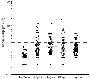
The distribution of hCGβ serum concentrations in controls and patients with various stages of RCC. The dashed line indicates the upper reference limit.
Clinical stage and grade were highly predictive of disease specific survival (P<0.0001 each) in univariate analysis. Patients with serum hCGβ concentrations above the median value (1.2 pmol l−1) had significantly shorter survival than those with lower levels (P=0.0029) (Figure 2A). A difference in survival time was observed also among patients with metastasized tumours (stage IV, Figure 2C) (P=0.06). When serum hCGβ concentration was compared as quartiles there was no difference in disease specific survival between the two lowest quartiles or between quartiles 3 and 4 (P>0.6). In multivariate analysis using the Cox regression model with age, gender, serum hCGβ, nuclear grade and stage as input variables, stage, grade and serum hCGβ concentration were independently associated with the disease specific survival (Table 3).
Figure 2.

Kaplan Meier cancer specific survival according to preoperative serum hCGβ concentration in patients with RCC. Results for patients with tumours of all stages (A), stages I-III (B) and stage IV (C). The median value of the patients (1.2 pmol l−1) was used as a cut off.
Table 3. Factors independently associated with decreased cumulative survival.

DISCUSSION
RCC is known for its unpredictable clinical behaviour. Recurrence can occur many years after surgery and metastases can spontaneously regress after removal of the primary tumour. In addition to clinical stage and grade, DNA ploidy has been found to be a prognostic factor (Ljungberg et al, 1996), but a marker available prior to surgery could be used to optimize treatment. Of the many serum markers studied, only a few, i.e. VEGF, interleukin-10, CA-125, and TATI are of potential prognostic value (Grankvist et al, 1997; Jacobsen et al, 2000; Paju et al, 2001; Wittke et al, 1999). Clinical parameters such as performance status, serum lactate dehydrogenase, hemoglobin, serum calcium and prior nephrectomy have also been used to predict survival of patients with metastatic RCC (Motzer et al, 1999).
HCG immunoreactivity occurs in many different molecular forms in serum and urine. During pregnancy, intact hCG is the main form in serum and the proportion of hCGβ is 1–8%. Various forms of hCG can also be detected in serum of non-pregnant women and men by highly sensitive methods. The concentrations of hCG in serum and hCG and hCGβ in urine increase with age, whereas those of hCGβ in serum are similar in men and women and change very little with age (Alfthan et al, 1992b). In addition to being a sensitive marker for trophoblastic tumours of placental or gonadal origin, hCGβ is also expressed by many non-trophoblastic tumours including ovarian, gastrointestinal and squamous cell carcinomas. Contrary to trophoblastic tumours, these very seldom express intact hCG. A very moderate (up to two-fold) increase of hCGβ in serum may occur in patients with benign pancreatic and hepatobiliary diseases, but this does not invalidate the use of hCGβ as a tumour marker (Alfthan et al, 1992a).
Expression of hCGβ has been observed in various tumours and cell lines on both at the protein and mRNA level (Alfthan et al, 1992a; Iles et al, 1989; Ind et al, 1997; Sheaff et al, 1996), but there is little data on the expression in renal tumours. Increased concentrations of hCGβ have been demonstrated by radioimmunoassay in concentrated urine specimens from RCC patients (Fukutani et al, 1983), but the study included very few cases. The present study shows that the concentrations of hCGβ in serum were elevated in 23% of the patients, and the median concentrations were significantly higher than in controls (P<0.0001). Patients with serum hCGβ levels above the median value (1.2 pmol l−1) had significantly shorter survival than those with lower levels (P<0.0029). In multivariate analysis serum hCGβ, tumour stage and grade were independent prognostic variables. The fairly low frequency (23%) of elevated serum hCGβ suggest that this marker is expressed by only a part of the RCCs. However, because normal levels between 1.2 and 2 pmol l−1 were associated with adverse prognosis, it is possible that part of the hCGβ in these patients is derived from the tumour. A possible correlation with the response to treatment could not be evaluated in this study. The absence of a correlation with stage indicates that elevated levels are not only a result of tumour burden, but hCGβ expression rather characterizes a subgroup of tumours. The lack of significant correlation with established prognostic variables such as grade, stage and DNA-ploidy demonstrates the independent character of this marker.
HCGβ expression has been regarded as a characteristic of malignant transformation or dedifferentiation of cells, and its association with aggressive disease in transitional cell carcinoma of the bladder is well documented (Iles et al, 1989). The association with poor prognosis in RCC is in line with the results for other tumours. Free hCGβ has no known hormonal activity, but it has been shown to stimulate phospholipid methylation in rat Leydig cells (Ronco et al, 1993), rendering it a potential biological role. The three dimensional structure of hCGβ resembles that of the cysteine knot growth factors (Lapthorn et al, 1994), and it has a stimulating effect on the growth of cancer cells in vitro, suggesting paracrine or autocrine growth factor-like activity. Recently hCGβ has been shown to confer immortality features to cells by inhibiting apoptosis (Butler et al, 2000). This is a potential explanation for aggressive tumour behaviour of hCGβ expressing tumours.
The clinical utility of hCGβ in the management of RCC is limited by the infrequent expression. About one fourth of the patients had elevated levels and in half of these the elevation was substantial. However, a prognostic value was observed even with normal levels exceeding the median value, and this was the case also in patients with advanced disease. Thus it appears worthwhile to study whether hCGβ could be used as an aid in the selection of therapy or in monitoring of the disease after primary therapy. In conclusion, our results indicate that hCGβ in serum is a prognostic factor independent of stage and grade that can be used to identify a subgroup of patients with increased risk of aggressive disease.
Acknowledgments
This study was supported by Lions Cancer Research Foundation of Umeå University, Finnish Cancer Foundation, Sigrid Juselius Foundation, Helsinki University Central Hospital, University of Helsinki, The Finska Läkaresällskapet, K Albin Johanssons stiftelse.
References
- AlfthanHHaglundCDabekJStenmanU-H1992bConcentrations of human choriogonadotropin, its beta-subunit, and the core fragment of the beta-subunit in serum and urine of men and nonpregnant women Clin Chem 3819811987 [PubMed] [Google Scholar]
- AlfthanHHaglundCRobertsPStenmanU-H1992aElevation of free β subunit of human choriogonadotropin and core β fragment of human choriogonadotropin in serum and urine of patients with malignant pancreatic and biliary disease Cancer Res 5246284633 [PubMed] [Google Scholar]
- AlfthanHSchröderJFraserRKoskimiesAHalilaHStenmanU-H1988Choriogonadotropin and its beta subunit separated by hydrophobic-interaction chromatography and quantified in serum during pregnancy by time-resolved immunofluorometric assays Clin Chem 3417581762 [PubMed] [Google Scholar]
- BahlOPCarlsenRBBellisarioRSwaminathanN1972Human chorionic gonadotropin, amino acid sequence of the alpha and beta subunits Biochem Biophys Res Commun 482416422 [DOI] [PubMed] [Google Scholar]
- ButlerSAIkramMSMathieuSIlesRK2000The increase in bladder carcinoma cell population induced by the free beta subunit of human chorionic gonadotropin is a result of an anti-apoptosis effect and not cell proliferation Br J Cancer 82915531556 [DOI] [PMC free article] [PubMed] [Google Scholar]
- FuhrmanSALaskyLCLimasC1982Prognostic significance of morphologic parameters in renal cell carcinoma Am J Surg Pathol 6655663 [DOI] [PubMed] [Google Scholar]
- FukutaniKLibbyJMPankoWBScardinoPT1983Human chorionic gonadotropin detected in urinary concentrates from patients with malignant tumors of the testis, prostate, bladder, ureter and kidney J Urol 1297477 [DOI] [PubMed] [Google Scholar]
- GelbABSudilovskyDWuCDWeissLMMedeirosLJ1997Appraisal of intratumoral microvessel density, MIB-1 score, DNA content, and p53 protein expression as prognostic indicators in patients with locally confined renal cell carcinoma Cancer 8017681775 [DOI] [PubMed] [Google Scholar]
- GolimbuMJoshiPSperberATesslerAAl-AskariSMoralesP1986Renal cell carcinoma, survival and prognostic factors Urology 27291301 [DOI] [PubMed] [Google Scholar]
- GrankvistKLjungbergBRasmusonT1997Evaluation of five glycoprotein tumour markers (CEA, CA-50, CA-19-9, CA-125, CA-15-3) for the prognosis of renal-cell carcinoma Int J Cancer 74233236 [DOI] [PubMed] [Google Scholar]
- GrignonDJAbdel-MalakMMertensWKosterJKeeneyMSakrWShepherdRR1995Prognostic significance of cellular proliferation in renal cell carcinoma, a comparison of synthesis-phase fraction and proliferating cell nuclear antigen index Mod Pathol 81824 [PubMed] [Google Scholar]
- IlesRKJenkinsBJOliverRTBlandyJPChardT1989Beta human chorionic gonadotropin in serum and urine. A marker for metastatic urothelial cancer Br J Urol 64241244 [DOI] [PubMed] [Google Scholar]
- IndTIlesRShepherdJChardT1997Serum concentrations of cancer antigen 125, placental alkaline phosphatase, cancer-associated serum antigen and free beta human chorionic gonadotropin as prognostic markers for epithelial ovarian cancer Br J Obst Gynaecol 10410241029 [DOI] [PubMed] [Google Scholar]
- JacobsenJRasmusonTGrankvistKLjungberbB2000Vascular endothelial growth factor as prognostic factor in renal cell carcinoma J Urol 163343347 [PubMed] [Google Scholar]
- KovacsGAkhtarMBeckwithBJBugertPCooperCSDelahuntBEbleJNFlemingSLjungbergBMedeirosLJMochHReuterVERitzERoosGSchmidtDSrigleyJRStorkeSvan den BergEZbarB1997The Heidelberg classification of renal cell tumours J Pathol 183131133 [DOI] [PubMed] [Google Scholar]
- LapthornAJHarrisDCLittlejohnALustbaderJWCanfieldREMachinKJMorganFJIsaacsNW1994Crystal structure of human chorionic gonadotropin Nature 369455461 [DOI] [PubMed] [Google Scholar]
- LjungbergBMehleCStenlingRRoosG1996Heterogeneity in renal cell carcinoma and its impact on prognosis – a flow cytometric study Br J Cancer 74123127 [DOI] [PMC free article] [PubMed] [Google Scholar]
- MarcillacITroalenFBidartJ-MGhillaniPRibragVEscudierBMalassagneBDrozJ-PLhomméCRougierPDuvillardPPradeMLugagneP-MRichardFPoynardTBohuonCWandsJBelletD1992Free human chorionic gonadotropin b subunit in gonadal and nongonadal neoplasms Cancer Res 5239013907 [PubMed] [Google Scholar]
- MotzerRJMazumdarMBacikJBergWAmsterdamAFerraraJ1999Survival and prognostic stratification of 670 patients with advanced renal cell carcinoma J Clin Oncol 1725302540 [DOI] [PubMed] [Google Scholar]
- PajuAJacobsenJRasmusonTStenmanU-HLjungbergB2001Tumor-associated trypsin inhibitor as a prognostic factor in renal cell carcinoma J Urol 165959962 [PubMed] [Google Scholar]
- ParkinDMPisaniPFerlayJ1999Global cancer statistics CA Cancer J Clin 493364 [DOI] [PubMed] [Google Scholar]
- RoncoAMLlanosMNValladaresLE1993Human chorionic gonadotropin and free beta subunits stimulate phospholipid methylation in intact rat Leydig cells Steroids 58314319 [DOI] [PubMed] [Google Scholar]
- SheaffMTMartinJEBadenochDFBaithunSJ1996Beta hCG as a prognostic marker in adenocarcinoma of the prostate J Clin Pathol 49329332 [DOI] [PMC free article] [PubMed] [Google Scholar]
- SkinnerDGColvinRBVermillionCDPfisterRCLeadbetterWF1971Diagnosis and management of renal cell carcinoma. A clinical and pathological study of 309 cases Cancer 2811651177 [DOI] [PubMed] [Google Scholar]
- SobinLHWittekindCH(eds)1997International union against cancer (UICC)inTNM classification of Malignant Tumors5th edpp 180182New York: Wiley-Liss [Google Scholar]
- VaitukaitisJLEbersoleER1976Evidence for altered synthesis of human chorionic gonadotropin in gestational trophoblastic tumors J Clin Endocrinol Metab 4210481055 [DOI] [PubMed] [Google Scholar]
- WittkeFHoffmannRBuerJDallmannIOevermannKSelSWandertTGanserAAtzpodienJ1999Interleukin 10 (IL-10), an immunosuppressive factor and independent predictor in patients with metastatic renal cell carcinoma Br J Cancer 7911821184 [DOI] [PMC free article] [PubMed] [Google Scholar]