Abstract
Passive stretching is commonly used to increase limb range of movement prior to athletic performance but it is unclear which component of the muscle–tendon unit (MTU) is affected by this procedure. Movement of the myotendinous junction (MTJ) of the gastrocnemius medialis muscle was measured by ultrasonography in eight male participants (20.5 ± 0.9 years) during a standard stretch in which the ankle was passively dorsiflexed at 1 deg s−1 from 0 deg (the foot at right angles to the tibia) to the participants' volitional end range of motion (ROM). Passive torque, muscle fascicle length and pennation angle were also measured. Standard stretch measurements were made before (pre-) and after (post-) five passive conditioning stretches. During each conditioning stretch the MTU was taken to the end ROM and held for 1 min. Pre-conditioning the extension of the MTU during stretch was taken up almost equally by muscle and tendon. Following conditioning, ROM increased by 4.6 ± 1.5 deg (17%) and the passive stiffness of the MTU was reduced (between 20 and 25 deg) by 47% from 16.0 ± 3.6 to 10.2 ± 2.0 Nm deg−1. Distal MTJ displacement (between 0 and 25 deg) increased from 0.92 ± 0.06 to 1.16 ± 0.05 cm, accounting for all the additional MTU elongation and indicating that there was no change in tendon properties. Muscle extension pre-conditioning was explicable by change in length and pennation angle of the fascicles but post-conditioning this was not the case suggesting that at least part of the change in muscle with conditioning stretches was due to altered properties of connective tissue.
Pre-exercise stretching is an integral part of many athletes' warm-up routines (Dadebo et al. 2004) and may be performed for many reasons but, probably, the most common is to increase flexibility. It is often assumed that these procedures change the physical properties of tendons (Witvrouw et al. 2004) but there is little objective evidence to support this view. Flexibility is usually quantified by measuring the maximum range of motion about a joint (ROM), such as in the sit and reach test (Pollock & Wenger, 1998). However, in this type of test a number of limitations have been reported; for example, the only measure is the end point of the movement which can be influenced by factors such as pain, stretch tolerance and reflex activation of the agonist muscle (Magnusson et al. 1996a; McHugh et al. 1998); nor does it give any information about the elastic properties of the muscle or tendon. An alternative approach is to determine the joint torque during a passive stretch (Sale et al. 1982) and the relationship between joint angle and torque is a measure of the overall stiffness of the muscle–tendon unit (MTU).
The stretching used by athletes usually involves taking the MTU to the end of the range of motion and holding it there for up to 1 min before relaxing and then repeating the procedure several times. There have been several reports that stretching of this nature reduces the slope of the relationship between joint angle and passive torque of the muscle–tendon unit, and consistently leads to an increase in the end range of motion (Wilson et al. 1992; Halbertsma et al. 1996; Evetovich et al. 2003; Witvrouw et al. 2004; Reisman et al. 2005). In none of these cases, however, was it possible to determine whether the change in MTU stiffness was due to alterations in the properties of the muscle or the tendon or some combination of the two.
Magnusson et al. (1997) proposed that the material properties of the muscle contribute to passive torque and Gajdosik (2001) suggested, more specifically, that the cytoskeleton of the sarcomere and intramuscular connective tissue constitute parallel elastic components that contribute to passive tension, modification of which could lead to a change in overall stiffness of the MTU. However, muscle fibres themselves are known to exhibit thixotropic properties although whether this short range elastic component resides in a small population of attached crossbridges or is a property of titin remains a matter of debate (Proske & Morgan, 1984; Rassier et al. 2005). Nevertheless changing the thixotropic state of the muscle fibres could account for the change in stiffness with stretching.
It was previously thought that the Achilles tendon contributed little to the increase in length of the MTU during stretch (Halar et al. 1978) but this now seems unlikely given the demonstration of the compliant nature of tendons in vivo (Fukunaga et al. 1996). Indeed, Herbert et al. (2002) report that during passive dorsiflexion and stretching of the gastrocnemius muscle only 27% of the overall length change was ‘seen’ as an increase in the muscle fascicle length and consequently the tendon, or other structures, must make the major contribution to the passive extension of the MTU.
The stiffness of the tendon can be estimated by ultrasonography, following the movement of the myotendinous junction during a ramped isometric contraction (Maganaris & Paul, 1999). Kubo et al. (2001) measured tendon stiffness in this manner before and after 5 min of passive stretching and reported an 8% decrease in stiffness and changes in the tendon hysteresis. However, the length–tension relationship of tendon is non-linear and the stiffness measured under the high forces generated during isometric contractions is unlikely to represent the stiffness of the tendon when subject to the relatively low forces involved in passive movement of the MTU (Ettema & Huijing, 1989; Lieber & Friden, 2000). It remains an open question therefore as to whether the changes in MTU properties as a result of stretching during warm-up are due to changes in the muscle or tendon.
There were three objectives of the work described here; the first was to make direct observations of the myotendinous junction to further the work of Herbert et al. (2002) and determine to what degree the muscle fascicles, distal tendon and aponeurosis contribute to the change in length of the MTU during passive stretching of the gastrocnemius medialis (GM). The second objective was to see if the conditioning achieved by passive stretch during warm-up could be replicated by a series of rapid short stretches that are known to reset the thixotropic properties of muscle. The third objective was to determine the extent to which muscle conditioning changes the properties of the tendon, muscle fascicles or other structures, to produce the change in overall stiffness of the MTU and increased flexibility sought after by many athletes.
Methods
Participants
Eight healthy males volunteered for the study (mean ± s.d.; age 20.5 ± 0.9 years, height 176 ± 7 cm, mass 78.4 ± 15.6 kg). All were recreationally active but not involved in any structured physical training regime and were free from lower limb injury. Written informed consent was obtained and all procedures were approved by the Local Ethics Committee of Manchester Metropolitan University. The study conformed to the principles set out in the Declaration of Helsinki.
Experimental set-up
Participants were secured about the hip in a prone position on a dynamometer (Cybex Norm, Cybex International Inc., NY, USA), with the knee in full extension and the right foot attached securely to the foot plate of the dynamometer so the ankle joint was aligned with the axis of the dynamometer. As the experiment involved an acute intervention, limb dominance was ignored and all procedures were performed on the right leg. An electrogoniometer (K100, Biometrics Ltd, UK) was attached at the ankle, and the foot was strapped to minimize heel displacement during dorsiflexion: all reported angle measurements refer to the ankle joint assessed with the goniometer, not the angle of the dynamometer foot plate. The passive range of motion (ROM) was determined using an approach similar to that adopted by others (Magnusson et al. 1997; McHugh et al. 1998); this involved passive isokinetic dorsiflexion at 1 deg s−1, starting from +10 deg (the foot at right angles to the leg being 0 deg), until discomfort caused the participant to stop the dynamometer by activating a safety trigger. The angle at which this occurred was taken as the end ROM. This procedure will be referred to below as ‘standard’ stretches. Six of these standard stretches were performed for data analysis, three prior (pre-) to the 5 min of conditioning stretches (for details see below) and three after (post-) the conditioning stretches. All standard stretches used for measurements of stiffness were performed up to 25 deg of dorsiflexion as this was the ROM that all participants could achieve; measurements were also made at end ROM if this was greater than 25 deg. Subjects were familiarized with the procedure and instructed to remain relaxed throughout the data collection. Electrical activity (EMG) was recorded from the medial head of the gastrocnemius muscle throughout the range of motion.
EMG, ankle joint angle and torque were sampled at 2 kHz using a multi-channel analog–digital converter (Biopac Systems Inc., USA), and analysed using the accompanying software (AcqKnowledge-MP100, Biopac Systems Inc.).
The effect of rapid mid-range stretches on MTU stiffness
To determine whether the stiffness of the MTU could be attributed to thixotropic properties of the muscle, the MTU was subjected to a series of 10 rapid stretches from 0 to 10 deg at a speed of 60 deg s−1. Standard stretches were carried out before and immediately after the series of rapid stretches, measuring the torque–angle relationship. The rapid stretches were carried out on a separate day to the conditioning stretches (see below) as we wanted to determine whether any change in MTU stiffness as a result of conditioning stretches could be attributed to a resetting of the thixotropic properties of the muscle.
Stretch conditioning
Five stretches were performed taking the muscle to the end ROM at 5 deg s−1 and holding it there for 1 min in maximum dorsiflexion before relaxing and repeating the procedure (McHugh et al. 1998). The maximum dorsiflexion angle was reassessed between each of the conditioning stretches. This procedure is referred to as ‘conditioning’ the muscle.
Movement of muscle and tendon and changes in fascicle length
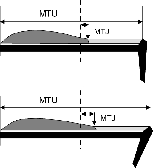
B-Mode ultrasonography (HDI-3000, ATL, Bothell, WA, USA) was used to determine the displacement of the myotendinous junction (MTJ) of the gastrocnemius medialis (GM) during the first two of the standard stretches pre- and post-conditioning. The MTJ was identified as described by Maganaris & Paul (1999) and visualized as a continuous sagittal plane ultrasound image using a 10 cm, 7.5 MHz linear-array probe, which was time locked with the torque and goniometer outputs. Displacement was measured relative to an acoustically reflective marker (a thin strip of micropore tape) secured to the skin proximal to the GM myotendinous junction (Fig. 1).
Figure 1. Schematic diagram showing the measurements made to determine the distal displacement of the myotendinous junction (MTJ) and to estimate changes in tendon length during dorsiflexion and stretch of the whole gastrocnemius medialis muscle–tendon unit (MTU).
The position of the MTJ (vertical arrow) was identified by ultrasound and the movement of this was related to a reflective surface marker, the position of which is indicated by the vertical dotted line. Change in total MTU length was estimated from the change in ankle angle (see Methods for explanation).
Images were recorded on digital tape at 30 Hz and analysed offline. Still ultrasound images were extracted every 5 deg of dorsiflexion, except for the last 5 deg, where they were examined every 1 deg. Images were also analysed at angles equivalent to the participant's pre- and post-end ROM.
The total MTU length was determined at the start of the experiment with an inextensible tape laid over the surface of the muscle and tendon and using ultrasound to identify the anatomical landmarks of the GM origin, the insertion at the MTJ and the insertion of the Achilles tendon. The change in MTU length at each joint angle was estimated using a cadaveric regression model (Grieve et al. 1978; Yuen & Orendurff, 2006). The percentage change in MTU length (ΔL) was calculated as follows:
where θ A is the ankle angle (deg), measured from the neutral position with the foot at right angles to the tibia, and estimating the change in length based on 5 deg increments of dorsiflexion. Using this approach the change in MTU length was found to be 0.78 mm deg−1, which is similar to the value of 0.83 mm deg−1 quoted by Herbert et al. (2002).
The extent of shortening of the tendon distal to the MTJ, and extension of the muscle proximal to the MTJ, were determined from the distal displacement of the MTJ (Fig. 1).
The lengths of three to five fascicles were measured in the belly of the medial gastrocnemius. The ultrasound probe was held at 50% of GM muscle length in the mid-sagittal line to record the movement of the muscle fascicles from the deep to superficial aponeuroses at 5 deg increments from 0 to 25 deg during the third of the standard stretches pre- and post-conditioning. The angle of insertion of the fascicles with the deep aponeurosis was also measured (pennation angle).
Ultrasound images of the MTJ and fascicles were quantified using open source digital measurement software (Image J, NIH, USA).
EMG
EMG activity of the GM muscle was recorded throughout the standard stretching manoeuvre using two pre-gelled, unipolar, 10 mm, Ag–AgCl percutaneous electrodes (Medicotest, Denmark). The electrodes were placed laterally to the mid-line of the muscle having defined the GM muscle boundaries using ultrasonography. Electrodes were placed at two thirds muscle length with 25 mm between the centres and a reference electrode placed over the lateral epicondyle of the femur. The root mean square of the raw signal was recorded for 0.5 s either side of the time when the ankle was at a specific angle of interest. Subjects were asked to make a maximum isometric contraction of the GM with the ankle at 0 deg and the electrical activity recorded during the standard passive stretches was expressed as a percentage of this maximum EMG activity.
Ultrasound validity
All measures of MTJ displacement were made relative to an acoustically reflective marker secured to the skin under the ultrasound probe (Maganaris, 2005). However, this common practice assumes that the skin, marker and ultrasound probe do not move during the stretching manoeuvre. To test this assumption four pins were glued to the skin of one participant over the MTJ spanning a distance equivalent to the length of the ultrasound probe and filmed with a digital camcorder fixed to an external tripod. The movement of these markers was then analysed at dorsiflexion angles of 0, 5, 10, 15, 20 and 25 deg during a standard stretch. Over four trials the skin markers translated distally by 1.8 ± 0.2 mm, with an approximately linear increase from 0 to 25 deg joint angle. There were no differences in the movements of the 1st and 4th markers.
Statistics
For all variables significant difference was determined pre- and post-conditioning using a repeated measures ANOVA; within-subject variables: angle (5 levels) and pre–post (2 levels). To determine differences between muscle and tendon stiffness an additional between-subject variable was included. If a significant interaction was found, differences between pre- and post-values were determined with paired t tests. Differences were considered significant at an alpha level of P < 0.05. Data are reported as mean ± s.e.m. unless otherwise indicated.
Results
Properties of pre-conditioned muscle and tendon
GM muscle length (the distance between muscle origin and MTJ) was 16.7 ± 0.7 cm and the ‘tendon’ length (the distance between the MTJ and the insertion of the Achilles tendon in the calcaneous) was 13.4 ± 0.9 cm.
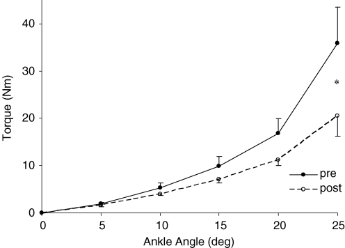
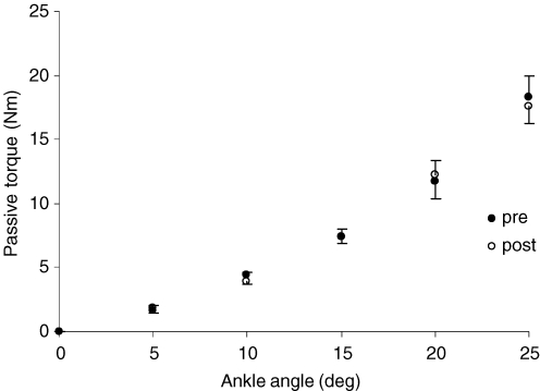
The relationship between ankle angle and passive torque during the standard passive isokinetic dorsiflexion stretches is illustrated in Fig. 2, showing the characteristic curvilinear relationship with passive torque increasing steeply towards the end ROM. Confirming reports from stretching the hamstring muscles (Magnusson et al. 1996b), in the present study the GM muscle was virtually electrically silent throughout the standard stretches and remained below 1% of the maximal value (data not shown), indicating that the measured torque was due to passive properties of the MTU and not the result of voluntary or reflex contractions.
Figure 2. Passive plantarflexion torque throughout a standard stretch pre- and post-conditioning with 5 min of passive stretching.
*Significant difference from pre-conditioning values, P < 0.05.
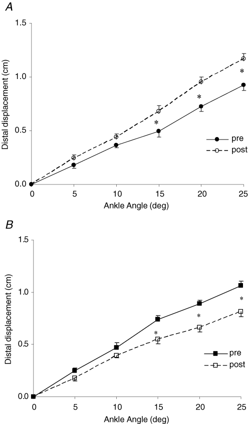
During the standard stretch to end ROM (28 deg), MTU length increased by 2.19 cm (Table 1). Ultrasound measurements showed that the distal displacement of the MTJ, relative to the acoustic marker on the skin, was 1.04 cm, thus accounting for 47% of the total elongation of the MTU. The remaining 1.15 cm was taken up by extension of the tendon, accounting for 53% of the overall change in MTU length. Extensions of both muscle and tendon components increased linearly with increasing ankle angle (Fig. 3) during the standard stretches.
Table 1.
Physical characteristics of the gastrocnemius medialis muscle–tendon unit (MTU) during a standard stretch, pre- and post-conditioning
| End ROM | Pre-conditioning | Post-conditioning |
|---|---|---|
| Ankle angle at end ROM (deg) | 28.1 ± 2.3 | 32.7 ± 2.4* |
| MTU elongation at end ROM (cm) | 2.19 ± 0.14 | 2.52 ± 0.14* |
| MTJ elongation at end ROM (cm) | 1.04 ± 0.08 | 1.38 ± 0.07* |
| Tendon elongation at end ROM (cm) | 1.15 ± 0.09 | 1.14 ± 0.10 |
| Passive torque at end ROM (Nm) | 45.6 ± 7.0 | 53.5 ± 10.8 |
Data presented include: ankle angle at the end of the range of motion (ROM); distal movement of the myotendinous junction (MTJ) and elongation of the tendon obtained by subtracting the elongation of the MTJ from that of the MTU; values are also given for the passive torque at end ROM. Significant difference from pre-conditioning (
P < 0.05).
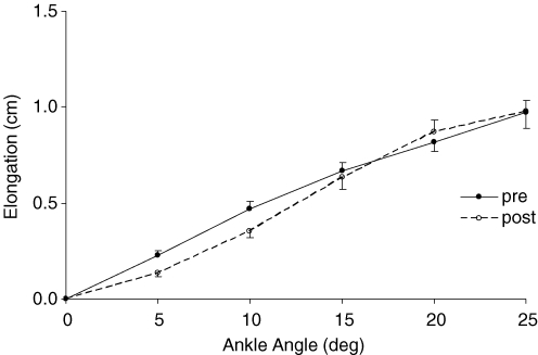
Figure 3. Distal displacement of the muscle (A) and tendon (B) during standard stretches, pre- and post-conditioning stretches.
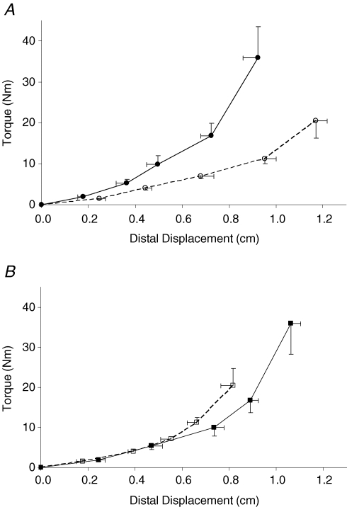
Figure 4 shows the torque–elongation relationship for the muscle and tendon. It is not possible to determine an absolute value for GM or tendon stiffness, since the contribution of the joint capsule to the measured plantarflexion torque is not known, and therefore the relative contribution of the GM cannot be calculated. Bearing this caveat in mind, however, the nominal values for stiffness averaged over the whole ROM was 38.8 ± 8.4 and 34.2 ± 7.5 Nm cm−1 for the muscle and tendon, respectively, although, clearly, the relationship was non-linear. Values for muscle and tendon were not significantly different pre-conditioning.
Figure 4. Passive plantarflexion torque–elongation properties for the GM muscle (A) and tendon (B) during standard stretches pre- (continuous line) and post-conditioning (broken line).
The effect of rapid stretches on MTU stiffness
To determine whether changing the putative thixotropic state of the muscle can alter the stiffness of the whole muscle–tendon unit, the MTU was subjected to a series of 10 rapid stretches from 0 to 10 deg at a speed of 60 deg s−1. The data for eight participants are shown in Fig. 5 and it is evident that the rapid stretches had no effect on the stiffness of the whole MTU, indicating that this procedure is not an effective method of warm-up. This suggests that changing the thixotropic properties of the muscle is unlikely to be the underlying mechanism to the reduction in MTU stiffness induced by the conditioning stretches.
Figure 5. Passive plantarflexion torque measured during standard stretches pre and post a series of 10 rapid isokinetic plantar and dorsiflexions (60 deg s−1).
Muscle and tendon properties following the conditioning stretches
Five conditioning stretches, each lasting 1 min, significantly increased the range of movement by approximately 4.6 deg (P < 0.05, Table 1). The increase in ROM was accompanied by an additional 0.33 cm extension of the whole muscle–tendon unit at the end ROM (P < 0.01). The distal displacement of the MTJ during a standard stretch to the end ROM increased by 0.34 cm (P < 0.01) and accounted entirely for the increased extension. The estimated tendon extension at end ROM remained unchanged as a result of the conditioning stretches (Table 1). Post-conditioning during a standard stretch between 0 and 25 deg, the muscle ‘saw’ an additional 12% of the total MTU extension compared with pre-conditioning (P < 0.05, Table 2).
Table 2.
Elongation of muscle, tendon and fascicles during a standard stretch from 0 to 25 deg, pre- and post-conditioning
| 0–25 deg | Pre-conditioning | Post-conditioning |
|---|---|---|
| MTU elongation (cm) | 1.98 ± 0.03 | 1.98 ± 0.03 |
| MTJ elongation (cm) | 0.92 ± 0.06 | 1.16 ± 0.05*† |
| Tendon elongation (cm) | 1.06 ± 0.04 | 0.82 ± 0.05* |
| Fascicle elongation (cm) | 0.97 ± 0.08 | 0.98 ± 0.05 |
Data shown include: distal muscle and tendon displacements (elongation) pre- and post-conditioning stretches. Gastrocnemius medialis muscle–tendon unit (MTU) elongation was calculated from changes in angle at the ankle; myotendinous junction (MTJ) movement detected by ultrasonography and tendon extension determined by subtracting MTJ movement from that of the MTU. Values are also given for change in muscle fascicle lengths.
Significant difference from pre-conditioning (P < 0.05).
Significant difference from tendon elongation (P < 0.05).
The conditioning stretches also reduced the passive torque at every angle measured during the standard stretch procedure, which reached significance at 25 deg (P < 0.05, Fig. 2) and MTU stiffness over the range 20 to 25 deg decreased from 16.0 ± 3.6 to 10.2 ± 2.0 Nm deg−1 as a result of the conditioning stretches (P < 0.05). For any given angle the displacement of the muscle was greater during a standard stretch after the conditioning while, conversely, the elongation of the tendon was reduced (Fig. 3). The relationships between extension and passive torque for the MTJ and tendon are shown in Fig. 4A and B, respectively, and demonstrate that the conditioning stretches reduced the stiffness of the muscle component of the MTU relatively more than that of the tendon. Bearing in mind the reservations mentioned above, the nominal value for muscle stiffness was 56% lower post-conditioning (17.2 ± 3.5 Nm cm−1 compared with 38.8 ± 8.4 Nm cm−1, P < 0.05), whereas there was no significant difference in tendon stiffness from pre- to post-stretching (26.0 ± 5.1 Nm cm−1 compared with 34.2 ± 7.5 Nm cm−1, respectively).
Clearly the major part of the change in stiffness and ROM of the MTU as a result of the conditioning stretches can be attributed to changes within the muscle proximal to the MTJ.
Muscle fascicle length and the effects of conditioning
During a standard stretch to 25 deg in the initial pre-conditioned state the muscle fascicles changed length in a very similar manner to the change in MTJ (Table 3). Consequently, the increase in length of the ‘muscle’ was entirely accounted for by the change in length of the muscle fascicles.
Table 3.
Fascicle length and its contribution to the elongation of the MTJ
| Pre-conditioning | Post-conditioning | |||
|---|---|---|---|---|
| Ankle angle | 0 deg | 25 deg | 0 deg | 25 deg |
| Fascicle length (cm) | 6.5 ± 0.3 | 7.5 ± 0.3 | 6.1 ± 0.3 | 7.1 ± 0.3 |
| Pennation angle (deg) | 18.3 ± 0.9 | 15.4 ± 0.8 | 21.7 ± 0.8 | 15.7 ± 0.6 |
| Resolved fascicle length (cm) | 6.2 ± 0.3 | 7.2 ± 0.3 | 5.7 ± 0.3 | 6.8 ± 0.3 |
| Change of resolved fascicle length (cm) | — | 1.03 ± 0.1 | — | 1.15 ± 0.04* |
| Corrected change in MTJ (cm) | — | 1.06 ± 0.07 | — | 1.34 ± 0.06* |
Fascicle length and pennation angle at ankle angles of 0 deg and 25 deg pre- and post-conditioning. Values are also given for the fascicle length resolved along the axis of the MTU, for the change in this length during a standard stretch pre- and post-conditioning and these values are compared with those for MTJ elongation taken from Table 2 and corrected for 15% underestimation (see text for details).
Significant difference from pre-conditioning (P < 0.05).
Following the conditioning stretches the fascicle extension during a standard stretch did not significantly change (Fig. 6, Table 3) even though the MTJ extension was greater (Table 2). The contribution of the fascicles to MTU length is proportional to the cosine of the angle of pennation (i.e. the fascicle length resolved along the axis of the muscle). The angle of pennation decreased during the stretch and consequently the relative contribution of the fascicles to the overall muscle length increased. Values for fascicle length, pennation angle and the resolved fascicle length are given in Table 3 showing changes from 0 to 25 deg both pre- and post-conditioning. When the angle of pennation and the fascicle length are taken into account it was not sufficient to explain the increased displacement of the MTJ after the conditioning stretches.
Figure 6. Muscle fascicle elongation during standard stretches pre- and post-conditioning.
Discussion
The findings reported here have confirmed the suggestion of Herbert et al. (2002) that in a resting pre- or non-conditioned MTU, such as the gastrocnemius medialis, a large proportion of the movement during passive stretch is taken up by the tendon. We have expanded these observations and those of others who have demonstrated a lower passive torque–angle relationship following stretching (Wilson et al. 1992; Halbertsma et al. 1996), by showing that the decrease in MTU stiffness following repeated stretches, such as those used by athletes during warm-up, is not due to a change in the tendon but rather to an increased compliance of the proximal, muscular, portion of the MTU. This increased compliance of the muscle is not, however, fully explained by a change in the extensibility of the muscle fascicles and we propose that connective tissue elements within the muscle change their elastic properties when subject to repeated stretches.
Stretch such as used during a warm-up is known to cause an acute increase in ROM which may in part be due to an increase in the subjects' tolerance of stretch (Magnusson et al. 2000) and also a decrease in the stiffness of the whole MTU, changing the passive torque–angle relationship of the hamstrings and gastrocnemius (Wilson et al. 1992; Evetovich et al. 2003). The changes we report in passive torque as a result of the conditioning stretches are very similar to these previous observations. However, the MTU is a complex structure consisting of tendon, muscle fibres and connective tissue and it is of interest to see how the different components respond following conditioning by repeated stretches. Ultrasound techniques allow measurements to be made of changes in fascicle length from which Herbert et al. (2002) made inferences about changes in tendon length. Here we have expanded these observations to include measurements of the displacement of the MTJ which allows a more direct estimation of changes in length of the tendon and other structures both before and after conditioning.
The movement of the MTJ of little more than a centimetre during the standard stretches is relatively small. To obtain an estimate of changes in tendon length the displacement of the MTJ has to be subtracted from the change in length of the whole MTU which in turn is estimated from the change in angle at the ankle joint. It is important therefore to assess the possibility of systematic errors in the measurements we have made. Observing markers placed on the skin it was found that there was a distal movement of the skin as the MTU was stretched, leading to a probable under-estimation of the displacement of MTJ and consequent over-estimation of the elongation of the tendon. The measurements were made pre-conditioning over four repeated stretches, but we have no reason to think that the movement of the skin would be any different after the five conditioning stretches. These observations were made in only one subject and so a correction has not been applied to the data presented here. However, the subject was typical in every respect so we assume that all MTJ movements may have been under-estimated by about 15%. This systematic error makes little or no difference to the conclusions related to our first and second objectives. Thus, we can confirm that during a standard passive stretch to 25 deg a substantial component of the MTU extension is taken up by the muscle and it makes very little difference whether this constitutes 46% or, after correction, 53% of the total movement. Likewise it does not affect our conclusions regarding the role of thixotropy (see below). In respect of the changes occurring as a result of the conditioning stretches, the error does not affect the qualitative observation that it is the muscle and not the tendon that is most affected. However, when it comes to quantitative assessments of the contribution of changes in fascicle length to the changes in stiffness of the distal, muscular, portion of the MTU it then becomes necessary to take into account the possible measurement errors.
The extension of the muscle fascicles during a standard stretch was slightly less than that of the MTJ (Table 1) and so accounted for a little less than 50% of the total MTU extension prior to conditioning stretches. This is considerably more than the 27% reported by Herbert et al. (2002) but is similar to values of 43–46% reported in the GM muscle by others (Kawakami et al. 1998; Maganaris, 2003). The most likely explanation is that in the study of Herbert et al. (2002) the subjects were tested with a flexed knee which would have shortened the gastrocnemius introducing some slack into the system which would be taken up before any extension of the fascicles occurred. In our experiments the knee was always fully extended. Nevertheless, the qualitative observation of the importance of tendon extensibility during passive movement is the same.
Our main concern was what would happen to the muscle when conditioned by repeated passive stretches, a type of warm-up that results in greater flexibility (ROM) and reduced overall MTU stiffness. Resting muscle fibres have thixotropic properties (Hill, 1968) and it is well known that muscle stiffness of this nature can be reduced by relatively small amounts of movement (Campbell & Lakie, 1998). Consequently we subjected the muscle to a number of quick stretches that would be expected to abolish the short range elastic component, or possibly change the conformation of titin, but this had no effect on the overall stiffness of the MTU (Fig. 5) or movement of the MTJ. We conclude that reducing the contribution of short-range elastic component of the muscle fibres is unlikely to be the mechanism leading to increased flexibility as a result of static stretching.
In contrast to the pre-conditioning state where the increase in total MTU length during a standard stretch was taken up almost equally between muscle and tendon, the proportion of the increase in MTU length due to changes in muscle length post-conditioning was increased while there was a decreased contribution from tendon elongation. It is tempting to calculate the stiffness of the various components of the MTU from data such as those illustrated in Fig. 4. The problem, however, is that the measured torque probably contains an appreciable component derived from frictional forces within the joint capsule making it impossible to know the actual torque experienced by the tendon and muscle and it is possible that compressing the ankle joint during the conditioning stretches may have altered its properties. Nevertheless, if it is assumed that the proportion of the measured torque contributed by the different elements remains the same pre- and post-conditioning then it is evident that the effect of the conditioning is to reduce the stiffness of the muscle by about half while the tendon shows no significant change following the conditioning stretches. Our conclusions contradict those of a number of authors who suggest that stretching primarily affects the stiffness of tendons (Wilson et al. 1992; Witvrouw et al. 2004). However, these latter observations were based on the reduction in the joint angle–torque relationships from which no direct conclusions could be made regarding the contribution of different components of the muscle–tendon unit.
Whilst the additional movement that occurred following the conditioning stretches was localized to the muscle it was surprising that this was not reflected in a change in the extension of the muscle fascicles during the standard stretch (Fig. 6; Table 2). In trying to account for changes in MTJ in terms of fascicle length it is necessary to take into account the angle of pennation with the relevant measurement being the fascicle length resolved along the axis of the MTU. Table 3 shows the change in the resolved length of the fascicles and these have been compared with the change in MTJ after it has been adjusted for the likely error arising from movement of the ultrasound markers on the skin. The effect of conditioning was to increase the angle of pennation with the ankle at 0 deg and the change in pennation angle during stretch to 25 deg was somewhat greater than in the initial pre-conditioning state. Pre-conditioning, the change in MTJ and resolved fascicle length were virtually identical while post-conditioning the movement of the MTJ was 0.19 cm greater than the change in resolved fascicle length. This suggests that the additional extension of the components of the MTU proximal to the MTJ was, at least in part, the result of changes in structures other than the muscle fibres. Muscle fibres are surrounded by a complex connective tissue network to which they are attached along the length of the fibre and not only at the ends. This connective tissue, particularly the perimysium, is considered to be a major extracellular contributor to passive stiffness (Purslow, 1989). Likewise, Gajdosik (2001) suggested that lengthening deformation of the connective tissues within the muscle belly (endomysium, perimysium and epimysium) could influence passive stiffness.
The increased ROM and flexibility resulting from the decreased stiffness of the muscle following conditioning stretches would clearly be beneficial to athletes competing in events where flexibility is essential. There is, however, evidence suggesting that static stretching could reduce muscle force and power output, with detrimental consequences to sporting performance (Gleim & McHugh, 1997). In the present investigation fascicle length was unchanged by stretching, which was probably the result of an increase in the aponeurosis compliance. More compliant connective tissue may reduce the sensitivity of muscle spindles (Avela et al. 1999), possibly reducing the speed of muscle activation and this may account for the reported reductions in power output during sprinting after stretching exercise (Nelson et al. 2005). Consequently in events such as gymnastics, high jumping and hurdling where both flexibility and high power are required, there may be a trade-off between the two effects of stretching during warm-up. However, as discussed by Magnusson et al. (2000) the time course of the in vivo physiological adaptations associated with static stretching remains unresolved.
The results presented here provide information about the behaviour of three components of the muscle–tendon complex when it is stretched and how these are affected by a series of conditioning stretches such as are commonly used during athletic warm-up. The conclusions are that pre-conditioning the MTU extension is taken up in nearly equal parts by the tendon and muscle fascicles but, post-conditioning, the anatomical muscle becomes less stiff and accounts for the decrease in overall stiffness of the MTU and the increased ROM. There are suggestions from our results that the changes within the muscle may be due, in part, to altered properties of connective tissue elements. The nature of these elements and how they are affected by conditioning stretches is largely unknown and clearly a topic of considerable future interest.
References
- Avela J, Kyrolainen H, Komi PV. Altered reflex sensitivity after repeated and prolonged passive muscle stretching. J Appl Physiol. 1999;86:1283–1291. doi: 10.1152/jappl.1999.86.4.1283. [DOI] [PubMed] [Google Scholar]
- Campbell KS, Lakie M. A cross-bridge mechanism can explain the thixotropic short-range elastic component of relaxed frog skeletal muscle. J Physiol. 1998;510:941–962. doi: 10.1111/j.1469-7793.1998.941bj.x. [DOI] [PMC free article] [PubMed] [Google Scholar]
- Dadebo B, White J, George KP. A survey of flexibility training protocols and hamstring strains in professional football clubs in England. Br J Sports Med. 2004;38:388–394. doi: 10.1136/bjsm.2002.000044. [DOI] [PMC free article] [PubMed] [Google Scholar]
- Ettema GJ, Huijing PA. Properties of the tendinous structures and series elastic component of EDL muscle-tendon complex of the rat. J Biomech. 1989;22:1209–1215. doi: 10.1016/0021-9290(89)90223-6. [DOI] [PubMed] [Google Scholar]
- Evetovich TK, Nauman NJ, Conley DS, Todd JB. Effect of static stretching of the biceps brachii on torque, electromyography, and mechanomyography during concentric isokinetic muscle actions. J Strength Cond Res. 2003;17:484–488. doi: 10.1519/1533-4287(2003)017<0484:eossot>2.0.co;2. [DOI] [PubMed] [Google Scholar]
- Fukunaga T, Ito M, Ichinose Y, Kuno S, Kawakami Y, Fukashiro S. Tendinous movement of a human muscle during voluntary contractions determined by real-time ultrasonography. J Appl Physiol. 1996;81:1430–1433. doi: 10.1152/jappl.1996.81.3.1430. [DOI] [PubMed] [Google Scholar]
- Gajdosik RL. Passive extensibility of skeletal muscle: review of the literature with clinical implications. Clin Biomech (Bristol, Avon) 2001;16:87–101. doi: 10.1016/s0268-0033(00)00061-9. [DOI] [PubMed] [Google Scholar]
- Gleim GW, McHugh MP. Flexibility and its effects on sports injury and performance. Sports Med. 1997;24:289–299. doi: 10.2165/00007256-199724050-00001. [DOI] [PubMed] [Google Scholar]
- Grieve DW, Cavanagh PR, Pheasent S. Prediction of gastrocnemius length from knee and ankle posture. In: Asmussen E, Jorgensen K, editors. Biomechanics. VI-A. Baltimore, MD, USA: University Park Press; 1978. pp. 405–412. [Google Scholar]
- Halar EW, Stolov WC, Venkatsh B, Brozovich FV, Harley JD. Gastrocnemius muscle belly and tendon length in stroke and able-bodied persons. Arch Phys Med Rehabilitation. 1978;59:476–484. [PubMed] [Google Scholar]
- Halbertsma JP, van Bolhuis AI, Goeken LN. Sport stretching: effect on passive muscle stiffness of short hamstrings. Arch Phys Med Rehabil. 1996;77:688–692. doi: 10.1016/s0003-9993(96)90009-x. [DOI] [PubMed] [Google Scholar]
- Herbert RD, Moseley AM, Butler JE, Gandevia SC. Change in length of relaxed muscle fascicles and tendons with knee and ankle movement in humans. J Physiol. 2002;539:637–645. doi: 10.1113/jphysiol.2001.012756. [DOI] [PMC free article] [PubMed] [Google Scholar]
- Hill DK. Tension due to interaction between the sliding filaments in resting striated muscles. The effect of stimulation. J Physiol. 1968;199:637–684. doi: 10.1113/jphysiol.1968.sp008672. [DOI] [PMC free article] [PubMed] [Google Scholar]
- Kawakami Y, Ichinose Y, Fukunaga T. Architectural and functional features of human triceps surae muscles during contraction. J Appl Physiol. 1998;85:398–404. doi: 10.1152/jappl.1998.85.2.398. [DOI] [PubMed] [Google Scholar]
- Kubo K, Kanehisa H, Kawakami Y, Fukunaga T. Influence of static stretching on viscoelastic properties of human tendon structures in vivo. J Appl Physiol. 2001;90:520–527. doi: 10.1152/jappl.2001.90.2.520. [DOI] [PubMed] [Google Scholar]
- Lieber RL, Friden J. Functional and clinical significance of skeletal muscle architecture. Muscle Nerve. 2000;23:1647–1666. doi: 10.1002/1097-4598(200011)23:11<1647::aid-mus1>3.0.co;2-m. [DOI] [PubMed] [Google Scholar]
- McHugh MP, Kremenic IJ, Fox MB, Gleim GW. The role of mechanical and neural restraints to joint range of motion during passive stretch. Med Sci Sports Exerc. 1998;30:928–932. doi: 10.1097/00005768-199806000-00023. [DOI] [PubMed] [Google Scholar]
- Maganaris CN. Force-length characteristics of the in vivo human gastrocnemius muscle. Clin Anat. 2003;16:215–223. doi: 10.1002/ca.10064. [DOI] [PubMed] [Google Scholar]
- Maganaris CN. Validity of procedures involved in ultrasound-based measurement of human plantarflexor tendon elongation on contraction. J Biomech. 2005;38:9–13. doi: 10.1016/j.jbiomech.2004.03.024. [DOI] [PubMed] [Google Scholar]
- Maganaris CN, Paul JP. In vivo human tendon mechanical properties. J Physiol. 1999;521:307–313. doi: 10.1111/j.1469-7793.1999.00307.x. [DOI] [PMC free article] [PubMed] [Google Scholar]
- Magnusson SP, Aagaard P, Nielson JJ. Passive energy return after repeated stretches of the hamstring muscle-tendon unit. Med Sci Sports Exerc. 2000;32:1160–1164. doi: 10.1097/00005768-200006000-00020. [DOI] [PubMed] [Google Scholar]
- Magnusson SP, Simonsen EB, Aagaard P, Boesen J, Johannsen F, Kjaer M. Determinants of musculoskeletal flexibility: viscoelastic properties, cross-sectional area, EMG and stretch tolerance. Scand J Med Sci Sports. 1997;7:195–202. doi: 10.1111/j.1600-0838.1997.tb00139.x. [DOI] [PubMed] [Google Scholar]
- Magnusson SP, Simonsen EB, Aagaard P, Kjaer M. Biomechanical responses to repeated stretches in human hamstring muscle in vivo. Am J Sports Med. 1996a;24:622–628. doi: 10.1177/036354659602400510. [DOI] [PubMed] [Google Scholar]
- Magnusson SP, Simonsen EB, Aagaard P, Sorensen H, Kjaer M. A mechanism for altered flexibility in human skeletal muscle. J Physiol. 1996b;497:291–298. doi: 10.1113/jphysiol.1996.sp021768. [DOI] [PMC free article] [PubMed] [Google Scholar]
- Nelson AG, Driscoll NM, Landin DK, Young MA, Schexnayder IC. Acute effects of passive muscle stretching on sprint performance. J Sports Sci. 2005;23:449–454. doi: 10.1080/02640410410001730205. [DOI] [PubMed] [Google Scholar]
- Pollock ML, Wenger NK. Physical activity and exercise training in the elderly: a position paper from the Society of Geriatric Cardiology. Am J Geriatr Cardiol. 1998;7:45–46. [PubMed] [Google Scholar]
- Proske U, Morgan DL. Stiffness of cat soleus muscle and tendon during activation of part of muscle. J Neurophysiol. 1984;52:459–468. doi: 10.1152/jn.1984.52.3.459. [DOI] [PubMed] [Google Scholar]
- Purslow PP. Strain-induced reorientation of an intramuscular connective tissue network: implications for passive muscle elasticity. J Biomech. 1989;22:21–31. doi: 10.1016/0021-9290(89)90181-4. [DOI] [PubMed] [Google Scholar]
- Rassier DE, Lee EJ, Herzog W. Modulation of passive force in single skeletal muscle fibres. Biol Lett. 2005;1:342–345. doi: 10.1098/rsbl.2005.0337. [DOI] [PMC free article] [PubMed] [Google Scholar]
- Reisman S, Walsh LD, Proske U. Warm-up stretches reduce sensations of stiffness and soreness after eccentric exercise. Med Sci Sports Exerc. 2005;37:929–936. [PubMed] [Google Scholar]
- Sale D, Quinlan J, Marsh E, McComas AJ, Belanger AY. Influence of joint position on ankle plantarflexion in humans. J Appl Physiol. 1982;52:1636–1642. doi: 10.1152/jappl.1982.52.6.1636. [DOI] [PubMed] [Google Scholar]
- Wilson GJ, Elliott BC, Wood GA. Stretch shorten cycle performance enhancement through flexibility training. Med Sci Sports Exerc. 1992;24:116–123. [PubMed] [Google Scholar]
- Witvrouw E, Mahieu N, Danneels L, McNair P. Stretching and injury prevention: an obscure relationship. Sports Med. 2004;34:443–449. doi: 10.2165/00007256-200434070-00003. [DOI] [PubMed] [Google Scholar]
- Yuen TJ, Orendurff MS. A comparison of gastrocnemius muscle-tendon unit length during gait using anatomic, cadaveric and MRI models. Gait Posture. 2006;23:112–117. doi: 10.1016/j.gaitpost.2004.12.007. [DOI] [PubMed] [Google Scholar]


