Abstract
Impaired nutrition of the intervertebral disc has been hypothesized to be one of the causes of disc degeneration. However, no causal relationship between decreased endplate perfusion and limited nutrient transport has been demonstrated to support this pathogenic mechanism. To determine the importance of endplate perfusion on solute diffusion into the nucleus pulposus and to show causality of endplate perfusion on intranuclear diffusion in large animal lumbar intervertebral discs, diffusive transport into ovine lumbar intervertebral discs was evaluated after inhibiting adjacent vertebral endplate perfusion. Partial perfusion blocks were created in vertebrae close and parallel to both endplates of lumbar discs of anaesthetized sheep. To assess diffusivity of small molecules through the endplate, N2O was introduced into the inhalation gas mixture and concentrations of intranuclear N2O were measured for 35 min thereafter. Post mortem, procion red was infused through the spinal vasculature and perfusion through the endplate was assessed by quantifying the density of dye-perfused endplate vascular buds in histology sections. Perfusion of the endplates overlying the nucleus pulposus was inhibited by almost 50% in the partially blocked discs relative to the control discs. There was also a nine-fold decreased transport rate of intranuclear N2O in partially blocked discs compared with control discs. The density of perfused endplate vascular buds correlated significantly to the amount of transported intranuclear N2O (r2 = 0.52, P = 0.008). The vertebral endplate was demonstrated to be the main route of intravascular solute transport into the nucleus pulposus of intervertebral discs, and inhibition of endplate perfusion can cause inhibited solute transport into the disc intranuclear tissue.
Keywords: diffusion, endplate, intervertebral disc, perfusion, sheep
Introduction
Low back pain is one of the major health problems in western society. About 85% of people at the age of 50 recall an episode of low back pain (Bigos et al. 1991). The costs are estimated to be $20 billion yearly in the USA alone (Cats-Baril & Frymoyer, 1991). In the Netherlands the direct and indirect costs in 1991 were estimated to be 1.7% of the gross national product (van Tulder et al. 1995). Low back pain is a multifactorial problem, which includes intervertebral disc degeneration (Adams, 2004). Besides other factors such as loading (Adams, 2004), heredity (Battie et al. 2004) and aging, it is believed that limited nutrition of the disc can be an aetiological factor in intervertebral disc degeneration.
The intervertebral disc is the largest avascular structure in the human body. Because of its avascularity, the cells inside the disc depend on diffusion for the transport of nutrients and waste products. Small capillary buds with a diameter of about 20–50 µm run through the pores of the bony endplate (Oki et al. 1994). Nutrients diffuse from these vascular buds and the small blood vessels in the outer part of the outer annulus to all cells inside the disc. Less permeable endplates could thus result in nutrient deficiency and very likely in cell death and intervertebral disc degeneration.
Almost 40 years ago, Nachemson et al. (1970) found a significant association between permeability of the endplate and intervertebral disc degeneration. In addition, Benneker et al. (2005) found a significant correlation between the amount of pores in the endplate and disc degeneration in a human cadaver study. Discs of grade 4 degeneration on the Thompson scale (Thompson et al. 1990) had about 50% fewer pores of 20–50 µm (corresponding in size to the capillary bud openings (Oki et al. 1994)) than discs of grade 1.
Magnetic resonance imaging has been used extensively to study diffusion into the disc. With the help of a contrast fluid, diffusion into the disc can be imaged and quantified. Most of these studies focused on the effect of maturation (Ibrahim et al. 1995) and degeneration (Nguyen-minh et al. 1997, 1998) on diffusion. But one study focused on the diffusion pathway and showed that diffusion of the contrast fluid into the nucleus pulposus derives mostly from the subchondral bone through the endplate (Rajasekaran et al. 2004).
Ogata & Whiteside (1981) showed that blockage of this endplate route had great effects on diffusion in a dog model. They disrupted the blood supply to the endplate by osteomization of the adjacent vertebrae close to the bone–disc interface and putting a metal plate into this slot. They found that this endplate blockage had a greater effect on the diffusion than disrupting the blood vessels over the annulus fibrosus, indicating that the endplate route is the most important.
So far, it is clear that nutrients reach the nucleus pulposus of the disc through diffusion, and that the endplate route is the most important route. However, the mechanistic relationship between endplate perfusion and diffusion remains unclear. Therefore, the goal of this study is to demonstrate the relationship between a decrease in functional capillary buds in the endplate and diffusion inhibition into the nucleus pulposus of intervertebral discs.
Materials and methods
Animals
The animal experiments were approved by the Animal Experimentation Commission of the Veterinary Office of the Canton of Grison, Switzerland, and followed the guidelines of the Swiss Federal Veterinary Office for the use and care of laboratory animals. Four Swiss Alpine sheep between 5 and 7 years old and approximately 50 kg body weight were sedated and anaesthetized with an inhalation mixture of oxygen and 2–3% isoflurane (Baxter, Volketswil, Switzerland). At the end of the experiment they were euthanized with an overdose of pentobarbital (100 mg kg−1, i.v. Pentobarbitalum natricum, Streuli & Co. AG, Uznach, Switzerland).
Surgical preparation
After the L1–L6 vertebrae were identified with a fluoroscope, the lateral aspect of the L2–L5 discs were exposed through a retro-peritoneal approach. A slot, approximately 2 cm deep and 1 cm wide and 0.6 mm thick was sawn in the vertebrae close and parallel to both endplates of the L2–L3 and L4–L5 intervertebral discs under fluoroscopic guidance (Fig. 1a). The slot was started on the antero-lateral aspect of the vertebral body approximately 1 mm from the edge of the endplate. The slot was oriented toward the opposite postero-lateral quadrant of the endplate to cover the central portion of the nucleus pulposus (Fig. 1b). A CP-titanium-foil (Allegheny Rodney Metals, Sprockhövel, Germany), approximately 25 mm long and 0.050 mm thick, (Fig. 1b) was inserted into the slot and the part extending from the slot was folded toward the bone and secured to the vertebrae with a cortical screw (1.5 × 6 mm, Synthes Inc., Oberdorf, Switzerland).
Fig. 1.
(a) Fluoroscope image during the surgery. The defects at the L2–L3 level have already been made. The saw blade is cutting the defect in the L5 vertebrae. (b) A schematic of the titanium foil overlaying the nucleus pulposus. Ti = titanium foil, CS = cortical screw, SB= saw blade, OA = outer annulus, IA = inner annulus, NP = nucleus pulposus.
Diffusion
Custom-made electrodes (Barron et al. 1997) were inserted into the centre of the nucleus pulposus of the L1–L2, L2–L3, L4–L5 and L5–L6 discs with fluoroscope guidance. On some occasions the L5–L6 disc could not be approached because the length of the needle did not allow clearance of the iliac crest, in which case diffusion measurements were taken in the L3–L4 disc. A reference electrode was inserted into the paraspinal musculature. Anaesthesia gas was then switched from O2 with 2–3% isoflurane to a mixture of 30%/70% O2/N2O with isoflurane, and amperometrical measurement of intranuclear concentrations of N2O (Urban et al. 2001) were started (t0). The measurements were done in triplicate and carried out every 5 min for 35 min.
The working electrodes used for the diffusion measurements were constructed with two silver wires (diameter = 0.125 mm with a 0.0125-mm coating) inserted into stainless steel tubes (outer diameter = 1.1 mm) separated by an insulating polymer. A silver/silver chloride electrode saturated with potassium chloride was used as combined reference and counter electrode. A multichannel potentiostat (Perkin Elmer Instruments VMP, Ametek, Illingen, Germany) connected to a laptop computer with complementary software was used for measurements. A potential from 0 to –1.5 V (at a rate of –40 mV/s) was applied and the current was measured at a frequency of 50 Hz. At –0.6 V and –1.2 V a plateau in the current-potential curve was observed. The first plateau was caused by the presence of O2, and the second by the presence of N2O (Urban et al. 2001). At these plateaux the current was averaged over five measured points. The calibration curves for each electrode were constructed afterward by measuring the currents in phosphate-buffered saline saturated with known gas concentrations (N2/O2 or N2O/O2 mixtures).
Perfusion
Five minutes after 25 000 IU of heparin (Fresenius Medical Care, Stans, Switzerland) were injected intravenously, the animal was euthanized. Post-mortem, femoral arteries were ligated and the abdominal aorta was cannulated as close to the diaphragm as possible in the abdominal cavity. The renal arteries as well as the thoracic aorta were ligated. Then, 50 000 IU of heparin in 1 L of normal saline was injected through the cannulated abdominal aorta at 100 mmHg to evacuate vasculature in the lumbar vertebrae. The vertebral vasculature was then infused with 800–1000 mL of 5% (w/w) procion red (BASF, H8BN) in water at 100 mmHg.
The lumbar spines were dissected into bone-disc-bone segments, snap frozen in liquid N2, freeze substituted in acetone to retain the water-soluble procion red in the specimens (Steinbrecht & Müller, 1987), and embedded in methylmethacrylate. Freeze substitution was done in four steps of a minimum of 2 days each at –80 ºC, –20 ºC, 4 ºC and room temperature, before embedding in methylmethacrylate. Sections of 200 µm were cut with a rotating blade diamond saw (Leitz 1600, Leica AG, Glattbrugg, Switzerland). From each bone-disc-bone segment the three most mid-sagittal sections overlying the nucleus pulposus were ground and polished on a micro grinding system (Typ AW-10, Exakt Apparatebau, Norderstedt, Germany) to approximately 100 µm thickness. Images, centred over the nucleus pulposus, were recorded on a fluorescent microscope (Zeiss Axioplan 2, Carl Zeiss AG, Feldbach, Switzerland) with three-band excitation (Zeiss filter set #25: exciter filter TBP 400/495/570, beam splitter FT 410/505/584, barrier filter TBP 460/530/610). Quantification of the density of patent endplate capillary buds was done semi-automatically with an image analysis program (axiovision, Carl Zeiss AG) and custom macro (excel, Microsoft, Microsoft, Redmond, WA, USA). Because of autofluorescence of the bone, the boundary between the bony and avascular cartilaginous endplates could be traced by hand. The area within 60 µm of the traced line over the bony endplate, extending between the inner annulus/nucleus pulposus borders, was set as the region of interest (ROI). Within this ROI the pixels were then thresholded for red values such that capillary buds were distinguished from variable amount of dye leakage from the vessel (threshold based initially on histogram and adjusted manually on each image). Based on the findings of Benneker et al. (2005) islands of threshold pixels larger than 15 µm2 were counted automatically.
Statistics
For each disc, the N2O concentrations per time point were normalized to the t0 level. This was done because not all measurements could be taken simultaneously, resulting in delay from change in anaesthesia gas mixture to initial measurement for some probes. The intervals between measurements were the same for all probes. Per time point, the triplicate values were averaged and then pooled into control and blocked discs.
The number of perfused vascular buds per mm of endplate was averaged per section (proximal and distal adjacent endplate) and pooled into control or blocked discs. Differences in diffusion between the control and blocked discs were tested with analysis of variance (anova) for repeated measures. Differences in perfusion between control and blocked discs were tested for significance using the independent Student t-test (spss v 13, SPSS AG, Zurich, Switzerland). A P-value lower than 0.05 was considered significant.
Results
Surgical procedure
No major complications occurred during the operations. In one animal, a caudal lumbar segmental artery was damaged during the approach. In this sheep, the L4–L5 disc was excluded from the analysis (n = 7 partially blocked discs and n = 8 control discs for diffusion data analysis). In two sheep the procion red infusion did not reach the most cranial vertebra of the lumbar spine, and in these levels no perfusion analysis could be performed (n = 21 partially blocked sections and n = 18 control sections for perfusion data analysis).
Diffusion
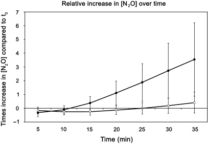
A steady increase in N2O concentration was measured in the control discs over time, whereas in the blocked discs this increase was less pronounced. The increase of N2O concentration in 35 min was on average 360% compared to the t0 level for control discs. In the experimental discs this increase was only 41%. The difference in increase between the control and blocked discs was significant (P = 0.005, Fig. 2), but there were no significant differences between the disc levels.
Fig. 2.
The increase in nuclear nitrous oxide concentration over time after the inhalation gas has been changed from 98% oxygen to 30% O2 and 70% N2O (t0). In the blocked discs this increase is significantly lower than in the control discs. The values have been normalized to t0. The increases are significantly different between the control and blocked discs (P = 0.005, n = 8 controls, and n = 7 partially blocked).
Perfusion
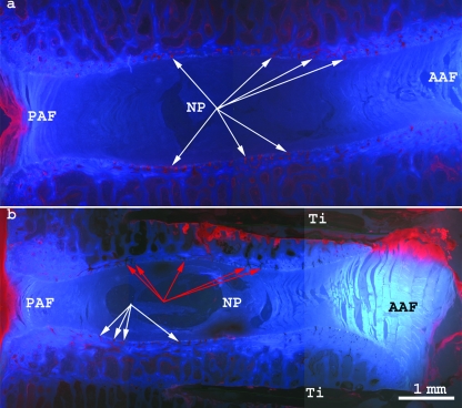
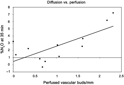
The histological sections clearly exhibited the procion red-infused capillary buds in the bony endplates (white arrows in Fig. 3). The difference between the blocked and normal discs was very clear. The perfusion block left many vascular buds unperfused (red arrows in Fig. 3). Over the nuclear region, in the control discs, 1.52 ± 0.99 vascular buds per mm were perfused, whereas in the blocked discs, this was only 0.78 ± 0.58 (P = 0.006). There was a weak (R2 = 0.52) but significant (P = 0.008) correlation between the amount of perfused vascular buds and the [N2O] diffused into the disc at 35 min (Fig. 4).
Fig. 3.
Procion red-filled endplate vascular buds (white arrows) and non-filled buds (red arrows). AAF anterior annulus fibrosus, PAF =posterior annulus fibrosus, NP = nucleus pulposus, Ti = titanium foil. (a) Control, (b) experimental.
Fig. 4.
Correlation between perfusion and diffusion measurements. The more vascular buds were perfused, the more N2O diffused into the disc at 35 min, after changing the inhalation gas mixture. Data points represent the average perfusion and diffusion of the 12 discs analysed both for diffusion and perfusion, r2 = 0.5229, P = 0.008.
Discussion
In this study the foil only covers about 30% of the disc surface, overlying the nucleus pulposus. Despite this small area of coverage, but because of its central location, endplate perfusion adjacent to the nucleus was decreased by almost 50% on average. The central part of the endplate is more permeable than the edge (Nachemson et al. 1970; Oki et al. 1996). Oki et al. (1996) suggest that this difference in permeability lies in the different structure of the vascular buds in the nucleus region of the endplate, where they are complex coils, compared with the region over the inner annulus, where they are single loops.
N2O was used as a tracer for small solutes instead of measuring glucose directly. Because N2O can be measured amperometrically and does not occur in the body naturally, the efficacy of the perfusion block could be measured. This would not be possible with glucose as all discs would have been saturated before the block would have been created. It is assumed that the inhibition of diffusion for N2O (mw 44) would be similar for other small nutrients such as glucose (mw 180) and oxygen (mw 32) as all three molecules are similar in size and have no charge. The technique used in this study for post mortem infusion of procion red may seem cumbersome, but it enabled us to infuse a large amount of the dye which would not be possible in vivo, hereby increasing the signal-to-noise ratio in the images.
For diffusion, the distance from the nearest blood supply to the cells is of great importance. For this reason a sheep model was chosen. The geometry of the sheep lumbar spine is comparable to that of humans and the discs are sufficiently large (Wilke et al. 1997a,b). Furthermore, the sheep has been generally accepted as a model in spinal research.
Diffusion was only measured for a very short time (35 min), therefore the measurements do not necessarily comply with steady state conditions. As can be seen in Fig. 3, the procion red stains the outer annulus quite intensively. This was also seen in a study by Brodin (1955) and is probably because of the small arteries in the outer part of the annulus fibrosus. One could argue that diffusion through the annulus could play a much bigger role under steady state conditions than in such a short-term study. However, literature on normal discs does not support this. Rajasekaran et al. (2004) have shown that 24 h after injection of contrast fluid the diffusion followed the pattern of the endplates, and no clear diffusion though the annulus was observed. An analytical study by Ferguson et al. (2004) also shows that after 24 h small nutrients do not diffuse though the annulus fibrosus into the nucleus pulposus. Furthermore, an in vitro study in which sheep intervertebral discs were cultured with endplates under static or diurnal loading showed that even after 4 days of culture with TMR-dextran (a fluorescent dye), the dye intensity dropped off steeply from the outer annulus into the inner annulus (Gantenbein et al. 2006). So, even with a blockage of the endplate the gradient from the inner annulus to the centre of the disc would be very small, which is not favourable for diffusion.
In the case of endplate blockage, fewer nutrients can diffuse from the capillary buds to the cells inside the disc (Roberts et al. 1996). Vice versa, waste products cannot be disposed of, resulting in a build up of lactic acid, thus lowering the pH in the centre of the disc. In vitro studies by Horner & Urban (2001) and Bibby et al. (2005) showed that cell viability and metabolism depend on the cell density, glucose, oxygen concentrations and pH. At high cell densities, the viable distance (distance from diffusion source and cell viability level of 95%) was much lower than under low cell densities. Under low oxygen conditions the cells survive up to 12 days but do not produce much proteoglycan; for that, oxygen is required (Horner & Urban, 2001). Under low glucose conditions or at a pH of 6 the cells do not survive for longer periods (Bibby et al. 2005). This supports the hypothesis that nutrient deficiency could result in intervertebral disc degeneration; however, in vivo studies should be done to test this.
This animal model is being developed to be used in investigating the effect of nutrient deprivation on cellular responses. To be able to exclude the effect of changes in the mechanics of the disc by creating such defects, this was tested. In previously frozen cadaver sheep lumbar motion segments the intranuclear pressure was measured before and after defect creation. Measurements were done with a needle pressure transducer described by Adams et al. (1996). There was no significant effect on intranuclear pressure.
Like the study by Ogata & Whiteside (1981) this study shows that a blockage of the endplates results in an inhibition of diffusion of small solutes into the nucleus pulposus. This indicates that this is the most important route for solute transport to the nucleus of the disc. The mechanism through which this happens is demonstrated by the correlation between the functional capillary buds and the diffusion. By creating such a partial perfusion block of the endplate, cellular responses to nutrient insufficiency can be investigated in vivo in longer term studies.
Acknowledgments
The authors thank S. Ohashi, P. Heil, I. Gröngröft, C. Sprecher, D. Arens, J. Urban and S. Smith for technical assistance. Funding was received from the AO Foundation, Switzerland.
References
- Adams M. Biomechanics of back pain. AcupunctMed. 2004;22:178–188. doi: 10.1136/aim.22.4.178. [DOI] [PubMed] [Google Scholar]
- Adams MA, McNally DS, Dolan P. ‘Stress’ distributions inside intervertebral discs. The effects of age and degeneration. J Bone Joint Surg Br. 1996;78:965–972. doi: 10.1302/0301-620x78b6.1287. [DOI] [PubMed] [Google Scholar]
- Barron DJ, Etherington PJ, Winlove CP, Pepper JR. Regional perfusion and oxygenation in the pedicled latissimus dorsi muscle flap: the effect of mobilisation and electrical stimulation. Br J Plast Surg. 1997;50:435–442. doi: 10.1016/s0007-1226(97)90331-3. [DOI] [PubMed] [Google Scholar]
- Battie M, Videman T, Parent E. Lumbar disc degeneration: epidemiology and genetic influences. Spine. 2004;29:2679–2690. doi: 10.1097/01.brs.0000146457.83240.eb. [DOI] [PubMed] [Google Scholar]
- Benneker L, Heini P, Alini M, Anderson S, Ito K. 2004 Young Investigator Award Winner: Vertebral endplate marrow contact channel occlusions and intervertebral disc degeneration. Spine. 2005;30:167–173. doi: 10.1097/01.brs.0000150833.93248.09. [DOI] [PubMed] [Google Scholar]
- Bibby SRS, Jones DA, Ripley RM, Urban JPG. Metabolism of the intervertebral disc: effects of low levels of oxygen, glucose, and pH on rates of energy metabolism of bovine nucleus pulposus cells. Spine. 2005;30:487–496. doi: 10.1097/01.brs.0000154619.38122.47. [DOI] [PubMed] [Google Scholar]
- Bigos SJ, Battié MC, Spengler DM, Fisher LD, Fordyce WE, Hansson TH, et al. A prospective study of work perceptions and psychosocial factors affecting the report of back injury. Spine. 1991;16:1–6. doi: 10.1097/00007632-199101000-00001. [DOI] [PubMed] [Google Scholar]
- Brodin H. Paths of nutrition in articular cartilage and intervertebral discs. Acta Orthop Scand. 1955;24:177–183. doi: 10.3109/17453675408988561. [DOI] [PubMed] [Google Scholar]
- Cats-Baril WL, Frymoyer JW. Identifying patients at risk of becoming disabled because of low-back pain. The Vermont Rehabilitation Engineering Center predictive model. Spine. 1991;16:605–607. doi: 10.1097/00007632-199106000-00001. [DOI] [PubMed] [Google Scholar]
- Ferguson SJ, Ito K, Nolte LP. Fluid flow and convective transport of solutes within the intervertebral disc. J Biomech. 2004;37:213–221. doi: 10.1016/s0021-9290(03)00250-1. [DOI] [PubMed] [Google Scholar]
- Gantenbein B, Grünhagen T, Lee CR, van Donkelaar CC, Alini M, Ito K. An in vitro organ culturing system for intervertebral disc explants with vertebral endplates: a feasibility study with ovine caudal discs. Spine. 2006;31:2665–2673. doi: 10.1097/01.brs.0000244620.15386.df. [DOI] [PubMed] [Google Scholar]
- Horner HA, Urban JP. 2001 Volvo Award Winner in Basic Science Studies: Effect of nutrient supply on the viability of cells from the nucleus pulposus of the intervertebral disc. Spine. 2001;26:2543–2549. doi: 10.1097/00007632-200112010-00006. [DOI] [PubMed] [Google Scholar]
- Ibrahim M, Haughton V, Hyde J. Effect of disk maturation on diffusion of low-molecular-weight gadolinium complexes: an experimental study in rabbits. AJNR Am J Neuroradiol. 1995;16:1307–1311. [PMC free article] [PubMed] [Google Scholar]
- Nachemson A, Lewin T, Maroudas A, Freeman MA. In vitro diffusion of dye through the end-plates and the annulus fibrosus of human lumbar inter-vertebral discs. Acta Orthop Scand. 1970;41:589–607. doi: 10.3109/17453677008991550. [DOI] [PubMed] [Google Scholar]
- Nguyen-minh C, Haughton V, Papke R, An H, Censky S. Measuring diffusion of solutes into intervertebral disks with MR imaging and paramagnetic contrast medium. AJNR Am J Neuroradiol. 1998;19:1781–1784. [PMC free article] [PubMed] [Google Scholar]
- Nguyen-minh C, Riley I L, Ho K, Xu R, An H, Haughton V. Effect of degeneration of the intervertebral disk on the process of diffusion. AJNR Am J Neuroradiol. 1997;18:435–442. [PMC free article] [PubMed] [Google Scholar]
- Ogata K, Whiteside LA. 1980 Volvo award winner in basic science. Nutritional pathways of the intervertebral disc. An experimental study using hydrogen washout technique. Spine. 1981;6:211–216. [PubMed] [Google Scholar]
- Oki S, Matsuda Y, Itoh T, Shibata T, Okumura H, Desaki J. Scanning electron microscopic observations of the vascular structure of vertebral end-plates in rabbits. J Orthop Res. 1994;12:447–449. doi: 10.1002/jor.1100120318. [DOI] [PubMed] [Google Scholar]
- Oki S, Matsuda Y, Shibata T, Okumura H, Desaki J. Morphologic differences of the vascular buds in the vertebral endplate: scanning electron microscopic study. Spine. 1996;21:174–177. doi: 10.1097/00007632-199601150-00003. [DOI] [PubMed] [Google Scholar]
- Rajasekaran S, Babu JN, Arun R, Armstrong BRW, Shetty AP, Murugan S. ISSLS prize winner: A study of diffusion in human lumbar discs: a serial magnetic resonance imaging study documenting the influence of the endplate on diffusion in normal and degenerate discs. Spine. 2004;29:2654–2667. doi: 10.1097/01.brs.0000148014.15210.64. [DOI] [PubMed] [Google Scholar]
- Roberts S, Urban JP, Evans H, Eisenstein SM. Transport properties of the human cartilage endplate in relation to its composition and calcification. Spine. 1996;21:415–420. doi: 10.1097/00007632-199602150-00003. [DOI] [PubMed] [Google Scholar]
- Steinbrecht R, Müller M. Freeze-substitution and freeze-drying. In: Steinbrecht R, Zierbold K, editors. Cryotechniques in Biological Electron Microscopy. Berlin: Springer-Verlag; 1987. pp. 149–172. [Google Scholar]
- Thompson JP, Pearce RH, Schechter MT, Adams ME, Tsang IK, Bishop PB. Preliminary evaluation of a scheme for grading the gross morphology of the human intervertebral disc. Spine. 1990;15:411–415. doi: 10.1097/00007632-199005000-00012. [DOI] [PubMed] [Google Scholar]
- Urban MR, Fairbank JC, Etherington PJ, Loh FL, Winlove CP, Urban JP. Electrochemical measurement of transport into scoliotic intervertebral discs in vivo using nitrous oxide as a tracer. Spine. 2001;26:984–990. doi: 10.1097/00007632-200104150-00028. [DOI] [PubMed] [Google Scholar]
- van Tulder M, Koes B, Bouter L. A cost-of-illness study of back pain in the Netherlands. Pain. 1995;62:233–240. doi: 10.1016/0304-3959(94)00272-G. [DOI] [PubMed] [Google Scholar]
- Wilke HJ, Kettler A, Claes LE. Are sheep spines a valid biomechanical model for human spines? Spine. 1997a;22:2365–2374. doi: 10.1097/00007632-199710150-00009. [DOI] [PubMed] [Google Scholar]
- Wilke HJ, Kettler A, Wenger KH, Claes LE. Anatomy of the sheep spine and its comparison to the human spine. Anat Rec. 1997b;247:542–555. doi: 10.1002/(SICI)1097-0185(199704)247:4<542::AID-AR13>3.0.CO;2-P. [DOI] [PubMed] [Google Scholar]


