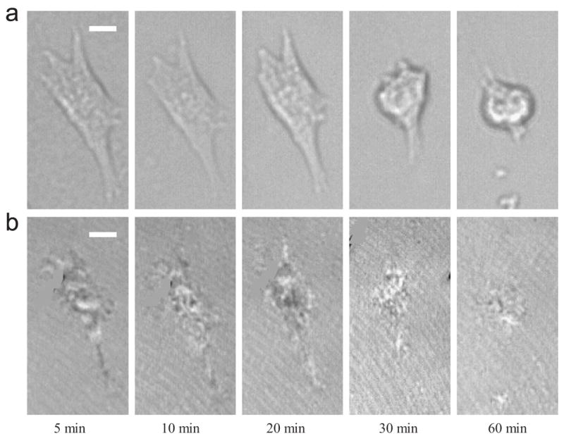
Fig. 5.

A series of (a) phase contrast images and (b) C-RICM images for a typical SMC on HBC 29 surface which has been pre-cultured at 37 °C for 24 h at different times during low-temperature (18 °C) incubation under 5% CO2. The scale-bar represents 10 μm.
