Abstract
The cytotoxic effect of Aplidin was investigated on fresh leukaemia cells derived from children with B-cell-precursor (BCP) acute lymphoblastic leukaemia (ALL) by using stromal-layer culture system and on four cell lines, ALL-PO, Reh, ALL/MIK and TOM-1, derived from patients with ALL with different molecular genetic abnormalities. In ALL cell lines Aplidin was cytotoxic at nanomolar concentrations. In the ALL cell lines the drug-induced cell death was clearly related to the induction of apoptosis and appeared to be p53-independent. Only in ALL-PO 20 nM Aplidin treatment caused a block of vascular endothelial growth factor (VEGF) secretion and downregulation of VEGF-mRNA, but Aplidin cytotoxicity does not seem to be related to VEGF inhibition since the sensitivity of ALL-PO cells to Aplidin is comparable to that observed for the other cells used. Aplidin induced a G1 and a G2 M block in ALL cell lines. In patient-derived leukaemia cells, Aplidin induced a strong cytotoxicity evidenced in a stroma-supported immunocytometric assay. Cells from children with genetic abnormalities such as t(9;22) and t(4;11) translocations, associated with an inferior treatment outcome, were sensitive to Aplidin to the same extent as that observed in other BCP-ALL cases. Aplidin exerted a strong cell killing effect (>88%) against primary culture cells from five relapsed ALL cases, at concentrations much lower than those reported to be achieved in plasma of patients receiving Aplidin at recommended doses. Taken together these data suggest that Aplidin could be a new anticancer drug to be investigated in ALL patients resistant to available therapy.
Keywords: marine natural compounds, apoptosis, cell cycle, stroma-supported immunocytometric assay
Acute lymphoblastic leukaemia (ALL) is the most common form of cancer in children. It arises in bone marrow from malignant B-lymphoid progenitors. Among the distinguishing cellular features of ALL, clonal abnormalities can be identified in approximately 65–70% of cases. Acute lymphoblastic leukaemia cells are sensitive to several currently used drug treatments, but in approximately one-third of the children, the disease recurs during, or following, therapy (Pui, 2000; Pui et al, 2001). Therefore, the identification of new antileukaemia drugs that are effective against ALL, particularly following a relapse, may help further improvement of treatment. Acute lymphoblastic leukaemia-derived primary cells are being used to improve the sensitivity of in vitro methods to measure new drug effectiveness–the goal of the work is presented here.
Aplidin (dehydro-didemnin B) is a marine depsipeptide isolated from the Mediterranean tunicate Aplidium albicans. It has shown activity against both human haematological and solid tumour cell lines growing in vitro (Urdiales et al, 1996; Lobo et al, 1997; Depenbrock et al, 1998; Geldof et al, 1999), in vivo murine B16 melanoma and in several human tumours xenografts (Faircloth et al, 1995, 1996).
Aplidin inhibits the progression of cells from G1 to the S phase of the cell cycle (Crampton et al, 1984). Recently, a G2 block was described for Aplidin in human leukaemia Molt-4 cell line (Erba et al, 2002). Inhibition of protein synthesis via GTP-dependent elongation factor 1-alpha in vitro (Crews et al, 1994), DNA and RNA syntheses in different cell lines (Crampton et al, 1984; Sakai et al, 1996) and inhibition of ornithine decarboxylase have been reported for Didemnins as well as for Aplidin (Urdiales et al, 1996; Erba et al, 2002).
In phase I and phase II clinical studies that are in progress, no neutropenia but a moderate lymphopenia has been observed (Raymond et al, 2000; Armand et al, 2001; Ciruelos Gil et al, 2002).
In this study, we assess the cytotoxic effect of Aplidin on fresh leukaemia cells derived from children with B-cell-precursor (BCP) ALL by using stromal-layer culture system, established by Campana and co-workers (Manabe et al, 1992; Campana et al, 1993, 1999; Kumagai et al, 1996). Aplidin effectiveness was also evaluated by assessing the cytotoxicity and the induction of apoptosis in four human leukaemic cell lines, ALL-PO, Reh, ALL/MIK and TOM-1, derived from patients with ALL with different molecular genetic abnormalities.
MATERIALS AND METHODS
Cell lines
The leukaemic cell lines, Reh, ALL/MIK, TOM-1 (Rosenfeld et al, 1977; Okabe et al, 1987; Higa et al, 1994) and ALL-PO (Gobbi et al, 1997), were derived from BCP-ALL patients and are all characterised by particular chromosomal translocations that are representative of the molecular genetic abnormalities most frequently found in childhood: two Philadelphia-positive cell lines TOM-1 and ALL/MIK cells with a t(9;22), Reh cells with a t(12;21) and ALL-PO cells with a t(4;11).
Cells were grown in an RPMI 1640 culture medium (Sigma-Aldrich, St Louis, MI, USA) with 10% Hyclone FBS (Hyclone Laboratories, Inc., Logan, UT, USA) and 4 mM glutamine at 37°C in a humidified atmosphere containing 5% CO2 in T25 cm2 tissue culture flasks (IWAKY, Bibby Sterilin, Staffordshire, UK).
Stroma-supported cultures of ALL cells
Leukaemic bone-marrow (BM) samples were collected from 14 patients with BCP-ALL aged less than 1–14 years (median, 6 years) either at the time of diagnosis (nine patients) or at relapse (five patients). The institutional review board approved this study and informed consent was obtained from patients and their guardians.
In all cases, more than 80% of the blasts were positive for CD19, class II antigens, terminal deoxynucleotidyl transferase (TdT), and lacked surface immunoglobulins (sIg). Karyotype features were determined by conventional banding techniques. Other clinical features of the patients are listed in Table 1 .
Table 1. Clinical features of the patients.
| Case (no) | Age (years) | Sex | WBC (l−1) | Phenotype | DNA index | Karyotype | Follow-up |
|---|---|---|---|---|---|---|---|
| 1 | 11 | F | 21 × 109 | cALL | 1,00 | 46,XX | +8 CR |
| 2 | 2 | M | 11 × 106 | cALL | 1,20 | 53, XY,+4,+6,+8,+12,+20+21+21+21 | +8 CR |
| 3 | 13 | F | 14 × 106 | Pre-Pre-B-ALL | 1,00 | 46,XX | +5 CR |
| 4 | 4 | F | 11 × 106 | cALL | 1,06 | 50,XX,+21[c] | +5 CR |
| 5 | <1 | F | 13 × 106 | Pre-B-ALL | 1,00 | 46,XX,+8,+19,+22 | +4 CR |
| 6 | 3 | M | 7,9 × 10⁶ | cALL | 1,00 | 47,XY,del(14q21),+marker | +5 CR |
| 7 | 4 | M | 350 × 106 | cALL | 1,00 | 46,XY t (9;22) | Resistant |
| 8 | 10 | F | 27 × 106 | cALL | 1,21 | 58,XX,+ × ,+1,+3,+8,+14,+16,+18,21,+19,+19,+21,+21,+21/46,XX | +30 CR |
| 9 | 9 | M | 140 × 106 | Pre-pre-B ALL | 1,00 | 46,XY t (4;11)/47,XY | +38 CR |
| 10a | F | 11 × 106 | cALL | ND | ND | Exitus | |
| 11a | 7 | M | 13 × 106 | Pre-B ALL | 1,00 | 46, XY | Exitus |
| 12a | 5 | F | 132 × 106 | LLA B mature | 1,00 | No mitosis | +2 TMO |
| 13a | 3 | M | 58 × 109 | cALL | 1,00 | ND | Exitus |
| 14a | 9 | M | 35 × 109 | Null ALL | ND | 46,XY | Exitus |
=relapse. ND=not determined.
Mononuclear cells (MNC) were obtained by centrifugation on a density gradient using Ficoll-Paque (Pharmacia LKB, Uppsala, Sweden). After washing with phosphate-buffered saline (PBS) ALL samples were cryopreserved in RPMI-1640 medium, with 50% heat-inactivated FBS Hyclone and 10% dimethyl sulphoxide (DMSO) and were stored in liquid nitrogen before use in the in vitro studies.
Previously frozen leukaemic cells were cultured and only cultures that had greater than 90% cell viability by trypan blue dye exclusion were used.
Bone marrow stromal layers were prepared as previously described by Campana and co-workers (Manabe et al, 1992; Campana et al, 1993, 1999). Briefly, normal BM MNC were resuspended in RPMI-1640 that contained 10% FCS, 1 μM hydrocortisone, 2 mM L-glutamine, and 1% penicillin–streptomycin. Cells were incubated at 37°C in 5% CO2 and 90% humidity in T 25 cm2 tissue culture flasks (IWAKY) and were fed every 7 days by replacing 50% of the supernatant with identical fresh medium.
After formation of confluent layers, cells were detached by trypsin, washed once with RPMI-1640 with 10% FBS and resuspended in fresh stroma culture medium. Cells were seeded in 96-well flat-bottom plates at 3 × 10⁴ cells well−1.
Drug treatment on cell lines
Aplidin was kindly supplied by PharmaMar SA (Madrid). The effect of drug treatment on cell lines was evaluated by a standard growth inhibition assay.
Leukaemia cells in the exponentially growing phase were treated for 1 h with different concentrations of Aplidin. After treatment, cells were washed with PBS and incubated with fresh drug-free medium; viable cells number was estimated by means of a Coulter Counter (Beckman Coulter Corp., Hialeath, FL, USA) at different time intervals after drug-washout.
Drug treatment on leukaemic BM cells
To determine the cytotoxicity of Aplidin in patient samples, we first established the cells in culture. The media from the BM stroma was removed and the adherent cells were washed seven times with AIM-V serum-free tissue culture medium (Gibco BRL). Resuspended leukaemia cells in AIM-V medium were aliquoted as 3 × 10⁵ cells on the stromal layers. Blast cells from individual patients were exposed for 7 days at 37°C in 5% CO2 and 90% humidity to different Aplidin concentrations in log increments that ranged from 0.005 to 5 nM.
Cell cycle
Monoparametric DNA analysis
Exponentially growing leukaemic cells were treated for 1 h with 0, 5, 10, and 20 nM Aplidin. After treatment, the drug-containing medium was removed, the cells were washed with PBS and were placed in fresh medium. At different time intervals after drug-washout, the number of cells was evaluated by a Coulter Counter and the cells were fixed in 70% ethanol and kept at 4°C before DNA staining (Erba et al, 2002).
Biparametric BrdUrd/DNA analysis
During the last 15 min of drug treatment, 20 μM bromodeoxyuridine (BrdUrd) was added to the cells. After treatment the drug-containing medium was removed, the cells were washed twice with PBS and fresh medium was provided. After 1 h treatment and at different time intervals after drug-washout, control and treated cells were fixed in 70% ethanol and stored at 4°C before staining. With this protocol it was also possible to obtain a distinct evaluation of cell cycle perturbations in cells which were in S phase (BrdUrd-positive cells), G1 phase or G2 M phase (BrdUrd-negative cells) during 1 h 10 nM Aplidin exposure (Erba et al, 2002).
Detection of apoptosis and caspase-3 activation in ALL cell lines
At different time intervals after drug-washout, the cells were fixed in 70% ethanol for terminal-dUTP-transferase (TdT) or in 1% paraformaldehyde for caspase analysis and stored at 4°C. The fixed cells were washed in cold PBS and incubated in 50 μl of TdT and FITC-conjugated dUTP deoxynucleotides solution (Roche Diagnostic SpA, Milan, Italy) or PE-conjugated rabbit anti-active caspase-3 (Becton Dickinson, San José, CA, USA) and analysed by flow cytometry (Erba et al, 2002).
Assessment of cytotoxicity and apoptosis on leukaemic BM cells
Before and after culturing on stroma, the ALL cell recovery and phenotype were determined as previously described (Manabe et al, 1992; Campana et al, 1993). Briefly, at termination of the cultures, cells were passed through a 19-gauge needle to disrupt clumps formed by stromal cells, washed with PBS that contained 0.2% bovine serum albumin and 0.2% sodium azide (PBSA) and incubated with Leu-12-PE (anti-CD19, Becton Dickinson, San Jose, CA, USA). After two washes in PBSA, the cells were resuspended in 0.5% paraformaldehyde and counted using a FACScan instrument and Cell Quest software (Becton Dickinson). Using the light-scatter dot plot, which depicts cell size and granularity, we identified the area where the vast majority of viable leukaemia cells were located at the beginning of cultures. This area was delimited with an electronic gate, which subsequently was used to enumerate leukaemia cells at the end of culture. The number of nonapoptotic lymphoid cells detected within a 30-s interval was corrected for the percentage of CD19+ cells present. The cell counts of drug-treated and control samples were compared to calculate the percentage of viable cells that remained after drug treatment. The following formula was used to calculate relative cell recovery after drug treatment: (no. cells recovered with drug)/(no. of cells recovered without drug) × 100.
VEGF RNAse protection
Exponentially growing cells were treated for 1 h with Aplidin (20 nM) and total RNA purified with the TRIZOL reagent (Gibco BRL) at 1, 6 and 24 h after drug-washout. VEGF and VEGFR-1 mRNA were measured by RNase protection assay using a commercially available kit (Becton Dickinson).
ELISA assay
The assay, aimed at evaluating the concentration of VEGF-A in the medium of cells, was performed on 96-well plates coated with an anti-VEGF antibody (Quantikine kit, R&D Systems Europe, Oxon, UK). Standards of VEGF protein ranging from 1000 to 31.2 pg ml−1 were prepared after reconstituting VEGF standard with 1 ml of calibrator diluent. To each well, 50 μl of Assay Diluent and 200 μl of medium (or standard) were added; after 2 h of incubation, wells were washed three times with wash buffer and 200 μl of VEGF-conjugated were added. After 2 h, wells were washed three times and 200 μl of Substrate Solution was added. After 20 min, 50 μl of stop solution was added to each well and optical density was evaluated by means of a plate-reader spectrophotometer (Labsystem Multiskan, Dasit, Italy) at 540 nm.
Liquid chromatography–tandem mass spectrometry analysis
Liquid chromatography–tandem mass spectrometry (LC–MS/MS) analyses of Aplidin in medium were performed using a method similar to that described for rat plasma (Celli et al, 1999).
RESULTS
Cytotoxicity, apoptosis and cell cycle perturbations induced by Aplidin on ALL cell lines
Figure 1A shows the growth inhibitory effect of 1 h treatment with different concentrations of Aplidin on different cell lines. Aplidin was active at nanomolar concentrations in all the cell lines and the cytotoxic effect was dose dependent. An irreversible growth inhibitory effect was observed after 1 h using 10 nM Aplidin treatment in All-PO, ALL/MIK and TOM-1 cells while in Reh cells Aplidin induced an irreversible growth inhibitory effect, but only at 20 nM. To assess the sensitivity of the leukaemic cell lines to Aplidin at different exposure times, cells were incubated with 20 nM Aplidin for 15 min, 1 or 24 h. As shown in Figure 1B the cytotoxicity against ALL-PO, Reh and TOM-1 cells was similar regardless of the incubation times. In contrast, ALL/MIK cells with 15 min of exposure to Aplidin survived longer than ALL/MIK cells after more prolonged treatments. Yet, an optimal effect was achieved by 1 h since similar effects were also seen at 24 h (Figure 1B).
Figure 1.
Effect of 1 h (A) or 15, 60 min 24 h (B) Aplidin exposure on cell growth evaluated at different time intervals after treatment and drug-washout on ALL-PO, Reh, ALL/MIK and TOM-1. Each point is the mean of three replicates; bars respresent the standard deviation.
The lack of increased cytotoxicity of Aplidin beyond 1 h compared to a short exposure time might have been due to a rapid decomposition of the peptide under cell culture conditions. However, this was not found to be the case. After 24 h incubation of Aplidin in medium at 37°C, 80% of the drug was detected as unchanged by HPLC–MS (data not shown).
Cell cycle perturbations induced on ALL cell lines
Figure 2 shows the effects on the cell cycle phase distribution caused by 1 h exposure with different concentrations of Aplidin in the different leukaemic cell lines evaluated at different time intervals after drug-washout. We found that in all the ALL cell lines Aplidin caused a block of the cells in the G1 phase of the cell cycle. In Reh, ALL/MIK and TOM-1 cells Aplidin induced also a G2 block.
Figure 2.
Cell cycle phase perturbations induced on (A) ALL-PO, (B) Reh, (C) ALL/MIK and (D) TOM-1 cells treated for 1 h with 0, 5, 10 or 20 nM Aplidin. Monoparametric DNA flow cytometric analysis were performed at different time intervals after drug-washout.
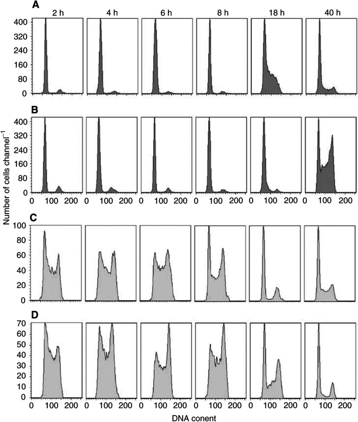
To better characterise the cell cycle phase perturbation induced by Aplidin the biparametric BrdU/DNA flow cytometric analysis were performed at different time intervals after drug-washout. Figure 3 reports, as an example, the DNA histograms of BrdU-negative and BrdU-positive cells obtained in Reh cells. Aplidin was found to delay those cells that were in the G1 phase (BrdU-negative cell population) during drug treatment from entering S phase. At 40 h after drug-washout, the BrdU-negative cells were accumulated in G1 and in G2. The cells that were in S phase (BrdU-positive cells) during Aplidin treatment progressed throughout this phase of the cell cycle more slowly than control cells. At 18h and UP 40h after; drug-washout, the BrdU-positive cells were accumulated in G1 and in G2.
Figure 3.
Biparametric BrdU/DNA analysis performed in Reh cells treated for 1 h with 10 nM Aplidin. During the last 15 min of drug treatment 20 μM BrdU was added to the cells, then the cells were washed with PBS and drug-free medium was provided. The flow cytometric analysis were performed at different time intervals after drug-washout. (A) DNA histograms of BrdU-negative control cells; (B) DNA histograms of BrdU-negative Aplidin-treated cells; (C) DNA histograms of BrdU-positive control cells; (D) DNA histograms of BrdU-positive Aplidin-treated cells.
Aplidin-induced apoptosis on ALL cell lines
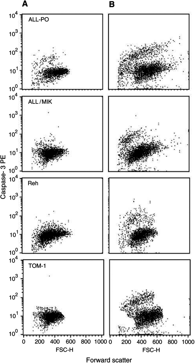
It has been reported that Aplidin acts in vitro by inducing apoptosis on different cells lines (Grubb et al, 1995; Erba et al, 2002). Aplidin induced apoptosis in all the cell lines used, as clearly seen by morphological examination by means of sulforhodamine/Dapi staining (data not shown). The level of apoptotic cells was different between the four cell lines as shown in Figure 4A, which shows an example of the TdT-dUTP flow cytometry analysis, and in Figure 4B, where the percentages of the fraction of apoptotic cells found at different times after drug-washout are reported. In Reh cells, Aplidin induced apoptosis only at the concentration of 20 nM. In ALL-PO, ALL/MIK and TOM-1 the amount of apoptotic cells increased dramatically when the cells were exposed to 10 or 20 nM Aplidin. In All-PO cells at 72 h after drug-washout 80% of the cells treated with 20 nM of Aplidin were apoptotic. As previously reported on other cell type (Garcia-Fernandez et al, 2002), Aplidin was found to induce apoptosis in a caspase-3-dependent manner (Figure 5).
Figure 4.
(A) Detection of apoptosis in ALL cells by TdT-dUTP flow cytometric analysis. Cells were treated with different concentrations of Aplidin and the biparametric FSC/TdT-dUTP analysis were performed after 72 h after drug-washout. (B) Percentage of apoptotic cells evaluated by TdT-dUTP flow cytometric analysis. Cells were treated with different concentrations of Aplidin and biparametric FSC/TdT-dUTP analysis were performed at different time intervals after drug-washout.
Figure 5.
Detection of active caspase-3 in ALL cells by flow cytometric analysis. Cells were treated with different concentrations of Aplidin and the biparametric FSC/caspase-3 analysis was performed at different times after drug-washout. In the figure are reported the analysis performed at 24 h after drug-washout. (A) control cells; (B) 20 nM Aplidin-treated cells.
Modulation of VEGF secretion by Aplidin on ALL cell lines
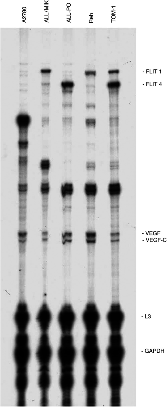
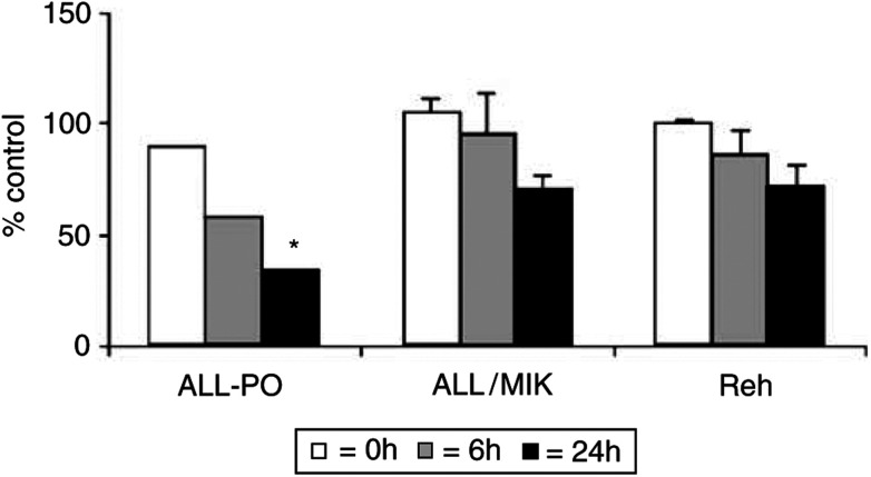
It has been reported by our group that Aplidin causes a strong block of VEGF secretion in Molt-4 cells with a subsequent downregulation of the transcription of VEGF and of its receptor VEGFR1 (Broggini et al, 2003). To test the hypothesis that Aplidin could exert its activity in ALL cell lines by inhibiting the VEGF/VEGFR-1 autocrine loop, we used an RNAse protection assay in each cell line. As shown in Figure 6, all the cell lines used expressed, at different levels, the VEGF mRNA and its receptors. ALL/MIK and Reh expressed the flt-1 receptor, ALL-PO only the flt-4 receptor while TOM-1 expressed both of them. Data reported in Figure 7 show that after different times from drug-washout, Aplidin downregulated the level expression of VEGF mRNA in ALL-PO cells. When tested in the other cell lines this observation was not further confirmed as shown in Figure 8. By using ELISA assay, we tested the level of VEGF secretion in the four leukaemia cell lines used and found that only ALL-PO secrete detectable amounts of the growth factor. Therefore, we evaluated whether a block in VEGF secretion in ALL-PO cells occurs as previously seen in Molt-4 cells (Broggini et al, 2003). We treated ALL-PO cells with 20 nM Aplidin for 1 h and tested the medium of the cells by means of an ELISA assay used to measure VEGF-A levels of secretion at 0, 6 and 24 h after drug-washout. As depicted in Figure 9 Aplidin was able to substantially abolish VEGF secretion in the medium at 6 and 24 h after drug-washout.
Figure 6.
Autoradiography of a typical RNAse protection assay on the four different human leukaemic cell lines. Human ovarian cancer A2780 cells were used as an internal control.
Figure 7.
Autoradiography of RNAse protection assay on ALL-PO cells treated with 20 nM Aplidin performed at different time intervals after drug-washout.
Figure 8.
Vascular endothelial growth factor mRNA levels in human leukaemic ALL-PO, ALL-MIK and Reh cell lines treated with 20 nM Aplidin and performed at different time intervals after drug-washout. Data have been obtained by densitometric analysis and expressend as % of control untreated cells. Each column represents the mean of three independent replicates. The bars represent s.d. *=P<0.05 (Duncan test).
Figure 9.
Vascular endothelial growth factor-A concentration in the medium of ALL-PO cells treated for 1 h with 20 nM Aplidin and evaluated at 0, 6 and 24 h after drug-washout. The values express the % of VEGF concentration in the medium of treated cells with regard to control cells.
Cytotoxicity and apoptosis induced by Aplidin on primary ALL cells
To assess the cytotoxic effect of Aplidin on fresh leukaemia cells obtained directly from patients affected by BCP-ALL, a stroma-supported immunocytometric assay was used. Previous studies have shown that, under these culture conditions, phenotypic and karyotype features of leukaemia cells are maintained even after several months of culture.
Table 1 shows the clinical features of the ALL patients at the time when the cells were derived. In nine out of 14 cases (nos. 1–9) the leukaemia cells were collected at the time of diagnosis, while in the other five cases (nos. 10–14), at the time of relapse. Among the 14 samples of ALL studied, the number of cells recovered after 7 days of stromal-supported culture ranged from 38 to 210% (median 83%) of those originally seeded.
It is known that changes in forward/side light-scatter parameters, consisting in a reduction in forward scatter, indicating a reduction in cell size, and an increase in side scatter, indicating an increase in cell granularity, are frequently associated with apoptosis. As detected on leukaemic cell lines Aplidin induced changes in the light-scattering properties by using flow cytometry forward/side light-scatter analysis on ALL cells too, suggesting an induction of apoptosis (Figure 10 shows a representative case).
Figure 10.
Example of light-scatter dot-plot analysis of blast cells growing on stroma feeder layer evaluated by flow cytometry. Forward and side scatter analysis were evaluated at the beginning of culture (A), after 7 days of culture (B, control cells) and after 7 days with 5 nM Aplidin (C, treated cells).
As shown in Table 2 , the cytotoxic effect of Aplidin on BCP-ALL cells was dose dependent. Aplidin at the concentration of 5 nM was strongly cytotoxic in all cases. The percentage of cell death ranged from 74 to 100% (median 97%). At the concentration of 0.5 nM Aplidin cell killing effect was not detectable in three cases (nos. 7, 8 and 9), while in the remaining 11 cases it ranged from nine to 99% (median 49%). Aplidin at 0.05 nM induced cell death in seven out of 13 cases (nos. 1–5 12 and 13) with a range from nine to 79% (median 20%). At 0.005 nM substantial cytotoxicity (47% compared to control cells) was observed only in one case (no. 13). Interestingly, the same level of cell kill (median 97%) was obtained with 5 nM Aplidin in cells taken either at the time of diagnosis (nos. 1–9) or at relapse (nos. 10–14).
Table 2. Stroma-supported immunocytometric assay % cell death after 7 days Aplidin exposure.
|
Aplidin (nM) |
||||
|---|---|---|---|---|
| Patient no | 0.005 nM | 0.05 nM | 0.05 nM | 5 nM |
| 1 | NT | 12 | 22 | 96 |
| 2 | NT | 79 | 79 | 100 |
| 3 | 14 | 20 | 40 | 99 |
| 4 | 16 | 36 | 60 | 100 |
| 5 | 12 | 19 | 46 | 100 |
| 6 | <1 | <1 | 9 | 96 |
| 7 | <1 | <1 | <1 | 74 |
| 8 | <1 | <1 | <1 | 97 |
| 9 | <1 | <1 | <1 | 90 |
| 10a | 31 | 3 | 35 | 82 |
| 11a | <1 | 1 | 49 | 98 |
| 12a | 10 | 9 | 99 | 88 |
| 13a | 47 | 33 | 55 | 97 |
| 14a | <1 | ND | 50 | 97 |
=relapse. ND=not determined.
The cytotoxicity of Aplidin extended to sample nos. 7 and 9, each of which carries adverse genetic features such as t(9;22) and t(4;11), respectively.
To investigate whether Aplidin cytotoxicity represents a direct effect on leukaemic cells or an indirect effect mediated by damage of the stroma layers, we incubated the stroma layers for 7 days with different Aplidin concentrations ranging from 0.005 to 5 nM. Then the cells were washed and seeded with leukaemic lymphoblasts in drug-free medium for 7 days. We found that the morphology and cell confluence were not affected by Aplidin treatment. In four out of five cases the percentage cell recovery obtained after 5 nM Aplidin exposure was between 78 and 92%. Only in one case did the ability of stromal cells to support leukaemia cell survival after Aplidin treatment not occur (data not shown).
DISCUSSION
This study shows that Aplidin is a potent antileukaemic agent against human lymphoblastic leukaemia cell lines, as well as fresh leukaemia cells derived directly from patients with childhood BCP-ALL. We found that on Philadelphia chromosome-positive TOM-1 and ALL/MIK with t(9;22), on ALL-PO with t(4;11) and on Reh t(12;21) cell lines, Aplidin induced a strong growth inhibition effect at nanomolar concentrations, the IC50 ranging from 5 to 20 nM.
The present study also shows that Aplidin is a strong inducer of apoptosis, a finding in keeping with previous data obtained on another human leukaemia Molt-4 cell line (Erba et al, 2002; Broggini et al, 2003). In all the cell lines investigated, the Aplidin-induced cell death was clearly related to the induction of apoptosis, even if in Reh cells the apoptosis was found only when the cells were exposed to high concentrations of Aplidin. ALL/MIK and TOM-1 cells express wt p53, whereas the other a mutated p53 (M Broggini, personal communication), thus indicating that Aplidin can induce apoptosis in a p53-independent manner.
We have recently reported that in Molt-4 cells Aplidin causes an inhibition of VEGF secretion and a downregulation of flt-1 (Broggini et al, 2003). The apoptosis induced by Aplidin in Molt-4 cell could be observed also by exposing the leukaemic cells to anti-VEGF antisense oligonucleotide (Gerber et al, 1998; Nor et al, 1999; Broggini et al, 2001, 2003). In these cells, the addition of VEGF could antagonise the apoptotic process induced by Aplidin. These data suggested that a potential mechanism of cytotoxicity of Aplidin was related to the blocking of an autocrine loop relevant for cell growth and survival. In the present study, however, we could not confirm this finding. In only one cell line, that is ALL-PO cells, we found evidence of the same phenomenon previously seen in Molt-4 cells. Since the sensitivity of ALL-PO cells to Aplidin is comparable to that observed for other ALL cell lines such as Reh, ALL/MIK and TOM-1, in which Aplidin was not inducing any effect on VEGF secretion, it should be concluded that Aplidin cytotoxicity against ALL cells is not related to VEGF inhibition. It may be speculated, however, that in the cell lines in which no block of VEGF loop was observed, Aplidin cytotoxicity is mediated by the inhibition of other growth factors. The evidence that Aplidin induces changes in the expression of genes involved in different cellular pathways (Marchini et al, 2002) is in line with this hypothesis. However, these aspects require to be further investigated.
The available data on the effects of didemnins on the cell cycle consistently indicate that these compounds cause a block of the cells in G1 (Crampton et al, 1984; Erba et al, 2002). It has been recently reported that in Molt-4 Aplidin mainly induces a G1 block, but a more sophisticated analysis revealed that the drug induced a G2 block too (Erba et al, 2002). In particular, by using a simulation program (Montalenti et al, 1998) suitable to describe drug-induced cell cycle block, delay, repair and death effects, it became apparent that a G2 blockade also occurs in Molt-4 cells exposed to Aplidin. In the present study we can confirm that in addition to a G1 arrest a G2 blockade was induced by Aplidin in ALL cell lines.
Aplidin was found to induce a strong cytotoxicity in patient-derived leukaemia cells evidenced in a stroma-supported immunocytometric assay. This system has been successfully used to investigate the antileukaemic activity of different compounds, such as 2-chloro-2-deoxyadenosine (Kumagai et al, 1994), Interferon-α (Manabe et al, 1993), Cyclosporin A (Ito et al, 1998), Taxol and Taxotere (Consolini et al, 1998), Vincristine, Teniposide and Ara C (Campana et al, 1993, 1999). Aplidin at 5 nM caused a dramatic cell death (median 97%) in all the 14 cases studied. At 0.5 nM cell death was still present in 11 out of 14 cases (median 49%). Although in some cases there might be discrepancies between the in vitro cytotoxic concentration and the active anticancer drug plasma levels, note that the Aplidin concentrations used are pharmacologically reasonable as Aplidin concentrations above 20 nM are achievable for several hours in the plasma of patients receiving the drug given as 24 h in a range doses much lower than the maximum tolerated dose of 6000 μg m−2 (Zucchetti, personal communication).
Cells from two children with genetic abnormalities such as t(9;22) and t(4;11) translocation, which are associated with an inferior treatment outcome, were sensitive to Aplidin to the same extent as that observed in other BCP-ALL cases. Likewise, the cell lines with t(9;22) (ALL/MIK and TOM-1) or t(4;11) (ALL-PO) were strongly sensitive to Aplidin at similar concentrations.
In relapsed ALL cases, Aplidin exerted a strong cell killing effect (97%) in all five primary cells indicating that Aplidin is a candidate antileukaemic agent in patients with ALL that are nonresponsive to standard chemotherapeutic agents.
The data obtained with ALL cell lines and on Molt-4 cells (Erba et al, 2002) clearly indicate a direct antileukaemic activity of Aplidin. However, in the stroma-supported cultures of BCP-ALL cells derived from patients, the Aplidin-induced apoptosis could be due to a toxic effect to stroma cells (Campana et al, 1993; Consolini et al, 1998; Ito et al, 1998). We did not find a decrease in the capacity of stroma pretreated with Aplidin, to support ALL cell viability. Recently (Albella et al, 2002), similar data have been reported on human bone haematopoietic progenitors treated by Aplidin. At concentrations similar to those used in this study Aplidin did not induce growth inhibition in the tested haematopoietic progenitors by using clonogenic assay. It must be taken into account that stroma is characterised by the presence of different cell types including endothelial, reticulo cells, macrophages, fibroblast and adipocytes. As the stroma layers used in this study were derived from different patients, the reduced survival of ALL cells found in one case after exposure to 5 nM Aplidin, could be related to biological variability in the susceptibility of the different cell types present in the stroma layer.
Although the treatment outcome of children affected by ALL showed marked improvements in the last decade, in one-third of the children, ALL is fatal. Identification of new antileukaemia agents is essential for improving the survival of patients with high-risk or refractory leukaemia. Clinical Phase I and II studies of Aplidin have shown antitumour activity in patients with neuroendocrine tumours and medullary thyroid carcinomas (Raymond et al, 2000; Armand et al, 2001; Ciruelos Gil et al, 2002). Since at the recommended doses for phase II studies Aplidin plasma levels are maintained for many hours in the range of 10–100 nM (Zucchetti, personal communication; Maroun et al, 2001, according to the results presented in this study it seems realistic to assume the drug has a potential for therapy of ALL patients resistant to or relapsing from the available chemotherapies.
Acknowledgments
This work was partially supported by a grant from the Italian Ministry of Health (Project No. ICS0301/RF00/192) and by a grant from CNR-MIUR ‘Progetti Strategici Oncologia'.
The generous contributions of the Fondazione Nerina e Mario Mattioli and of the Fondazione M Tettamanti are gratefully acknowledged.
References
- Albella B, Faircloth G, Lopez-Lazaro L, Guzman C, Jimeno J, Bueren JA (2002) In vitro toxicity of ET-743 and aplidine, two marine-derived antineoplastics, on human bone marrow haematopoietic progenitors, comparison with the clinical results. Eur J Cancer 38: 1395–1404 [DOI] [PubMed] [Google Scholar]
- Armand JP, Ady-Vago N, Faivre S, Chieze S, Baudin E, Ribrag V, Lecot F, Iglesias L, Lopez-Lazaro L, Guzman C, Jimeno J, Ducreux M, Le Chevalier T, Raymond E (2001) Phase I and pharmacokinetic study of aplidine (APL) given as a 24-hour continuous infusion every other week (q2w) in patients (pts) with solid tumor (ST) and lymphoma (NHL)[abstract]. Proceedings 37th ASCO Annual Meeting. San Francisco 12–15 May, Vol. 20, p 120a [Google Scholar]
- Broggini M, Marchini S, Galliera E, Borsotti P, Taraboletti G, Erba E, Sironi M, Giavazzi R, Jimeno J, Faircloth G, D'Incalci M (2003) Aplidine, a new anticancer agent of marine origin, inhibits vascular endothelial growth factor (VEGF) secretion and blocks VEGF-VEGF-RI (flt-1) autocrine loop in human leukemic cells MOLT-4. Leukemia 17: 52–59 [DOI] [PubMed] [Google Scholar]
- Broggini M, Marchini S, Galliera E, D'Incalci M, Taraboletti G, Giavazzi R, Faircloth GT, Jimeno J (2001) Aplidine-induced apoptosis in MOLT-4 cells is mediated by its ability to block VEGF secretion [abstract]. Proceedings AACR- NCI-EORTC International Conference: Molecular targets and cancer therapeutics, Miami, 22 October–2 November, p 79
- Campana D, Houghton PJ, Rivera GK (1999) Testing antileukemic drugs. In Childhood Leukemias, Ching-Hong Pui (ed) pp 393–412. Cambridge [Google Scholar]
- Campana D, Manabe A, Evans WE (1993) Stroma-supported immunocytometric assay (SIA): a novel method for testing the sensitivity of acute lymphoblastic leukemia cells to cytotoxic drugs. Leukemia 7: 482–488 [PubMed] [Google Scholar]
- Celli N, Gallardo AM, Rossi C, Zucchetti M, D'Incalci M, Rotilio D (1999) Analysis of aplidine (dehydrodidemnin B), a new marine-derived depsipeptide, in rat biological fluids by liquid chromatography–tandem mass spectrometry. J Chromatogr B Biomed Sci Appl 731: 335–343 [DOI] [PubMed] [Google Scholar]
- Ciruelos Gil EM, Twelves C, Dominguez MJ, McKay H, Anthony A, Castellanos D, Bezares S, Ruiz A, Lopez-Lazaro L, Jimeno J, Celli N, Cotes-Funes H, Paz-Ares L (2002) Phase I clinical and pharmacokinetic study of the marine compound aplidine (APL) administered as a 3 h infusion every 2 weeks. Proceedings 38th ASCO Annual Meeting, Orlando, 18–21 May, Vol. 21, p 106a
- Consolini R, Pui CH, Behm FG, Raimondi SC, Campana D (1998) In vitro cytotoxicity of docetaxel in childhood acute leukemias. J Clin Oncol 16: 907–913 [DOI] [PubMed] [Google Scholar]
- Crampton SL, Adams EG, Kuentzel SL, Li LH, Badiner G, Bhuyan BK (1984) Biochemical and cellular effects of didemnins A and B. Cancer Res 44: 1796–1801 [PubMed] [Google Scholar]
- Crews CM, Collins JL, Lane WS, Snapper ML, Schreiber SL (1994) GTP-dependent binding of the antiproliferative agent didemnin to elongation factor 1alpha. J Biol Chem 269: 15411–15414 [PubMed] [Google Scholar]
- Depenbrock H, Peter R, Faircloth GT, Manzanares I, Jimeno J, Hanauske AR (1998) In vitro activity of aplidine, a new marine-derived anti-cancer compound, on freshly explanted clonogenic human tumour cells and haematopoietic precursor cells. Br J Cancer 78: 739–744 [DOI] [PMC free article] [PubMed] [Google Scholar]
- Erba E, Bassano L, Di Liberti G, Muradore I, Chiorino G, Ubezio P, Vignati S, Codegoni A, Desiderio MA, Faircloth G, Jimeno J, D'Incalci M (2002) Cell cycle phase perturbations and apoptosis in tumour cells induced by aplidine. Br J Cancer 86: 1510–1517 [DOI] [PMC free article] [PubMed] [Google Scholar]
- Faircloth G, Perez J, Fernandez JLSP, Avila J, Garcia M, Erba E, D'Incalci M, Canedo A, Garcia de Quesada T, Jimeno J (1995) Marine depsipeptides with activity against solid tumour models [abstract]. Proceedings 8th ECCO Congress, Paris, 29 October–2 November, p 529
- Faircloth JG, Rinehart K, Nunez de Castro I, Jimeno J (1996) Dehydrodidemnin B (DDB) a new marine derived antitumour agent (MDA) with activity against experimental tumour models [abstract]. Ann Oncol 7 (Suppl 1): 34 [Google Scholar]
- Garcia-Fernandez LF, Losada A, Alcaide V, Alvarez AM, Cuadrado A, Gonzalez L, Nakayama K, Nakayama KI, Fernandez-Sousa JM, Munoz A, Sanchez-Puelles JM (2002) Aplidin induces the mitochondrial apoptotic pathway via oxidative stress-mediated JNK and p38 activation and protein kinase C delta. Oncogene 21: 7533–7544 [DOI] [PubMed] [Google Scholar]
- Geldof AA, Mastbergen SC, Henrar RE, Faircloth GT (1999) Cytotoxicity and neurocytotoxicity of new marine anticancer agents evaluated using in vitro assays. Cancer Chemother Pharmacol 44: 312–318 [DOI] [PubMed] [Google Scholar]
- Gerber HP, McMurtrey A, Kowalski J, Yan M, Keyt BA, Dixit V, Ferrara N (1998) Vascular endothelial growth factor regulates endothelial cell survival through the phosphatidylinositol 3′-kinase/Akt signal transduction pathway. Requirement for Flk-1/KDR activation. J Biol Chem 273: 30336–30343 [DOI] [PubMed] [Google Scholar]
- Gobbi A, Di Berardino C, Scanziani E, Garofalo A, Rivolta A, Fontana G, Rambaldi A, Giavazzi R, Biondi A (1997) A human acute lymphoblastic leukemia line with the T(4;11) translocation as a model of minimal residual disease in SCID mice. Leuk Res 21: 1107–1114 [DOI] [PubMed] [Google Scholar]
- Grubb DR, Wolvetang EJ, Lawen A (1995) Didemnin B induces cell death by apoptosis: the fastest induction of apoptosis ever described. Biochem Biophys Res Comm 215: 1130–1136 [DOI] [PubMed] [Google Scholar]
- Higa T, Okabe M, Kunieda Y, Kodama S, Itaya T, Kurosawa M, Sakurada K, Maekawa I, Shoji M, Kasai M, Miyazaki T (1994) Establishment and characterization of a new Ph1-positive ALL cell line (ALL/MIK) presenting bcr gene rearrangement on bcr-2 and ALL-type bcr/abl transcript: suggestion of in vitro differentiation to monocytoid lineage. Leuk Lymphoma 12: 287–296 [DOI] [PubMed] [Google Scholar]
- Ito C, Ribeiro RC, Behm FG, Raimondi SC, Pui CH, Campana D (1998) Cyclosporin A induces apoptosis in childhood acute lymphoblastic leukemia cells. Blood 91: 1001–1007 [PubMed] [Google Scholar]
- Kumagai M, Manabe A, Coustan-Smith E, Blakley RL, Beck WT, Santana VM, Behm FG, Raimondi SC, Campana D (1994) Use of stroma-supported cultures of leukemic cells to assess antileukemic drugs. II. Potent cytotoxicity of 2-chloro-deoxyadenosine in acute lymphoblastic leukemia. Leukemia 8: 1116–1123 [PubMed] [Google Scholar]
- Kumagai M, Manabe A, Pui CH, Behm FG, Raimondi SC, Hancock ML, Mahmoud H, Crist WM, Campana D (1996) Stroma-supported culture in childhood B-lineage acute lymphoblastic leukemia cells predicts treatment outcome. J Clin Invest 97: 755–760 [DOI] [PMC free article] [PubMed] [Google Scholar]
- Lobo C, Garcia-Pozo SG, De Castro IN, Alonso FJ (1997) Effect of dehydrodidemnin B on human colon carcinoma cell lines. Anticancer Res 17: 333–336 [PubMed] [Google Scholar]
- Manabe A, Coustan-Smith E, Behm FG, Raimondi SC, Campana D (1992) Bone marrow-derived stromal cells prevent apoptotic cell death in B-lineage acute lymphoblastic leukemia. Blood 79: 2370–2377 [PubMed] [Google Scholar]
- Manabe A, Yi T, Kumagai M, Campana D (1993) Use of stroma-supported cultures of leukemic cells to assess antileukemic drugs. I. Cytotoxicity of interferon alpha in acute lymphoblastic leukemia. Leukemia 7: 1990–1995 [PubMed] [Google Scholar]
- Marchini S, Chiorino G, Faircloth GT, D'Incalci M (2002) Changes in gene expression profile induced by the anticancer agent Aplidine in Molt-4 leukemic cell lines. J Biol Regul Homeost Agents 16: 241–248 [PubMed] [Google Scholar]
- Maroun JA, Goel R, Stewart DJ, Tomiak E, Belanger K, Soulieres D, Charpentier D, Seymour L, Matthews S, Jimeno J, Guzman C (2001) Phase I study of aplidine in a 5 day bolus Q 3 weeks in patients with solid tumors and lymphomas. Proceedings 37th ASCO Annual Meeting, San Francisco, 12–15 May, Vol. 20, p 83b
- Montalenti F, Sena G, Cappella P, Ubezio P (1998) Simulating cancer-cell kinetics after drug treatment: application to cisplatin on ovarian carcinoma. Phys Rev E 57: 5877–5887 [Google Scholar]
- Nor JE, Christensen J, Mooney DJ, Polverini PJ (1999) Vascular endothelial growth factor (VEGF)-mediated angiogenesis is associated with enhanced endothelial cell survival and induction of Bcl-2 expression. Am J Pathol 154: 375–384 [DOI] [PMC free article] [PubMed] [Google Scholar]
- Okabe M, Matsushima S, Morioka M, Kobayashi M, Abe S, Sakurada K, Kakinuma M, Miyazaki T (1987) Establishment and characterization of a cell line, TOM-1, derived from a patient with Philadelphia chromosome-positive acute lymphocytic leukemia. Blood 69: 990–998 [PubMed] [Google Scholar]
- Pui CH (2000) Acute lymphoblastic leukemia in children. Curr Opin Oncol 12: 3–12 [DOI] [PubMed] [Google Scholar]
- Pui CH, Campana D, Evans WE (2001) Childhood acute lymphoblastic leukaemia–current status and future perspectives. Lancet Oncol 2: 597–607 [DOI] [PubMed] [Google Scholar]
- Raymond E, Ady-Vago N, Baudin E, Ribrag V, Faivre SLF, Wright T, Lopez Lazaro L, Guzman C, Jimeno J, Ducreux M, Chevalier T, Armand JP (2000) A phase I and pharmacokinetic study of aplidine (APL) given as a 24-hour continuous infusion every other week (q2w) in patients (pts) with solid tumor (ST) and lymphoma (NHL) [abstract]. Proceedings 11th NCI-EORTC-AACR Symposium, Amsterdam, 7–10 November, p 86
- Rosenfeld C, Goutner A, Choquet C, Venuat AM, Kayibanda B, Pico JL, Greaves MF (1977) Phenotypic characterisation of a unique non-T, non-B acute lymphoblastic leukaemia cell line. Nature 267: 841–843 [DOI] [PubMed] [Google Scholar]
- Sakai R, Rinehart KL, Kishore V, Kundu B, Faircloth G, Gloer JB, Carney JR, Namikoshi M, Sun F, Hughes Jr RG, Gravalos DG, De Quesada TG, Wilson GR, Heid RM (1996) Structure–activity relationships of the didemnins. J Med Chem 39: 2819–2834 [DOI] [PubMed] [Google Scholar]
- Urdiales JL, Morata P, Nunez de Castro I, Sanchez-Jimenez F (1996) Antiproliferative effect of dehydrodidemnin B (DDB), a depsipeptide isolated from Mediterranean tunicates. Cancer Lett 102: 31–37 [DOI] [PubMed] [Google Scholar]


