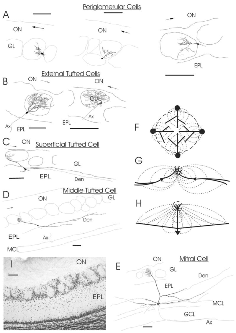
Fig. 6.

(A–E) Reconstructions of biocytin-labeled neurons having dendritic arborization in a single glomerulus. (A) Three PG cells with a branching primary dendrite (Den) within the glomerulus. (B) Two ET cells with the somata in the glomerular shell and an extensive dendritic tuft occupying entire glomerulus. (C) Superficial medium-tufted cell having long primary Den terminating with a tuft, cell body in the upper EPL and a few long secondary Dens parallel to GL/EPL border. (D) Medium-tufted cell with the soma deep in the EPL. (E) Mitral cell with a tufted primary Den and long lateral Dens. (F–H) Diagrams of current flow: (F) for a group of JG cells surrounding a glomerulus, (G) for tufted cells, (H) for mitral cell. Here dashed circle designates a glomerulus, thick lines are Dens and thin dashed lines are directions of currents. Note convergence of currents from broadly distributed sources of M/T cells into a comparatively small sink in the glomerular core. In contrast, somata of JG cells create a source distributed primarily in the glomerular shell and Dens give rise to a sink in the core. In the reconstructions Ax stands for an axon; arrow in the ON shows rostro-caudal direction. Scale bars = 100 μm in all drawings and on the photo. (I) photograph of a fragment from 60 μm-thick section immunolabeled with NeuN for mature neurons.
