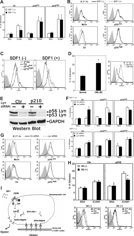
Figure 2.
BCR-ABL1 alters adhesive responses to SDF-1 through a LFA-1 integrin-mediated mechanism. (A) Attachment of SDF-1–stimulated or –unstimulated M-07e cells to surfaces coated with ICAM-1 or BSA (control) after transfection with p210BCR-ABL1 or control vector (Document S1). See Figure 1A for the BCR-ABL1 Western blot. Values are means plus or minus SEM (n = 4); *P < .05. (B) FACS analysis of β2 integrin-activation epitope expression in control and BCR-ABL1–transfected M-07e cells stimulated with SDF-1. Cells were stained with anti-human integrin monoclonal antibody 24 (which recognizes a conformationally dependent high-affinity epitope on β2 integrins), followed by secondary antibodies and FACS analysis (Document S1). (C) FACS analysis showing increased expression of β2 integrin-activation epitope 24 in BCR-ABL+ M-07e cells compared with BCR-ABL− M-07e cells. (D) Adhesion of primary human CML blast crisis CD34+ cells or normal CD34+ cells to stromal marrow cells obtained from a healthy individual. FACS analysis shows the expression level of the β2 integrin-activation epitope. Cells were stained with anti–human integrin monoclonal antibody 24, followed by secondary antibodies and FACS analysis. Values in adhesion assays are means plus or minus SEM (n = 3); *P < .05. (E) The expression levels of Lyn in control and BCR-ABL+ M-07e cells were determined by Western blotting 48 hours after nucleoporation with Lyn siRNA or control siRNA (Document S1). The blot was stripped and reprobed with an anti-GAPDH antibody. (F) Attachment of SDF-1–stimulated or –unstimulated BCR-ABL+ or control M-07e cells to surfaces coated with ICAM-1 or BSA (control) at 48 hours after transfection with Lyn siRNA or control siRNA. Values in adhesion assays are means plus or minus SEM (n = 3). (G) FACS analysis of β2 integrin-activation epitope 24 expression in SDF-1–stimulated BCR-ABL+ or control M-07e cells at 48 hours after nucleoporation with Lyn siRNA or control siRNA. We were unable to use primary blasts in these experiments because of apoptosis. As we previously showed, Lyn siRNA–treated BCR-ABL1+ primary blasts underwent apoptosis, whereas the same treatment inhibited the growth of several BCR-ABL1+ cell lines; however, they remained viable.8 (H) Effect of imatinib (IM) on the BCR-ABL1 kinase-dependent LFA-1 activity was measured by both adhesion assay and FACS analysis. M-07e cells transfected with control (Ctr) or p210-BCR-ABL (p210) were treated with or without IM (1 μM) for 16 to 18 hours; we then measured either attachment to surfaces coated with ICAM-1 and BSA (control) or β2 integrin-activation epitope 24 expression by FACS analysis. Values in adhesion assays are means plus or minus SEM (n = 3); *P < .05. (I) We propose that BCR-ABL1 alters SDF-1–mediated cell adhesion and movement via constitutively activating LFA-1 and inhibiting SDF-1–induced inside-out signaling involving CXCR4 and Lyn.

