

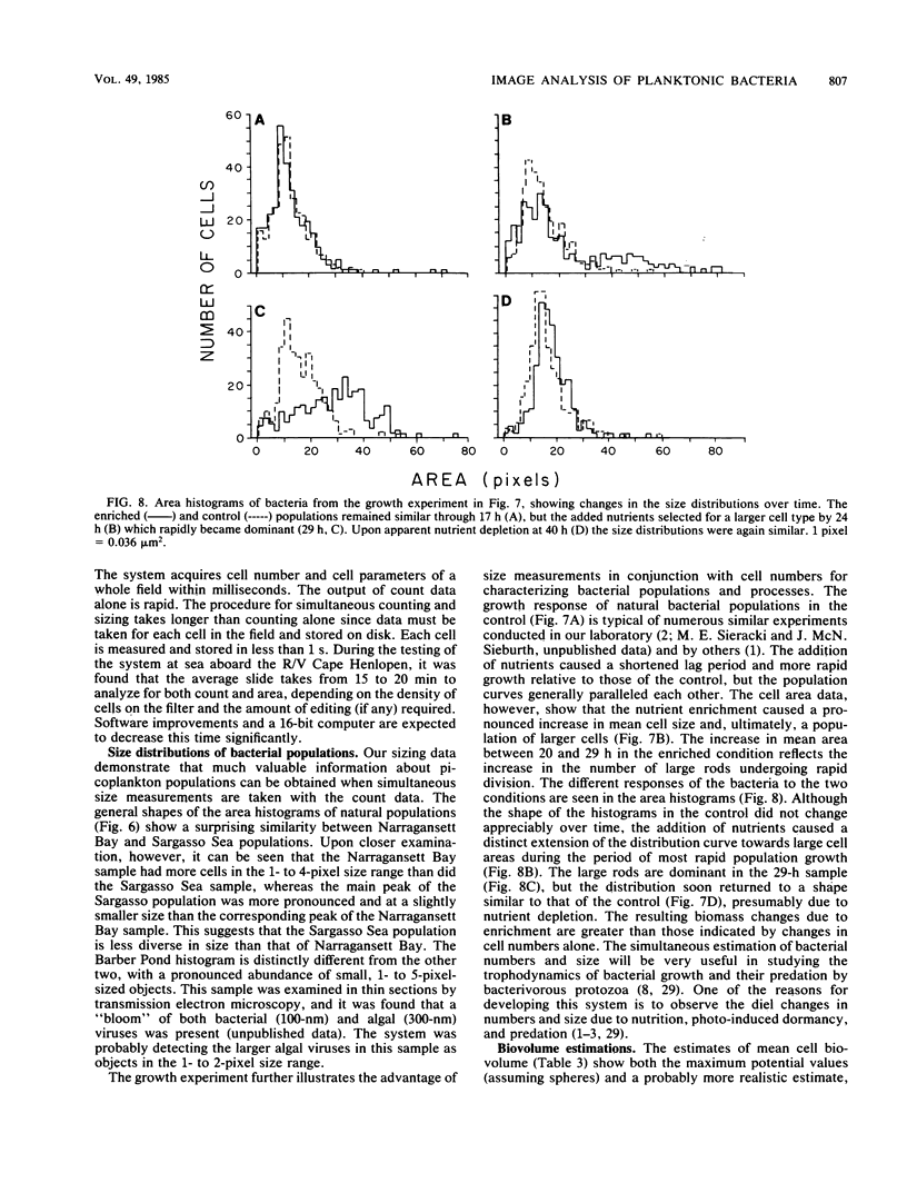
Abstract
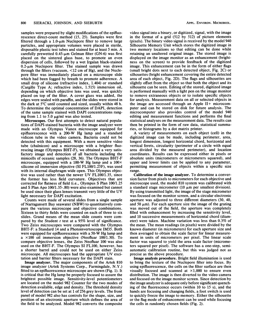
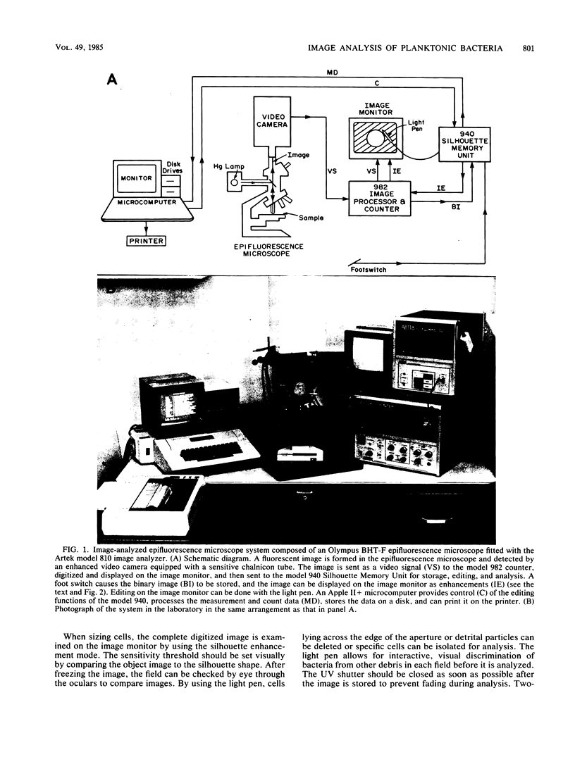
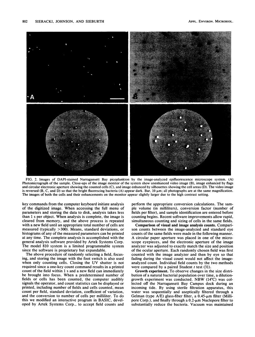
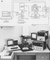
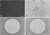
Epifluorescence microscopy is now being widely used to characterize planktonic procaryote populations. The tedium and subjectivity of visual enumeration and sizing have been largely alleviated by our use of an image analysis system consisting of a modified Artek 810 image analyzer and an Olympus BHT-F epifluorescence microscope. This system digitizes the video image of autofluorescing or fluorochrome-stained cells in a microscope field. The digitized image can then be stored, edited, and analyzed for total count or individual cell size and shape parameters. Results can be printed as raw data, statistical summaries, or histograms. By using a stain concentration of 5 micrograms of 4'6-diamidino-2-phenylindole per ml of sample and the optimal sensitivity level and mode, counts by image analysis of natural bacterial populations from a variety of habitats were found to be statistically equal to standard visual counts. Although the time required to prepare slides, focus, and change fields is the same for visual and image analysis methods, the time and effort required for counting is eliminated since image analysis is instantaneous. The system has been satisfactorily tested at sea. Histograms of cell silhouette areas indicate that rapid and accurate estimates of bacterial biovolume and biomass will be possible with this system.
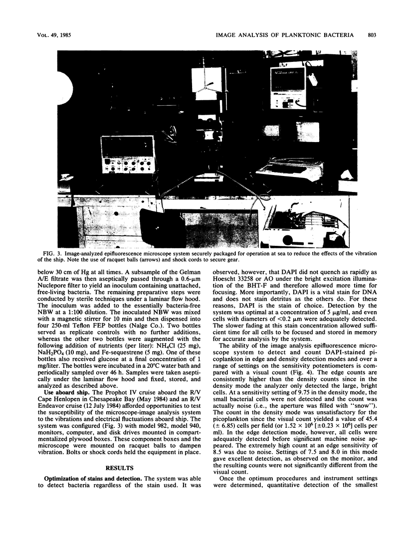
Full text
PDF
Images in this article
Selected References
These references are in PubMed. This may not be the complete list of references from this article.
- Baxter M., Sieburth J. M. Metabolic and ultrastructural response to glucose of two eurytrophic bacteria isolated from seawater at different enriching concentrations. Appl Environ Microbiol. 1984 Jan;47(1):31–38. doi: 10.1128/aem.47.1.31-38.1984. [DOI] [PMC free article] [PubMed] [Google Scholar]
- Campbell L., Carpenter E. J., Iacono V. J. Identification and enumeration of marine chroococcoid cyanobacteria by immunofluorescence. Appl Environ Microbiol. 1983 Sep;46(3):553–559. doi: 10.1128/aem.46.3.553-559.1983. [DOI] [PMC free article] [PubMed] [Google Scholar]
- Caron D. A. Technique for enumeration of heterotrophic and phototrophic nanoplankton, using epifluorescence microscopy, and comparison with other procedures. Appl Environ Microbiol. 1983 Aug;46(2):491–498. doi: 10.1128/aem.46.2.491-498.1983. [DOI] [PMC free article] [PubMed] [Google Scholar]
- Christian R. R., Hanson R. B., Newell S. Y. Comparison of methods for measurement of bacterial growth rates in mixed batch cultures. Appl Environ Microbiol. 1982 May;43(5):1160–1165. doi: 10.1128/aem.43.5.1160-1165.1982. [DOI] [PMC free article] [PubMed] [Google Scholar]
- Dahle A. B., Laake M. Diversity dynamics of marine bacteria studied by immunofluorescent staining on membrane filters. Appl Environ Microbiol. 1982 Jan;43(1):169–176. doi: 10.1128/aem.43.1.169-176.1982. [DOI] [PMC free article] [PubMed] [Google Scholar]
- Ferguson R. L., Buckley E. N., Palumbo A. V. Response of marine bacterioplankton to differential filtration and confinement. Appl Environ Microbiol. 1984 Jan;47(1):49–55. doi: 10.1128/aem.47.1.49-55.1984. [DOI] [PMC free article] [PubMed] [Google Scholar]
- Hagström A., Larsson U., Hörstedt P., Normark S. Frequency of dividing cells, a new approach to the determination of bacterial growth rates in aquatic environments. Appl Environ Microbiol. 1979 May;37(5):805–812. doi: 10.1128/aem.37.5.805-812.1979. [DOI] [PMC free article] [PubMed] [Google Scholar]
- Hobbie J. E., Daley R. J., Jasper S. Use of nuclepore filters for counting bacteria by fluorescence microscopy. Appl Environ Microbiol. 1977 May;33(5):1225–1228. doi: 10.1128/aem.33.5.1225-1228.1977. [DOI] [PMC free article] [PubMed] [Google Scholar]
- Ingram M., Preston K., Jr Automatic analysis of blood cells. Sci Am. 1970 Nov;223(5):72–82. doi: 10.1038/scientificamerican1170-72. [DOI] [PubMed] [Google Scholar]
- Kirchman D., Sigda J., Kapuscinski R., Mitchell R. Statistical analysis of the direct count method for enumerating bacteria. Appl Environ Microbiol. 1982 Aug;44(2):376–382. doi: 10.1128/aem.44.2.376-382.1982. [DOI] [PMC free article] [PubMed] [Google Scholar]
- Krambeck C., Krambeck H. J., Overbeck J. Microcomputer-assisted biomass determination of plankton bacteria on scanning electron micrographs. Appl Environ Microbiol. 1981 Jul;42(1):142–149. doi: 10.1128/aem.42.1.142-149.1981. [DOI] [PMC free article] [PubMed] [Google Scholar]
- Li W. K., Rao D. V., Harrison W. G., Smith J. C., Cullen J. J., Irwin B., Platt T. Autotrophic picoplankton in the tropical ocean. Science. 1983 Jan 21;219(4582):292–295. doi: 10.1126/science.219.4582.292. [DOI] [PubMed] [Google Scholar]
- Newell S. Y., Christian R. R. Frequency of dividing cells as an estimator of bacterial productivity. Appl Environ Microbiol. 1981 Jul;42(1):23–31. doi: 10.1128/aem.42.1.23-31.1981. [DOI] [PMC free article] [PubMed] [Google Scholar]
- Paul J. H. Use of hoechst dyes 33258 and 33342 for enumeration of attached and planktonic bacteria. Appl Environ Microbiol. 1982 Apr;43(4):939–944. doi: 10.1128/aem.43.4.939-944.1982. [DOI] [PMC free article] [PubMed] [Google Scholar]
- Reed W. M., Dugan P. R. Study of Developmental Stages of Methylosinus trichosporium with the Aid of Fluorescent-Antibody Staining Techniques. Appl Environ Microbiol. 1979 Dec;38(6):1179–1183. doi: 10.1128/aem.38.6.1179-1183.1979. [DOI] [PMC free article] [PubMed] [Google Scholar]
- WALTON W. H. Automatic counting of microscopic particles. Nature. 1952 Mar 29;169(4300):518–520. doi: 10.1038/169518a0. [DOI] [PubMed] [Google Scholar]
- Ward B. B., Perry M. J. Immunofluorescent Assay for the Marine Ammonium-Oxidizing Bacterium Nitrosococcus oceanus. Appl Environ Microbiol. 1980 Apr;39(4):913–918. doi: 10.1128/aem.39.4.913-918.1980. [DOI] [PMC free article] [PubMed] [Google Scholar]
- Watson S. W., Novitsky T. J., Quinby H. L., Valois F. W. Determination of bacterial number and biomass in the marine environment. Appl Environ Microbiol. 1977 Apr;33(4):940–946. doi: 10.1128/aem.33.4.940-946.1977. [DOI] [PMC free article] [PubMed] [Google Scholar]