Abstract
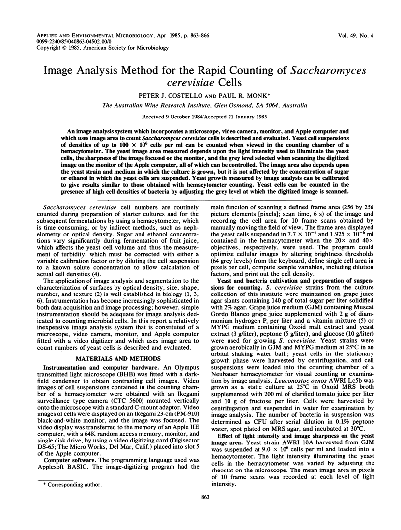
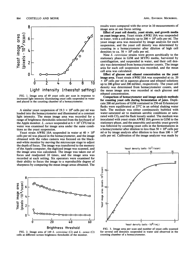
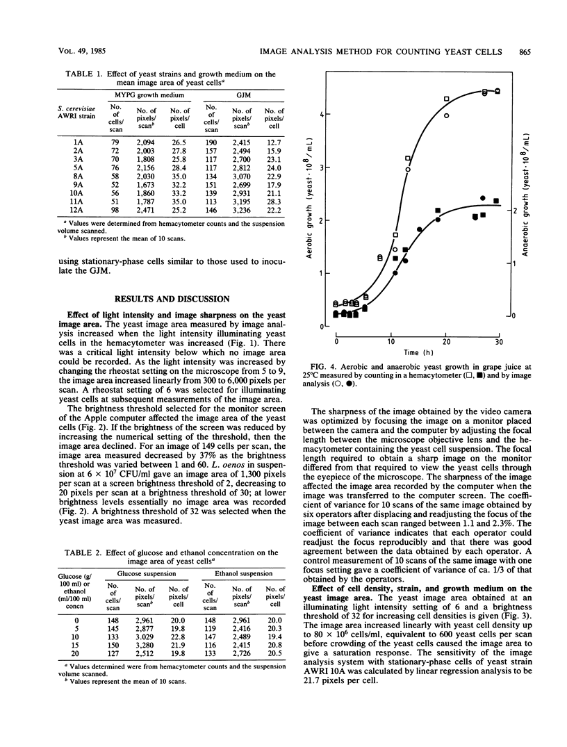
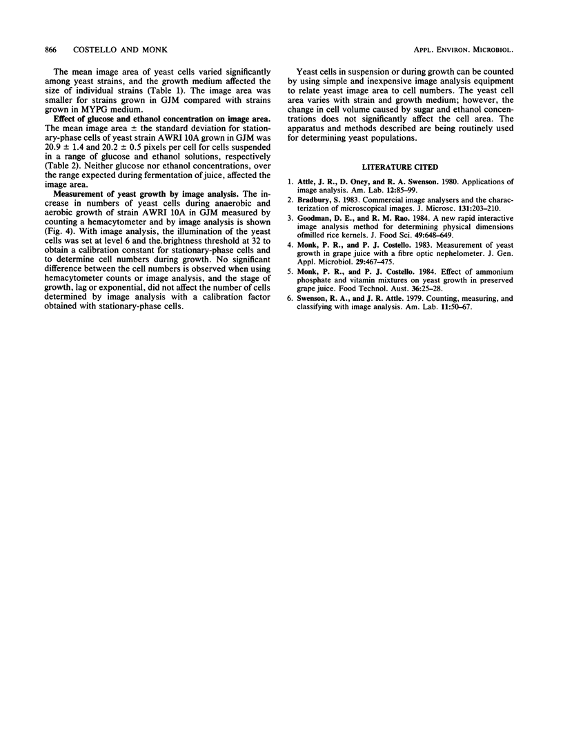
An image analysis system which incorporates a microscope, video camera, monitor, and Apple computer and which uses image area to count Saccharomyces cerevisiae cells is described and evaluated. Yeast cell suspensions of densities of up to 100 X 10(6) cells per ml can be counted when viewed in the counting chamber of a hemacytometer. The yeast image area measured depends upon the light intensity used to illuminate the yeast cells, the sharpness of the image focused on the monitor, and the grey level selected when scanning the digitized image on the monitor of the Apple computer, all of which can be controlled. The image area also depends upon the yeast strain and medium in which the culture is grown, but it is not affected by the concentration of sugar or ethanol in which the yeast cells are suspended. Yeast growth measured by image analysis can be calibrated to give results similar to those obtained with hemacytometer counting. Yeast cells can be counted in the presence of high cell densities of bacteria by adjusting the grey level at which the digitized image is scanned.
Full text
PDF


Selected References
These references are in PubMed. This may not be the complete list of references from this article.
- Bradbury S. Commercial image analysers and the characterization of microscopical images. J Microsc. 1983 Aug;131(Pt 2):203–210. doi: 10.1111/j.1365-2818.1983.tb04246.x. [DOI] [PubMed] [Google Scholar]


