This report describes the occurrence of a secondary chrondrosarsoma in the cuboid bone, a most unusual location. All practitioners, especially residents and trainees, should be aware of this condition. Of interest is the increased risk of malignant change in patients with multiple exostoses. We describe a simple and biological approach to reconstruction after excision of the tumour.
Case report
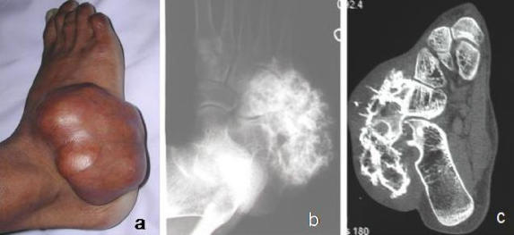
A 30-year-old man was seen because of swelling in the right foot that had been present for several years but was rapidly increasing in size. On examination, there was a bony hard swelling measuring 13 cm × 10 cm, on the lateral aspect of the foot (Fig. 1a). The swelling was continuous with underlying bone, but the overlying skin was free. Examination revealed multiple bony swellings at the metaphyseal ends of all long bones. These had been present in childhood but had stopped increasing in size 10–12 years earlier.
FIG. 1. (a) Preoperative photograph shows a large swelling on the dorsolateral aspect of the right foot. (b) A radiograph of the foot shows a bony tumour with probable origin from the cuboid bone. (c) A CT scan shows the osteochondroma originating from the cuboid and with probable malignant change.
Radiographs suggested a bony lesion probably arising from the cuboid bone (Fig. 1b). Skeletal survey revealed typical osteochondromas bilaterally around the knees and upper end of the humeri. CT suggested osteochondroma of the cuboid bone with probable malignant change (Fig. 1c). Investigations such as chest radiography and abdominal ultrasonography to look for metastases gave negative results.
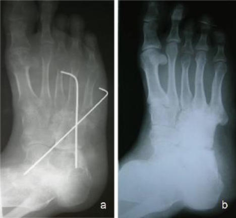
Multiple osteochondromatosis with probable sarcomatous change in osteochondroma of the cuboid was diagnosed. Excision of the tumour with biopsy was planned. Intraoperatively, the swelling was seen to originate from the cuboid. Wide surgical excision was performed, and the defect was reconstructed with use of a rectangular iliac crest graft and fixed with crossed Kirschner wires (Fig. 2a).
FIG. 2. (a) Immediate postoperative view after excision of the tumour and fixation with crossed Kirschner wires. (b) At 2-year follow-up, there is full incorporation of the graft and reconstruction of the lateral longitudinal arch.
Microscopic examination of the specimen revealed grade 1 chondrosarcoma. Postoperatively, there was superficial necrosis of the skin flaps that healed satisfactorily with antibiotics and dressing changes. A below-knee plaster cast was applied with no weight bearing for 3 months. The Kirschner wires were removed 3 months postoperatively at which time the foot was mobilized with use of an ankle foot orthosis. By 6 months, the graft was fully incorporated (Fig. 2b), the orthosis was discarded and full weight bearing was allowed. When the patient was last seen, 3 years postoperatively, there was no local recurrence.
Discussion
Osteochondroma is the most frequent tumour of bone,1 having a predilection for appendicular skeleton but also involving flat bones. It is very rare in bones of the hands and feet.2 The risk of malignant change is 1%–2% in a solitary exostosis and 5%–25% in multiple exostoses.1
Secondary chondrosarcomas have a predilection for flat bones2 and show peak incidence in the third decade of life, compared with chondrosarcoma arising de novo, in which the peak incidence is from the fourth to the sixth decade.1
The pelvis is the most common site, followed by the femur and shoulder girdle.1 Chondrosarcomas are rare in the foot. Of 83 cases of foot tumours reported by Kinoshita and associates,3 36 (43%) involved bone. These tumours included osteochondroma (52%), enchondroma (20%), solitary cyst (10%), chondroblastoma (5%), lipoma (2%), osteoma (2%), ganglion (2%,) chondrosarcoma (2%) and metastatic disease 4%. The majority involved the metatarsals (61%) and the hind foot (33%).
Secondary chondrosarcoma constitutes about 1% of all malignant bone tumours and 11.4% of all chondrosarcomas in small bones of the hands and feet.2 In a large series of hand and foot tumours, Ogose and associates4 reported 75 cases of chondrosarcoma of the foot and the distribution of the tumour as phalanges (53%), calcaneus (28%), metatarsals (16%), talus (13%), cuboid (2.6%) and cuneiforms (2.6%).
The most frequent presenting feature of secondary chondrosarcoma is a mass present for a long period with a recent history of sudden increase in size with or without pain.1 Symptoms may also result from the growing tumour mass and impingement on adjacent structures.
Plain radiography may be a valuable adjunct to diagnosis.1 However, in flat bones and small bones of the hands and feet, this investigation may not reveal findings typically associated with malignant change. CT may be required to establish the origin of swelling in the foot. In our case the bone of origin could be clearly identified only on CT. Histologically, chondrosarcomas are categorized as grades 1, 2 and 3 with most being grade 1.1
The treatment of chondrosarcomas is wide surgical excision.4 No report of efficient adjuvant chemotherapy has been published. Radiotherapy has a limited role — treatment of inoperable disease or recurrence. A grade 1 tumour has a better prognosis than tumours of grades 2 and 3.1 Adequate removal of the tumour is the mainstay of treatment and prevention of local recurrence.1,2 In foot surgery, the wound margins may have healing problems due to poor vascularity as demonstrated in our case. En bloc removal of the tumour creates a defect that needs to be reconstructed to restore the continuity of the longitudinal arch of the foot. We used a large rectangular autograft harvested from the iliac crest to successfully reconstruct the lateral longitudinal arch as biologically as possible for a good functional outcome.
Competing interests: None declared.
Accepted for publication June 19, 2006
Correspondence to: Dr. Manish Chadha, C-A/16, Tagore Garden, New Delhi-110027; mchadha@hotmail.com
References
- 1.Garrison RC, Unni KK, Mcleod RA, et al. Chondrosarcoma arising in osteochondroma. Cancer 1982;49:1890-7. [DOI] [PubMed]
- 2.Ahmed AR, Tan TS, Unni KK, et al. Secondary chondrosarcoma in osteochondroma: report of 107 patients. Clin Orthop Relat Res 2003;411:193-206. [DOI] [PubMed]
- 3.Kinoshita G, Matsumoto M, Maruoka T, et al. Bone and soft tissue tumours of the foot: review of 83 cases. J Orthop Surg (Hong Kong) 2002;10:173-8. [DOI] [PubMed]
- 4.Ogose A, Unni KK, Swee RG, et al. Chondrosarcoma of small bones of the hands and feet. Cancer 1997;80:50-9. [PubMed]


