Adenoid cystic carcinomas (ACCs) constitute 0.1%–1% of all malignant breast tumours. They have better prognosis than other breast masses: they usually do not metastasize, and treatment is usually followed by prolonged survival. We present a case of multifocal, focally infiltrating ACC of the breast. A search of the English literature failed to reveal any other such case.
Case report
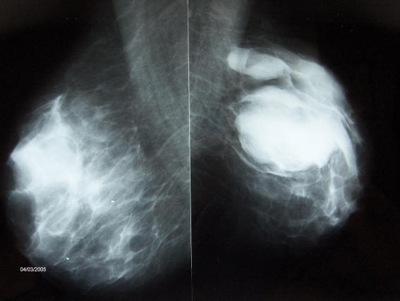
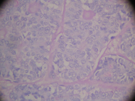
An 83-year-old woman was seen with a mass in her left breast that had been present for 20 years. She complained of rapid enlargement of this mass over the previous 2 months. She had undergone total abdominal hysterectomy and bilateral salpingo-oophorectomy 45 years earlier. Her medical history revealed that for the last 20 years she had controlled diabetes and coronary artery disease. Breast examination revealed 2 palpable masses in the upper lateral quadrant measuring 8 × 7 cm and 4 × 3 cm. No axillary lymphadenopathy was detected. Ultrasonography identified 3 similar macrolobulated solid lesions with irregular contours (7 × 6 × 3 cm, 4 × 2 cm and 2 × 1 cm in dimension, respectively). Adjacent to these masses were several hypoechoic masses, the largest having a diameter of 10 mm. We could not determine whether these were round satellite lesions or metastatic intramammarian lymph nodes. Mammography revealed 3 macrolobulated masses with irregular contours: a 7 × 6-cm mass located in the upper lateral quadrant of the left breast, a 4 × 2-cm mass superior to this and adjacent to the axillary tail, and adjacent to the latter, another 3 × 2-cm mass that could not be clearly evaluated owing to superposition (Fig. 1). Pathological examination of a tru-cut biopsy specimen identified an invasive ductal carcinoma with focal intraductal carcinoma. A modified left radical mastectomy was performed. Pathological examination of the surgical specimen revealed 2 multifocal adenoid cystic carcinomas, 5.5 cm and 2.5 cm, respectively, in greatest diameter (Fig. 2). Resection borders were free of carcinoma, 16 axillary lymph nodes showed only reactive hyperplasia and testing for both estrogen-progesterone receptor and c-erbB2 gave negative results. Findings on follow-up examination at 6 and 12 months were unremarkable.
FIG. 1. Mammographic appearance showing a mass in the left breast.
FIG. 2. Pathological appearance of the resected specimen (hematoxylin–eosin stain, original magnification 400×).
Discussion
ACC is the most frequent tumour of minor salivary canals; however, it constitutes only 0.1%–1% of all breast tumours. McClenathan and de la Roza1 reviewed a national health database for the years 1960–2000 and reported only 22 ACCs among 27 970 cases of breast cancer. So far, globally, about 200 cases of ACC of the breast have been reported. Although it is known as a disease of women, breast ACC may be seen in men.2 Although patients with ACC are usually between 35 and 94 years old, a 19-year-old girl with ACC of the breast was recently reported.3 Histologically, ACC arises from myoepithelium-like cells and ducts. Dense mucoid material ultrastructurally resembling dense lamina of basal membrane is found in glandular structures. Solid areas constitute less than 10% of tumour tissue. Histologically, ACC is usually identified by staining with hematoxylin and eosin.4 Despite a characteristic histologic appearance, ACC may be confused with other pathologic conditions of the breast, particularly intraductal carcinoma and the cribriform type of invasive ductal carcinoma.5 From examination of a biopsy specimen, our patient was initially misdiagnosed as having invasive ductal carcinoma.
ACC may be seen in salivary glands, lacrimal glands, the outer ear, upper respiratory tract, esophagus, Bartholin gland, cervix uteri and prostate gland.6 When it is localized in the breast, it is characteristically a well-circumscribed, mobile mass. When localized adjacent to the peripheral neural network, it will be tender on palpation. Invasion of the pectoralis muscle and retraction of the nipple and breast skin are uncommon. This tumour is most frequently located behind the nipple-areola complex.2 In our case, the tumour was multifocal and showed focal infiltrations. We could find no other reported case with multifocality. Fine-needle aspiration biopsy is important for the early identification of breast lesions.7 Axillary and distant metastases are rare. Distant metastasis (most frequently to lungs) may be seen before axillary metastasis develops.1,2
Because axillary metastasis is not expected, breast-conserving surgery (simple mastectomy or modified radical mastectomy) is recommended for the treatment of breast ACC. Ro and colleagues,8 using a classification based on salivary gland ACCs, suggested local excision without axillary dissection for grade 1 tumours, simple mastectomy for grade 2 tumours and modified radical mastectomy for grade 3 tumours. We performed modified radical mastectomy on our patient. Because of the rarity of breast ACCs, there are no studies comparing adjunctive radiotherapy or chemotherapy. However, the need for radiotherapy after breast-conserving surgery is emphasized. Conversely, some investigators advocate local excision or simple mastectomy.1,2
Several previous series reported a 15%–31% recurrence within 2.3–11.9 years and a 9%–10% mortality for breast ACCs.1,2 No local reccurrence or regional or distant metastasis was found at the 12-month follow-up of our patient.
ACC is a disease with a good response to conservative treatment and a favourable prognosis, and relapses are characterized by local recurrence.
Competing interests: None declared.
Correspondence to: Dr. H. Alis, Imrahorcesme sk, Yunus Apt, No:2, D:5 Uskudar, Istanbul, Turkey; halilalis@gmail.com
References
- 1.McClenathan JH, de la Roza G. Adenoid cystic breast cancer. Am J Surg 2002;183:646-9. [DOI] [PubMed]
- 2.Page DL. Adenoid cystic carcinoma of breast, a special histopathologic type with excellent prognosis [comment]. Breast Cancer Res Treat 2005;93:189-90. Comment on: 2004;87:225-32. [DOI] [PubMed]
- 3.Delanote S, Van den Broecke R, Schelfhout VR, et al. Adenoid cystic carcinoma of the breast in a 19-year-old girl. Breast 2003;12:75-7. [DOI] [PubMed]
- 4.Due W, Herbst WD, Loy V, et al. Characterisation of adenoid cystic carcinoma of the breast by immunohistology. J Clin Pathol 1989;42:470-6. [DOI] [PMC free article] [PubMed]
- 5.Khalbuss WE. Cytomorphology of rare malignant tumors of the breast. Clin Lab Med 2005;25:761-75. [DOI] [PubMed]
- 6.Blanco M, Egozi L, Lubin D, et al. Adenoid cystic carcinoma arising in a fibroadenoma. Ann Diagn Pathol 2005;9:157-9. [DOI] [PubMed]
- 7.Saqi A, Mercado CL, Hamele-Bena D. Adenoid cystic carcinoma of the breast diagnosed by fine-needle aspiration. Diagn Cytopathol 2004;30:271-4. [DOI] [PubMed]
- 8.Ro JY, Silva EG, Gallager HS. Adenoid cystic carcinoma of the breast. Hum Pathol 1987;18:1276-81. [DOI] [PubMed]


