Contrary to the standard presentation, a Meckel's diverticulum unusually located and wedged into the mesenteric side of the ileum was detected in a 7-month-old boy. The case is presented to discuss the embryologic basis and clinical importance of this entity.
Case report
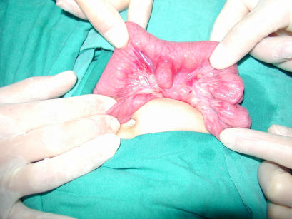
A 7-month-old boy was admitted to our clinic with apparently painless rectal bleeding. No abnormality was detected on physical examination. Erythrocyte and technetium-99m scintigraphy gave negative results. The first episode lasted about 18 hours. A second episode occurred a month later. This time both scintigraphic studies were positive for bleeding Meckel's diverticulum. Laparotomy was performed electively. During exploration a diverticulum was detected about 40 cm from the ileocecal valve, and multiple mesenteric lymphadenopathies were present. The diverticulum was a Meckel's diverticulum except that it was located on the mesenteric border of the ileum; the antimesenteric border of ileum was normal (Fig. 1). Since it was located at the base of the mesentery, resection and anastomosis was preferred instead of a wedge resection. Pathological examination revealed a free antimesenteric border of ileum and a Meckel's diverticulum, the base of which was at the mesenterolateral junction of the ileum. It had a base of about 2 cm and a direct wide connection with the adjacent ileum. On microscopic examination there was congestion and fibrin accumulation. Heterotopic gastric mucosa was detected but no Helicobacter pylori. Postoperative recovery was uncomplicated.
FIG. 1. Intraoperative view of Meckel's diverticulum wedged into the mesenteric border of the ileum.
Discussion
The antimesenteric location is emphasized as one of the cardinal findings in defining the Meckel's diverticulum.1,2 The first description in a different location other than mesenteric location was reported in 1941. Segal and colleagues3 recently described a case of Meckel's diverticulum in a mesenteric location that presented as an inflammatory mass. They considered enterogenous cyst in the differential diagnosis and favoured the diagnosis of Meckel's diverticulum.3 They emphasized that the mesenteric location of Meckel's diverticulum is a forgotten entity.
In the English literature we encountered another case of Meckel's diverticulum wedged into the mesenterium. A patent omphalomesenteric canal detected during the newborn period was reported to have disappeared when the infant was 3 months old, leaving a Meckel's diverticulum adherent to the mesentery with no mesodiverticular band.4 Kurzbart and associates4 commented on the unexpected disappearance of the patent omphalomesenteric canal but not on the unusual location and the sequencing of these 2 conditions. This is an interesting case not only because patency of the omphalomesenteric canal disappeared in 3 months but also because it left behind a Meckel's diverticulum wedged into the mesenterium. The most distinguished difference between a Meckel's diverticulum in the mesenteric location and ileal duplication is the fact that the former is a remnant of the omphalomesenteric canal. This case proves that Meckel's diverticulum attached to the mesenterium is a distinct variant of Meckel's diverticulum.
Donellan2 based his definition of this condition on an early description but did not state its frequency in his series. He offered the possible explanation that the etiology of the anomaly was due to congenital and inflammatory adhesions and focused on the difficulty of its differentiation from duplication.
In our case, the Meckel's divertuculum was located at the mesenterolateral junction of the ileum. A possibility is the persistence of a very short vitelline artery that creates a mesodiverticular band from the mesentery to the tip of the diverticulum, which diverts the diverticulum away from the antimesenteric border during rapid growth. In our case, pathological examination did not reveal a mesodiverticular band or vitelline artery within the fused portion of the mesentery of the ileum but did support chronic inflammation. In general, ileal duplications share the wall and the blood supply of the ileum and the Meckel's diverticulum has its own artery. However, this is still not sufficient for a differential diagnosis because the vitelline artery is present in about 10% of cases.5 The anomaly presented could have been due to a short vitelline artery that disappeared without leaving a remnant or to an intrauterine adhesion between the mesentery of the ileum and the omphalomesenteric canal. Thus, during the elongation and growing process, the “stuck” diverticulum might have been diverted from the antimesenteric border of the ileum.
Although the treatment of a symptomatic diverticulum is straightforward, the removal of asymptomatic Meckel's diverticulum detected incidentally during laparotomy is still controversial.5 This rare location deserves more attention and is more alarming than a usual antimesenteric location because it may erode mesentery and rupture into the mesenteric vasculature during the inflammatory process. Therefore, we suggest that the surgical decision should be standard resection even if this lesion is incidentally detected during laparotomy.
We believe that Meckel's diverticulum in a mesenteric location is a distinct variant that is forgotten or underestimated and difficult to distinguish from an ileal duplication. It is possible that this entity is accepted as an ileal duplication by many authors because it is not reported in large series of Meckel's diverticula.
Presented at the 5th Congress of the Mediterranean Association of Pediatric Surgeons, Marseille, France, Oct. 14–16, 2004.
Competing interests: None declared.
Accepted for publication July 6, 2007
Correspondence to: Dr. Akile Sarioglu-Buke, Department of Pediatric Surgery, Pamukkale University, Denizli, Turkey; fax 90 258 2410034; akilebuke@hotmail.com
References
- 1.McVay CB. Abdominal wall. In: Anson BJ, McVay CB, editors. Anson and McVay surgical anatomy. 6th ed. Philadelphia: WB Saunders; 1984. p. 484-584.
- 2.Donellan WL. Meckel's diverticulum and related anomalies. In: Donellan WL, Burrington JD, Kimura K, et al, editors. Abdominal surgery of infancy and childhood. Luxembourg: Harwood Academic Publishers; 2001. p. 41/1-41/12.
- 3.Segal SD, Albrecht DS, Belland KM, et al. Rare mesenteric location of Meckel's diverticulum, a forgotten entity: a case study aboard USS Kitty Hawk. Am Surg 2004;70:985-8. [PubMed]
- 4.Kurzbart E, Zeitlin M, Feigenbaum D, et al. Rare spontaneous regression of patent omphalomesenteric duct after birth. Arch Dis Child Fetal Neonatal Ed 2002;86:F63. [DOI] [PMC free article] [PubMed]
- 5.Kusumoto H, Yoshida M, Takahashi I, et al. Complications and diagnosis of Meckel's diverticulum in 776 patients. Am J Surg 1992;164:382-3. [DOI] [PubMed]


