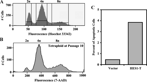
Figure 4.
Isolated tetraploid cells are viable but have higher spontaneous apoptotic rates compared with diploid cells. (A) Viable diploid G1 (2n) cells were separated from tetraploid G2 (8n) cells using Hoechst Staining and flow cytometry and propagated in culture. (B) After 10 passages in culture, the ploidy of the tetraploid cells was measured by 7-AAD staining for total DNA content and flow cytometry. (C) The percentage of apoptotic cells (y-axis) in SF4068-Vector (Vector) and isolated tetraploid cells from SF4068-HES1 (HES1-T) are shown.

