Abstract
The proto-oncogene c-Myc is overexpressed in 70% of colorectal tumours and can modulate proliferation and apoptosis after cytotoxic insult. Using an isogenic cell system, we demonstrate that c-Myc overexpression in colon carcinoma LoVo cells resulted in sensitisation to camptothecin-induced apoptosis, thus identifying c-Myc as a potential marker predicting response of colorectal tumour cells to camptothecin. Both camptothecin exposure and c-Myc overexpression in LoVo cells resulted in elevation of p53 protein levels, suggesting a role of p53 in the c-Myc-imposed sensitisation to the apoptotic effects of camptothecin. This was confirmed by the ability of PFT-α, a specific inhibitor of p53, to attenuate camptothecin-induced apoptosis. p53 can induce the expression of p21Waf1/Cip1, an antiproliferative protein that can facilitate DNA repair and drug resistance. Importantly, although camptothecin treatment markedly increased p21Waf1/Cip1 levels in parental LoVo cells, this effect was abrogated in c-Myc-overexpressing derivatives. Targeted inactivation of p21Waf1/Cip1 in HCT116 colon cancer cells resulted in significantly increased levels of apoptosis following treatment with camptothecin, demonstrating the importance of p21Waf1/Cip1 in the response to this agent. Finally, cDNA microarray analysis was used to identify genes that are modulated in expression by c-Myc upregulation that could serve as additional markers predicting response to camptothecin. Thirty-four sequences were altered in expression over four-fold in two isogenic c-Myc-overexpressing clones compared to parental LoVo cells. Moreover, the expression of 10 of these genes was confirmed to be significantly correlated with response to camptothecin in a panel of 30 colorectal cancer cell lines.
Keywords: camptothecin, apoptosis, microarray, p21Waf1/Cip1
The tumour-suppressor gene p53 plays a pivotal role in determining cell fate after cytotoxic insult with DNA-damaging agents. p53 is a transcription factor that can either trigger an apoptotic cell death by transcriptionally modulating multiple target genes, or promote cell cycle arrest, and thus facilitate DNA damage repair, through the upregulation of the cyclin-dependent kinase (cdk) inhibitor p21Waf1/Cip1 (el-Deiry et al, 1993; Miyashita et al, 1994). Recently, levels of the proto-oncogene c-Myc have been shown to be critical in switching the p53-dependent response from cell cycle arrest to apoptosis after gamma radiation or treatment with daunorubicin (Seoane et al, 2002), a topoisomerase II inhibitor that is not frequently used for the treatment of colorectal malignancies (Harvey et al, 1985). These effects are mediated through the ability of c-Myc to interact with Miz-1 and downregulate the expression of p21Waf1/Cip1, thus favouring the proapoptotic activities of p53. Previously, we demonstrated the clinical value of c-myc as a marker that predicts response to treatment with 5-fluorouracil (5FU), the standard chemotherapeutic agent used in the treatment of colorectal cancer (O'Dwyer and Stevenson, 1998). Low-level amplification of c-myc, together with a wild-type p53 gene, identified a subset of patients with locally advanced colorectal cancer showing increased disease-free and overall survival in response to 5FU-based adjuvant therapy (Augenlicht et al, 1997; Arango et al, 2001).
Camptothecin is a topoisomerase I inhibitor that interferes with DNA replication and transcription by stabilising the covalent complex formed between Topoisomerase I and DNA. The camptothecin derivative CPT-11 (Irinotecan) has been shown to be a useful chemotherapeutic agent for the treatment of colorectal cancer patients, improving response rates and survival when used in combination with 5FU, and has also been shown to be effective in 5FU-resistant tumours (Cunningham et al, 1998; Douillard et al, 2000; Arango and Augenlicht, 2001). Recently, there has been significant progress in the identification of genetic markers that allow prediction of response to 5FU and/or oxaliplatin, which together with CPT-11 are the main chemotherapeutic agents used in the treatment of colorectal cancer (Augenlicht et al, 1997; Salonga et al, 2000; Arango et al, 2001, 2003; Park et al, 2001; Shirota et al, 2001). However, there is great need for markers predicting the efficacy of CPT-11 treatment. In this study, we hypothesised that the levels of c-Myc could modulate the cellular response to camptothecin. Using an in vitro isogenic system, we demonstrated the important role of c-Myc in the apoptotic response of colon cancer cells to camptothecin. Understanding of the molecular mechanisms underlying this observation would allow significant insight to be gained into the key determinants of response to chemotherapy and could identify new targets for intervention. Here we demonstrate a p53-dependent component in the c-Myc-imposed sensitisation to camptothecin-induced apoptosis. Moreover, forced expression of c-Myc resulted in reduced levels of p21Waf1/Cip1 despite elevated levels of p53, and targeted inactivation of p21Waf1/Cip1 resulted in increased sensitivity to apoptosis induced by this agent.
Finally, to identify additional markers capable of predicting apoptotic response to this agent, we used a cDNA microarray analysis approach. The levels of expression of 9216 sequences were assessed in camptothecin-resistant LoVo colon carcinoma cells and camptothecin-sensitive isogenic derivatives overexpressing c-Myc. Thiry-four sequences were identified exhibiting over 4-fold difference in expression. The potential of 10 of these genes as markers predicting response to camptothecin was confirmed by the significant correlation observed between the levels of expression of these genes and the extent of apoptosis induced by camptothecin in a panel of 30 different colorectal cancer cell lines.
MATERIALS AND METHODS
Cell lines and culture conditions
Colon adenocarcinoma LoVo cells and two isogenic clones overexpressing c-Myc (L2 and L3) have been extensively characterised and were maintained as described (Arango et al, 2001). TGR1 rat fibroblasts, isogenic HO15-19 cells with targeted disruption of both alleles of c-myc, and HOmyc3 cells in which c-Myc expression has been restored in the knockout cells (Mateyak et al, 1997) were kindly provided by Dr Sedivy (Brown University, RI, USA) and maintained with DMEM supplemented with 10% FBS and 1 × antibiotic/antimycotic (Invitrogen, Carlsbad, CA, USA). HCT116, a colon carcinoma cell line with a functional p53 gene, and an isogenic line with a targeted inactivation of p21Waf1/Cip1 (Bunz et al, 1998) were kind gifts of Dr Vogelstein (Johns Hopkins University School of Medicine). The panel of colorectal cancer cell lines used was: Caco-2, Colo201, Colo205, Colo320, DLD-1, HCT116, HCT-15, HCT-8, LoVo, LS174T, RKO, SK-CO-1, SW1116, SW403, SW48, SW480, SW620, SW837, SW948, T84 and WiDr (all from the American Type Culture Collection, Manassas, VA, USA), HT29, HT29-Cl.16E, HT29-Cl.19A (from Dr Laboisse, Institut National de la Sante et de la Recherche Medicale U539, France (Augeron and Laboisse, 1984)), LIM1215, LIM2405 (from Dr Whitehead, Vanderbilt University, Nashville, TN, USA (Whitehead et al, 1985; Devine et al, 1992)), HCC2998, KM12 (from the NCI-Frederick Cancer DCT tumour repository), RW2982 and RW7213 (Tibbetts et al, 1988). All the colorectal cancer cell lines were maintained in MEM supplemented with 10% FBS and 1 × antibiotic/antimycotic (Invitrogen, Carlsbad, CA, USA).
Quantification of apoptosis
1 × 10⁵ cells were seeded in triplicate in six-well plates and 24 h later treated with the indicated concentrations of camptothecin (Calbiochem, La Jolla, CA, USA). In some experiments, Pifithrin-α (PFT-α; Calbiochem, La Jolla, CA, USA), a specific p53 inhibitor (Komarov et al, 1999), was also added at time 0 (15–30 μM). The apoptotic response to camptothecin treatment was quantified after 72 h by propidium iodide (PI) staining and assessment of the proportion of cells with a subdiploid content of DNA by FACS analysis, as described (Arango et al, 2001).
Western blot analysis
Both untreated and camptothecin-treated cultures (0.1 or 0.5 μM for 24 h) growing in T75 flasks were rinsed twice with PBS, harvested and the pellet resuspended in 300 μl of RIPA buffer (1% NP-40, 1% sodium deoxycholate, 0.1% SDS, 0.15 M NaCl, 0.01 M sodium phosphate pH 7.2, 2 mM EDTA, 50 mM sodium fluoride, 0.2 mM sodium vanadate and 100 U ml−1 aprotinin), lysed for 30 min on ice, and cleared by centrifugation. The appropriate volume of Laemmli loading buffer (6 ×) was added to 100 μg aliquots and fractionated in 15% SDS–polyacrylamide gels. Proteins were transferred to a PVDF membrane (Amersham, Piscataway, NJ, USA), blocked with 10% nonfat milk for 1 h and then probed at room temperature with the appropriate primary antibody in 5% nonfat milk for 1 h. The following antibodies and dilutions were used: anti-p53 and anti-p21Waf1/Cip1 (DO-1, 1/7000 and H-164, 1/200, respectively; Santa Cruz Biotechnology, Santa Cruz, CA, USA), anti-β-actin (clone AC74, 1/1000; Sigma, Saint Louis, MO, USA). Membranes were washed three times with washing buffer (PBS with 0.1% Tween 20) and then probed with a peroxidase-conjugated secondary antibody for 1 h (Boehringer Mannheim, Indianapolis, IN, USA). After washing three times with washing buffer, the signal was detected using ECL plus (Amersham, Piscataway, NJ, USA) and a Storm PhosphorImager (Molecular Dynamics, Sunnyvale, CA, USA). The signal from the β-actin probe was used as a loading control.
p21Waf1/Cip1 promoter activity
p21Waf1/Cip1 promoter activity was measured in parental LoVo cells and c-Myc-transfected L2 and L3 cells using a transient transfection assay. The p21P construct (Datto et al, 1995) has a 2.4 kb insert containing the p21Waf1/Cip1 promoter sequences in pGL2-basic (Promega, Madison, WI, USA), driving transcription of a firefly luciferase reporter gene. LoVo, L2 and L3 cells (5 × 10⁴ per well) were seeded in 24-well plates and cotransfected with p21P and TK-Renilla (Promega, Madison, WI, USA) 24 h later using GenePORTER II (Gene Therapy Systems, San Diego, CA, USA). After 48 h, cells were harvested, and firefly and Renilla luciferase activity levels assessed using the Dual Luciferase kit (Promega, Madison, WI, USA) according to the manufacturer's specifications. In addition, parental LoVo cells were cotransfected with both p21P and TK-Renilla as well as either p290-Myc(2,3), a c-Myc expression vector (Reed et al, 1989) or the empty p290 vector, to assess the effects of exogenous c-Myc on p21Waf1/Cip1 promoter activity. In all cases, values from TK-Renilla luciferase activity were used to correct for differences in transfection efficiency.
Microarray analysis
Parental LoVo cells as well as L2 and L3 c-Myc transfectants (5 × 10⁶ cells) were seeded in T150 flasks, and harvested after 96 h. Total RNA was prepared using the RNeasy Midi kit (Qiagen, Valencia, CA, USA). RNA aliquots (100 μg) were reverse-transcribed and labelled with Cy5 as described (Mariadason et al, 2000). All experimental samples were compared to a common reference RNA resulting from mixing equal amounts of total RNA from 12 different colon carcinoma cell lines. Reference RNA aliquots (100 μg) were reverse-transcribed and labelled with Cy3 in parallel with the corresponding experimental samples, and then combined and hybridised to a 9216-sequence cDNA microarray from the Albert Einstein Cancer Center Microarray Facility, as described (Mariadason et al, 2000). Microarray slides were scanned and GenePix Pro 3.0 (Axon Instruments, Foster City, CA, USA) was used to quantify signal and background in the Cy5 and Cy3 channels for each spot, as well as the Cy5/Cy3 signal ratio and a normalisation factor that centred the average ratios over the slide on 1. These data were transferred to a Microsoft Excel spreadsheet, and normalised among arrays by multiplying the Cy5/Cy3 ratio by the normalisation factor, thereby allowing interarray comparison. All experiments were done in duplicate, beginning with different RNA preparations. Data from both hybridisations were averaged and used for subsequent analysis if there was a significant level of expression (defined as signal>background plus two standard deviations in the Cy5 and/or Cy3 channel). Relative expression between LoVo and either L2 or L3 cells was calculated by dividing the corresponding average values for each sequence. These values were entered into Microsoft Access and sequences with a difference in expression greater than four-fold between LoVo, and both L2 and L3 were identified. GeneCluster and TreeView software (Stanford University) were used to visualise the results.
Quantitative real-time RT–PCR
The expression levels of 14 genes randomly selected from those identified as differentially expressed (over four-fold) in microarray experiments were confirmed using quantitative real-time RT–PCR. RNA aliquots (5 μg) from the same preparations used for the microarray experiments were reverse transcribed using SuperScript II (Invitrogen, Carlsbad, CA, USA). PCR primers for specific target genes were designed using Primer Express software (Applied Biosystems, Foster City, CA, USA) and used to quantify the relative gene expression (see Table 1 ; primer sequences available at www.realtimeprimers.org). 10 ng cDNA aliquots from LoVo, L2 and L3 cells were amplified with specific primers using the SYBR green Core Reagents Kit and a 7900HT real-time PCR instrument (Applied Biosystems, Foster City, CA, USA). Expression of each gene was standardised using GAPDH as a reference, and relative levels in LoVo, L2 and L3 cells were quantified calculating 2ΔΔCT, where ΔΔCT is the difference in CT (cycle number at which the amount of amplified target reaches a fixed threshold) between target and reference, relative to parental LoVo cells.
Table 1. Genes differentially expressed in parental LoVo cells and c-Myc-overexpressing L2 and L3 isogenic cells.
|
Microarraya |
RT–PCRb |
Correlationc |
||||||
|---|---|---|---|---|---|---|---|---|
| Accession | Gene name | Function | L2/L | L3/L | L2/L | L3/L | r | P |
| AA088861 | Cadherin 17, LI cadherin (liver–intestine) | Cell adhesion | 0.23 | 0.18 | 0.006 | 0.023 | −0.16 | |
| AA130579 | Lectin, galactoside-binding, soluble, 4 (galectin 4) | Cell adhesion | 7.14 | 10.89 | 0.33 | |||
| N30868 | Integrin, alpha 4 | Cell adhesion | 4.75 | 6.87 | 0.48 | 0.016 | ||
| R60995 | Coagulation factor C homologue, cochlin | Cell communication | 0.12 | 0.17 | −0.54 | 0.002 | ||
| AA453783 | Mal, T-cell differentiation protein 2 | Development–differentiation | 6.26 | 4.5 | 0.35 | |||
| H17696 | Myelin basic protein | Development–differentiation | 12.03 | 10.41 | 0.260 | 0.350 | −0.09 | |
| AA290737 | Glutathione S-transferase M1 | Drug metabolism/resistance | 0.17 | 0.25 | 0.002 | 0.012 | −0.43 | 0.019 |
| H29521 | ATP-binding cassette 3 | Drug metabolism/resistance | 0.14 | 0.19 | 0.003 | 0.001 | −0.09 | |
| H88329 | Calbindin 1, (28 kDa) | Drug metabolism/resistance | 0.11 | 0.1 | 0.004 | 0.001 | 0.00 | |
| AA664101 | Aldehyde dehydrogenase 1 family, member A1 | Drug metabolism/resistance | 44.09 | 28.97 | 832 | 1137 | 0.49 | 0.006 |
| AA031513 | Matrix metalloproteinase 7 (matrilysin, uterine) | Extracellular matrix | 20.68 | 17.23 | −0.06 | |||
| AA056013 | Microfibril-associated glycoprotein-2 | Extracellular matrix | 0.19 | 0.2 | 0.11 | |||
| AA479199 | Nidogen 2 | Extracellular matrix | 0.21 | 0.22 | 0.004 | 0.002 | −0.14 | |
| AA458965 | Natural killer cell transcript 4 | Immune/inflammatory | 0.08 | 0.16 | −0.07 | |||
| AA682631 | Calcineurin A alpha | Kinase/phosphatase | 0.23 | 0.24 | −0.28 | |||
| AA431773 | Fatty acid desaturase 1 | Lipid metabolism | 0.18 | 0.21 | 0.005 | 0.007 | −0.59 | 0.001 |
| AA432066 | Sarcoglycan, epsilon | Metabolism | 0.23 | 0.2 | −0.12 | |||
| AA434024 | Lanosterol synthase | Metabolism | 0.23 | 0.24 | 0.101 | 0.210 | −0.14 | |
| AA401137 | Lipocalin 2 (oncogene 24p3) | Oncogene | 10.98 | 8.89 | 0.12 | |||
| AA045436 | Basic leucine zipper transcription factor MafG | Oncogene | 5.07 | 4.33 | 1.370 | 4.820 | −0.15 | |
| N63943 | Lysozyme (renal amyloidosis) | Antimicrobial | 10.01 | 8.18 | 2161.8 | 1817.5 | 0.55 | 0.002 |
| AA134814 | TRAF family member-associated NFKB activator | Signal transduction | 4.83 | 4.44 | 0.55 | 0.013 | ||
| AA598567 | Myelin gene expression factor 2 | Transcriptional coactivator | 5.02 | 4.85 | −0.05 | |||
| AA055486 | Tripartite motif-containing 29 | Transcription factor | 5.35 | 4.84 | −0.10 | |||
| AI017703 | Eukaryotic translation initiation factor 3, subunit 3 | Translation/AA biochemistry | 5.11 | 4.25 | 0.45 | 0.013 | ||
| R38343 | Protein tyrosine phosphatase, receptor type, G | Transmembrane receptor | 0.23 | 0.18 | −0.16 | |||
| T89391 | Caveolin 2 | Tumour suppressor | 0.19 | 0.22 | 0.025 | 0.067 | −0.01 | |
| AA112057 | KIAA0143 protein | Unknown | 6.73 | 5.57 | 0.40 | 0.031 | ||
| AA282134 | OK/SW-cl.68 mRNA, complete cds | Unknown | 16.06 | 10.51 | 774.2 | 568.4 | 0.12 | |
| AA702350 | Autism-related protein 1 | Unknown | 0.06 | 0.08 | −0.04 | |||
| AA702949 | KIAA0443 gene product | Unknown | 0.11 | 0.16 | −0.06 | |||
| R40481 | cDNA FLJ34699 fis, clone MESAN2002186 | Unknown | 6.57 | 4.55 | 0.39 | 0.037 | ||
| T62854 | Hypothetical protein FLJ22662 | Unknown | 0.14 | 0.19 | 0.06 | |||
| W16832 | Muscleblind-like protein MBLL39 | Unknown | 4.48 | 4.93 | 39.1 | 87.0 | 0.19 | |
Genes with significant correlations between their expression levels and apoptotic response to camptothecin in a panel of 30 colorectal cancer cell lines are shown in bold font.
The relative mRNA levels are shown for the 34 genes with a difference in expression greater than four-fold in LoVo vs L2 and L3 in the microarray experiments (mean of two experiments).
The relative expression levels are measured using quantitative real-time RT–PCR for 14 of these 34 genes.
Pearson's correlation for gene expression and response to camptothecin in 30 colorectal cancer cell lines.
RESULTS
Overexpression of c-Myc sensitises colon carcinoma cells to apoptosis induced by camptothecin
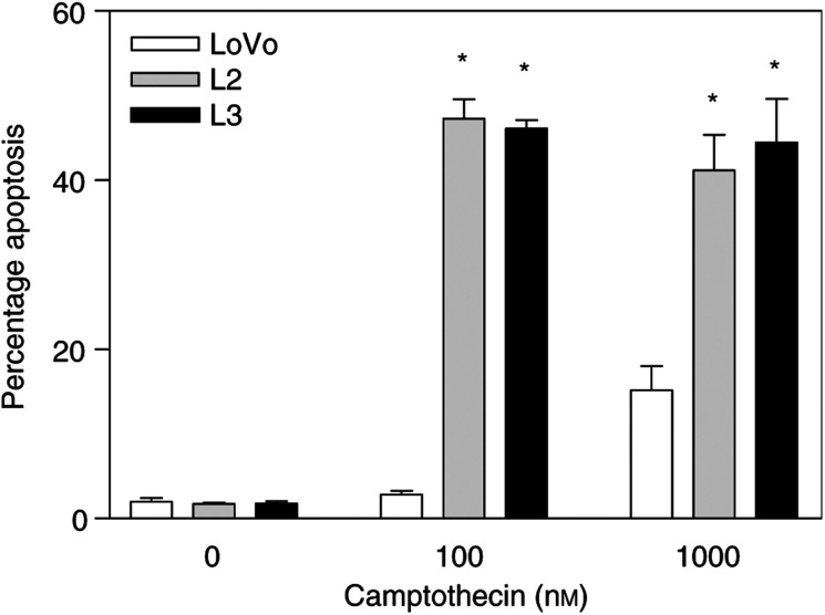
To investigate the possible role of c-Myc in determining the apoptotic response of colon cancer cells to camptothecin, we used an isogenic in vitro system engineered by stably introducing a c-Myc expression vector into the LoVo colon cancer cell line (Arango et al, 2001). Two different derivative clones, L2 and L3, have been extensively characterised and shown to have c-Myc levels three- and eight-fold higher than parental LoVo cells, respectively, and correspondingly increased c-Myc transactivation activity levels (Arango et al, 2001). Quantification of the number of apoptotic cells after exposure to 100 or 1000 nM camptothecin for 72 h demonstrated that the higher c-Myc levels in L2 and L3 cells resulted in significantly (P<0.0001) increased apoptosis induced by this agent (Figure 1).
Figure 1.
c-Myc overexpression sensitises colon cancer cells to camptothecin-induced apoptosis. The higher c-Myc levels and activity found in LoVo cells transfected with c-Myc (L2 and L3 cells) resulted in significant sensitisation to camptothecin-induced apoptosis, as demonstrated by PI staining and FACS analysis of cells exposed to 100 or 1000 nM camptothecin. The mean of three experiments±standard error of the mean is shown. Asterisks indicate significant differences (Student's t-test; P<0.0001) between L2 or L3 cells and parental LoVo cells.
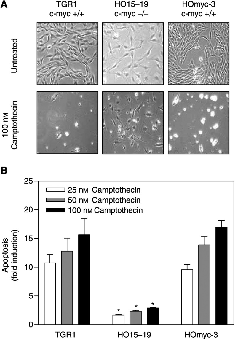
To further confirm the observed c-Myc-induced sensitisation to the apoptotic effects of camptothecin, we used TGR1 rat fibroblasts and their derivative, HO15-19 cells, which contain a homozygous deletion of the c-myc gene (Mateyak et al, 1997). In agreement with the results obtained using the LoVo system, the apoptotic response of TGR1 cells to camptothecin was significantly (P<0.025) reduced in c-Myc-deficient HO15-19 isogenic cells exposed for 72 h to concentrations of camptothecin ranging from 25 to 100 nM (Figure 2A, B). Moreover, when c-Myc expression was rescued in HO15-19 cells (HOmyc3 cells), their response to camptothecin returned to that observed in parental TGR1 cells (Figure 2A, B). Collectively, these results demonstrate an important role for c-Myc in the apoptotic response to camptothecin.
Figure 2.
c-Myc levels modulate the apoptotic response to camptothecin in TGR1 cells. Panel (A) shows representative phase-contrast micrographs of TGR1 cells and derivatives exposed to 100 nM camptothecin for 72 h. In panel (B), the apoptotic response to camptothecin was quantified by PI staining and FACS analysis. Targeted deletion of the c-Myc gene in TGR1 cells (HO15-19 cells) results in a significant reduction in camptothecin-induced apoptosis (A, B). Restoring c-Myc expression in c-Myc-deficient HO15-19 cells (HOmyc-3 cells) results in a significant increase in apoptosis. The mean of three experiments±standard error of the mean is shown in panel (B). Asterisks indicate significant differences (Student's t-test; P<0.025) between c-Myc null HO15-19 cells and both parental TGR1 cells and c-Myc rescue HOmyc-3 cells for a given dose.
Role of p53 in c-Myc-imposed sensitisation to camptothecin-induced apoptosis
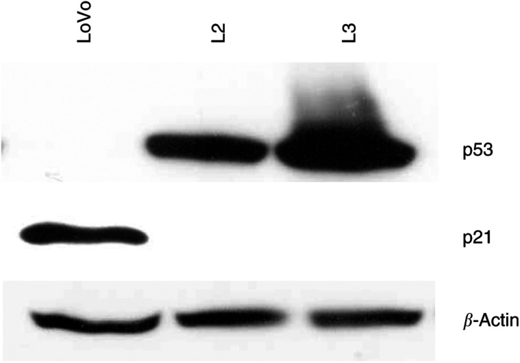
c-Myc has been shown to regulate p53 levels both directly and through modulation of p19ARF (Reisman et al, 1993; Tavtigian et al, 1994; Zindy et al, 1998). In addition, p53 has been reported to modulate the cellular response to several chemotherapeutic agents, including camptothecin and its derivatives (Gupta et al, 1997; Yang et al, 1996). Therefore, to investigate the possible role of p53 in the increased apoptotic response to camptothecin imposed by c-Myc overexpression, we assessed the relative levels of this tumour suppressor in p53 wild-type LoVo cells and c-Myc-transfected clones L2 and L3. c-Myc overexpression in L2 and L3 cells resulted in markedly increased p53 protein levels (Figure 3), consistent with the chemosensitive phenotype of these cells. Moreover, treatment of parental LoVo cells with 0.1 or 0.5 μM camptothecin for 24 h lead to modestly increased p53 levels (Figure 4). However, Figure 4 shows that p53 levels in camptothecin-treated parental LoVo cells remained significantly lower than in untreated L2 and L3 cells. To directly assess the functional role of p53 in camptothecin-induced apoptosis, c-Myc-overexpressing L2 and L3 cells were exposed to camptothecin in the presence of doses of PFT-α, a specific inhibitor of p53 (Komarov et al, 1999), that we have previously shown to inhibit 5FU-induced apoptosis in this system (Arango et al, 2001). Here, inhibition of p53 function resulted in over 50% reduction in the apoptotic response to 0.5 μM camptothecin, demonstrating a p53-dependent component in the c-Myc-dependent increase in apoptosis following treatment with this agent (Figure 5).
Figure 3.
Effects of c-Myc overexpression on p53 and p21Waf1/Cip1 levels. Western blot analysis demonstrated that forced overexpression of c-Myc in LoVo cells (L2 and L3 cells) results in increased p53 and reduced p21Waf1/Cip1 protein levels.
Figure 4.
Effects of camptothecin treatment on p53 and p21Waf1/Cip1 levels. Western blot analysis demonstrated that exposure to 0.1–0.5 μM camptothecin for 24 h results in a modest induction of p53 and significant accumulation of p21Waf1/Cip1 in parental LoVo cells. Overexpression of c-Myc in L2 and L3 cells completely abrogates upregulation of p21Waf1/Cip1 levels in response to camptothecin, despite the elevated p53 levels.
Figure 5.
Role of p53 in the c-Myc imposed sensitisation to camptothecin-induced apoptosis. Exposure of L2 and L3 cells to camptothecin for 72 h in the presence of the specific inhibitor of p53 PFT-α demonstrated a p53-dependent component in their increased apoptotic response to this agent compared to parental LoVo cells. Mean of three experiments±standard error of the mean.
Role of p21Waf1/Cip1 in c-Myc-imposed sensitisation to camptothecin-induced apoptosis
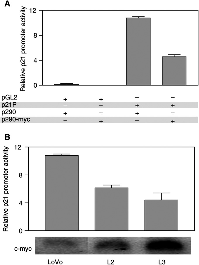
p53 can strongly induce apoptosis by transcriptionally regulating the expression of a number of key players in the apoptotic cascade (Miyashita et al, 1994). However, p53 can alternatively induce cell cycle arrest and promote DNA damage repair through upregulation of the cdk inhibitor p21Waf1/Cip1 (el-Deiry et al, 1993). c-Myc overexpression in LoVo cells resulted in significantly increased p53 levels, which could lead to increased p21Waf1/Cip1 levels. However, c-Myc has been shown to directly downregulate p21Waf1/Cip1 levels (Mitchell and El-Deiry, 1999; Gartel et al, 2001). Therefore, we decided to assess the overall effects of c-Myc overexpression on the transcriptional activity of the p21Waf1/Cip1 promoter. First, we investigated whether c-Myc can reduce p21Waf1/Cip1 promoter activity in parental LoVo cells. Cotransfection of a c-Myc expression vector and a construct containing the p21Waf1/Cip1 promoter region upstream of a luciferase reporter gene (p21P plasmid), demonstrated that c-Myc could effectively reduce the promoter activity of p21Waf1/Cip1 in LoVo cells (Figure 6A). Next, we compared p21Waf1/Cip1 promoter activity in parental LoVo cells and L2 and L3 cells showing elevated levels of c-Myc and p53. The promoter activity of p21Waf1/Cip1 was found to be significantly reduced in L2 and L3 cells compared to parental LoVo cells (1.75- and 2.5-fold downregulation, respectively; Figure 6B). Consistent with the reduced transcriptional activity of the p21Waf1/Cip1 promoter, Western blot analysis demonstrated that the higher c-Myc levels in LoVo transfectants (L2 and L3 cells) resulted in a significant reduction in p21Waf1/Cip1 protein (Figure 3), despite the higher p53 levels.
Figure 6.
effects of c-Myc on p21Waf1/Cip1 promoter activity. (A) Parental LoVo cells were transfected with a vector containing the p21Waf1/Cip1 promoter sequences upstream of a Firefly Luciferase reporter gene (p21P) and a c-Myc expression vector (p290-Myc). This demonstrated a c-Myc-dependent reduction in p21Waf1/Cip1 promoter activity when compared to cells transfected with p21P and p290 empty vector (two-fold). Additional controls showed that transfection of LoVo cells with pGL2 (p21P without the p21Waf1/Cip1 promoter sequence) and either p290 or p290-Myc resulted in minimal luciferase activity. (B) LoVo, L2 and L3 cells were transfected with p21P to measure the differences in promoter activity. The increased c-Myc levels in L2 and L3 resulted in a c-Myc dose-dependent reduction of p21Waf1/Cip1 promoter activity. Relative c-Myc mRNA levels are shown underneath the histogram. Cotransfection with the plasmid TK-Renilla was used to correct for differences in transfection efficiency. The experiments were done three times in triplicate, and results of a representative experiment are shown.
Importantly, although exposure of parental LoVo cells to 0.1 or 0.5 μM camptothecin for 24 h resulted in upregulation of p53 and the p53 target gene p21Waf1/Cip1, c-Myc-overexpressing L2 and L3 cells failed to upregulate p21Waf1/Cip1 protein levels in response to camptothecin treatment (Figure 4), strongly suggesting that p21Waf1/Cip1 levels could modulate apoptosis induced by camptothecin. To directly investigate this possibility, we used an engineered in vitro system where both alleles of the p21Waf1/Cip1 gene have been inactivated by targeted deletion in HCT116 cells, a colon cancer cell line with a wild-type p53 gene (Bunz et al, 1998). Inactivation of p21Waf1/Cip1 in these cells resulted in significantly (P<0.004) increased sensitivity to apoptosis induced by camptothecin compared to parental wild-type p21Waf1/Cip1 HCT116 cells (Figure 7), further demonstrating a role for p21Waf1/Cip1 in regulating response of colon cancer cells to camptothecin.
Figure 7.
Role of p21 in the apoptotic response to camptothecin. Targeted inactivation of both alleles of p21Waf1/Cip1 in HCT116 colon cancer cells resulted in significant sensitisation to camptothecin-induced apoptosis. The mean of four different experiments±standard error of the mean is shown. Asterisks indicate significant differences (Student's t-test, P<0.004) between apoptotic levels in HCT116 p21+/+ and −/− for a given dose.
Identification of additional markers predicting response to camptothecin
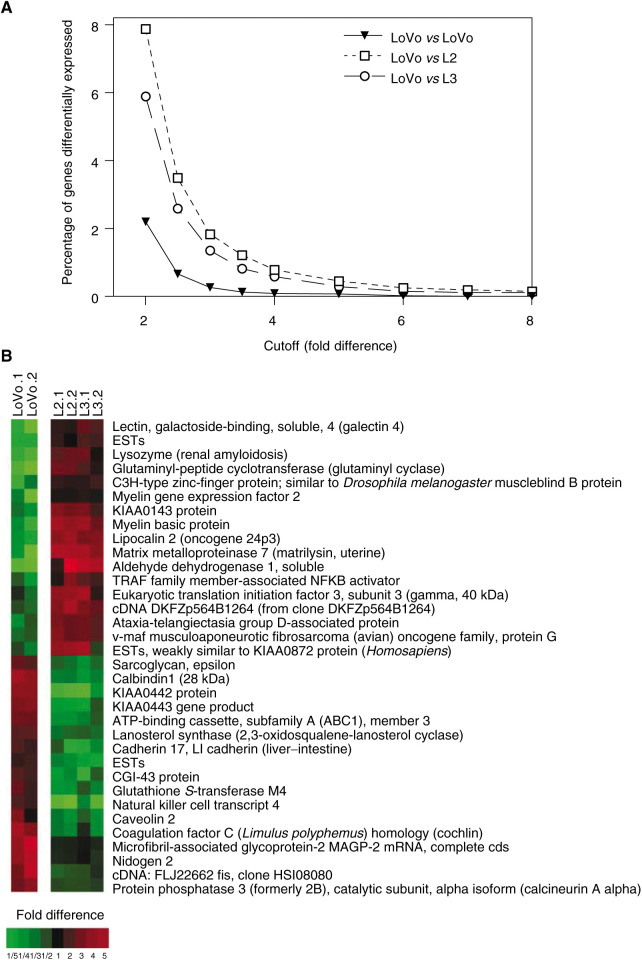
To identify additional genes that could serve as markers predicting apoptotic response to camptothecin, we measured the relative expression of 9216 sequences, in duplicate, in resistant LoVo cells, as well as in sensitive c-Myc-overexpressing L2 and L3 isogenic cells, using cDNA microarray analysis. The complete databases are available at http://sequence.aecom.yu.edu/b ioinf/Augenlicht/default.html. First, to assess the background variability due to methodological and biological factors, we averaged the values for the two LoVo replicas and compared them to the average of another two different replicates of LoVo cells, and quantified the number of genes identified as differentially expressed as a function of selected cutoff values (Figure 8A). Using a conservative cutoff value of four-fold, there were six sequences that would be considered differentially expressed between the two pairs of LoVo replicas. However, using the same four-fold cutoff, LoVo vs L2 and LoVo vs L3 differed in the expression of 63 and 47 genes, respectively. Moreover, 34 of those genes were differentially expressed over four-fold in both L2 and L3 compared to parental LoVo cells (Figure 8B; Table 1).
Figure 8.
cDNA microarray analysis of LoVo and isogenic L2 and L3 cells overexpressing c-Myc. (A) The number of genes identified as differentially expressed is a function of the cutoff used. Selection of a stringent cutoff value of four-fold reduces the number of false positives expected when assessing differences in gene expression between LoVo and L2/L3 cells. (B) TreeView image showing the expression profile of the 34 genes with over four-fold expression difference between LoVo and L2/L3 that characterise camptothecin-resistant and -sensitive cells. Both replicas are shown. Red and green indicate genes that are over-represented and under-represented, respectively, relative to the reference RNA used (see Materials and Methods).
Utilisation of a four-fold change in expression for gene selection greatly minimises the probability of false positives. This was confirmed by quantitative real-time RT–PCR analysis of the relative expression levels of 14 of the 34 genes identified as differentially expressed by the microarray analysis. Good qualitative agreement was observed in changes in the levels of expression assessed by microarray and real-time RT–PCR for 13 out of the 14 genes tested by the two methods (Table 1). However, as observed in other studies (Sgroi et al, 1999; Menssen and Hermeking, 2002; Reinhold et al, 2003), the magnitude of the differences in gene expression assessed by quantitative real-time RT-PCR was greater than those observed using a cDNA microarray approach (Table 1).
Although a subset of the 34 genes differentially expressed over four-fold in LoVo cells and c-Myc-overexpressing L2 and L3 cells is likely to play a role in their differential apoptotic response to camptothecin, elevated c-Myc levels have been shown to affect other phenotypic characteristics of colon cancer cells, including cell cycle progression and basic metabolism (Tikhonenko et al, 1996; Dang, 1999; Menssen and Hermeking, 2002). Therefore, the subset of genes that may be specific markers of response to camptothecin is not clear. To identify this subset of genes, we screened the expression of these 34 sequences in a separate microarray database containing the expression profile of 30 different colorectal cancer lines assessed using the same 9216-sequence cDNA chips (Mariadason et al, 2003; the full database is available at http://sequence.aecom.yu.edu/b ioinf/Augenlicht/default.html). In addition, we have determined the relative sensitivity of these cell lines to camptothecin-induced apoptosis (percent apoptosis after 72 h exposure to 1 μM camptothecin; Mariadason et al, 2003). Of the 34 genes that varied in expression over four-fold in parental LoVo cells and c-Myc-overexpressing L2 and L3 cells, 10 showed a significant correlation (R>0.39; P<0.037) between expression levels and response to camptothecin in each of the 30 colorectal cancer cell lines in our panel (Table 1). As a control, we selected the 34 genes showing the least difference in expression between resistant LoVo cells and sensitive L2 and L3 cells. None of these genes showed a significant correlation between expression levels and apoptotic response to camptothecin in the panel of 30 colorectal cancer cell lines (not shown), highlighting the potential of the genes modulated by c-Myc as biomarkers of response to camptothecin.
DISCUSSION
The proto-oncogene c-Myc is overexpressed in approximately 70% of colorectal tumours (Erisman et al, 1985). Deregulation and overexpression of c-Myc and other oncogenes, in addition to having proliferative effects, is frequently associated with an apoptosis-prone phenotype, thus opening the possibility of therapeutic intervention (Prendergast, 1999). This study demonstrates that upregulation of c-Myc levels and activity in colon cancer cells results in markedly increased sensitivity to apoptosis induced by camptothecin. Forced overexpression of c-myc in LoVo colon cancer cells, induced significant accumulation of p53, and the increased apoptotic response to camptothecin was at least partially dependent upon p53 function. However, p53 can induce cell cycle arrest and facilitate DNA damage repair through the transcriptional activation of the cdk inhibitor p21Waf1/Cip1. The factors determining a p53-dependent apoptotic or cytostatic response to cytotoxic insult remain unclear. Recently, elevated c-Myc levels have been identified as one such factor favouring a p53-dependent apoptotic response (Seoane et al, 2002). Here we show that despite the elevated p53 levels in c-Myc-overexpressing L2 and L3 cells compared to parental LoVo cells, c-Myc directly repressed the transcriptional activity of p21Waf1/Cip1 and resulted in reduced p21Waf1/Cip1 protein levels. Moreover, although camptothecin treatment resulted in p21Waf1/Cip1 accumulation in resistant LoVo cells, sensitive L2 and L3 cells failed to upregulate p21Waf1/Cip1 levels in the presence of camptothecin. The functional relevance of this finding was further demonstrated using an isogenic in vitro system in which both alleles of p21Waf1/Cip1 were inactivated by homologous recombination in HCT116 cells. Inactivation of p21Waf1/Cip1 in HCT116 cells resulted in significantly increased apoptosis following camptothecin treatment compared to p21Waf1/Cip1 wild-type parental HCT116 cells. Collectively, these studies identified the c-Myc-imposed reduction in p21Waf1/Cip1 levels as an important factor determining the increased sensitivity to camptothecin-induced apoptosis associated with elevated c-Myc levels.
Despite significant progress in the identification of markers predicting response to other chemotherapeutic agents commonly used in the treatment of colorectal malignancies, namely, 5FU and oxaliplatin (Augenlicht et al, 1997; Salonga et al, 2000; Arango et al, 2001, 2003; Park et al, 2001; Shirota et al, 2001), there is a great need for clinical predictors of response to camptothecin derivatives. The capability of predicting response to the chemotherapeutic agents available for the treatment of colorectal cancer would allow tailoring of treatment to individual patients, thus maximising the probability of optimal response to therapy. Here we identified c-Myc as a marker predicting response to camptothecin. Moreover, dissection of the molecular mechanisms responsible for the c-Myc-imposed sensitisation to camptothecin demonstrated the key role of p21Waf1/Cip1 in determining response of colon cancer cells to this agent, and pointed at this cdk inhibitor as an additional predictor of response.
To gain further insight into the differences between colon cancer cells that vary in their apoptotic response to camptothecin, and to identify additional markers that could help recognise tumours that vary in their response to this agent, we assessed the level of expression of 9216 sequences in resistant LoVo cells, and in sensitive L2 and L3 derivatives, using cDNA microarray analysis. This identified 34 genes that varied in expression over four-fold in parental LoVo cells and c-Myc-overexpressing L2 and L3 cells. To determine which of these 34 genes are effective predictors of the apoptotic response to camptothecin, we utilised a database containing the levels of expression of the same 9216 sequences in a panel of 30 different colorectal cancer cell lines using cDNA microarray analysis. In addition, we have also determined the relative sensitivity of these 30 cell lines to camptothecin. To identify those sequences associated with differences in sensitivity to camptothecin, we assessed whether each one of the 34 genes differentially expressed in resistant LoVo and sensitive L2/L3 cells showed a significant correlation between expression levels and camptothecin-induced apoptosis in the 30 cell lines in our panel. This identified 10 genes with significant expression/response correlations (R>0.39 and P<0.037; Table 1). This list of candidate genes capable of predicting response to camptothecin contains genes involved in drug metabolism/resistance, signal transduction, protein translation and general metabolism (see Table 1). At least some of these genes have been shown to vary in expression in colorectal tumours, including GST-M1, ALDH and lysozyme (Yuen et al, 1998; Saadat and Saadat, 2001), and could play a functional role in the cellular response to camptothecin and its derivatives. Therefore, in addition to their value as markers predicting response to camptothecin, each of these genes could represent a new target for therapeutic intervention.
Acknowledgments
The plasmids p21P, p290-Myc(2,3) and TGR1 cells and derivatives were kindly provided by Dr Wang (Duke University, NC, USA), Dr Dalla-Favera (Columbia University, NY, USA) and Dr Sedivy (Brown University, RI, USA), respectively. We also thank Aldo Massimi and Dr Geoff Childs of the Albert Einstein Microarray Facility for their assistance, and Dr Barbara Heerdt and Dr Edward Schwartz for critical reading of the manuscript. This work was supported in part by National Cancer Institute grants CA81328, CA88104 and P30-13330.
References
- Arango D, Augenlicht LH (2001) New approaches to colorectal cancer treatment. In Recent Research Developments in Cancer, Vol. 3 pp 385–395. Trivandrum: Transworld Research Network [Google Scholar]
- Arango D, Corner GA, Wadler S, Catalano PJ, Augenlicht LH (2001) c-myc/p53 interaction determines sensitivity of human colon carcinoma cells to 5-fluorouracil in vitro and in vivo. Cancer Res 61: 4910–4915 [PubMed] [Google Scholar]
- Arango D, Wilson AJ, Mariadason JM, Corner GA, Arañes MJ, Nicholas C, Augenlicht LH (2003) Molecular mechanisms of action and prediction of response to oxaliplatin in colorectal cancer cells. (submitted) [DOI] [PMC free article] [PubMed]
- Augenlicht LH, Wadler S, Corner G, Richards C, Ryan L, Multani AS, Pathak S, Benson A, Haller D, Heerdt BG (1997) Low-level c-myc amplification in human colonic carcinoma cell lines and tumors: a frequent, p53-independent mutation associated with improved outcome in a randomized multi-institutional trial. Cancer Res 57: 1769–1775 [PubMed] [Google Scholar]
- Augeron C, Laboisse CL (1984) Emergence of permanently differentiated cell clones in a human colonic cancer cell line in culture after treatment with sodium butyrate. Cancer Res 44: 3961–3969 [PubMed] [Google Scholar]
- Bunz F, Dutriaux A, Lengauer C, Waldman T, Zhou S, Brown JP, Sedivy JM, Kinzler KW, Vogelstein B (1998) Requirement for p53 and p21 to sustain G2 arrest after DNA damage. Science 282: 1497–1501 [DOI] [PubMed] [Google Scholar]
- Cunningham D, Pyrhonen S, James RD, Punt CJA, Hickish TF, Heikkila R, Johannesen TB, Starkhammar H, Topham CA, Awad L (1998) Randomised trial of irinotecan plus supportive care versus supportive care alone after fluorouracil failure for patients with metastatic colorectal cancer. The Lancet 352: 1413–1418 [DOI] [PubMed] [Google Scholar]
- Dang CV (1999) c-Myc target genes involved in cell growth, apoptosis, and metabolism. Mol Cell Biol 19: 1–11 [DOI] [PMC free article] [PubMed] [Google Scholar]
- Datto MB, Yu Y, Wang XF (1995) Functional analysis of the transforming growth factor beta responsive elements in the WAF1/Cip1/p21 promoter. J Biol Chem 270: 28623–28628 [DOI] [PubMed] [Google Scholar]
- Devine PL, Birrell GW, Whitehead RH, Harada H, Xing PX, McKenzie IF (1992) Expression of MUC1 and MUC2 mucins by human tumor cell lines. Tumour Biol 13: 268–277 [DOI] [PubMed] [Google Scholar]
- Douillard JY, Cunningham D, Roth AD, Navarro M, James RD, Karasek P, Jandik P, Iveson T, Carmichael J, Alakl M, Gruia G, Awad L, Rougier P (2000) Irinotecan combined with fluorouracil compared with fluorouracil alone as first-line treatment for metastatic colorectal cancer: a multicentre randomised trial. Lancet 355: 1041–1047 [DOI] [PubMed] [Google Scholar]
- el-Deiry WS, Tokino T, Velculescu VE, Levy DB, Parsons R, Trent JM, Lin D, Mercer WE, Kinzler KW, Vogelstein B (1993) WAF1, a potential mediator of p53 tumor suppression. Cell 75: 817–825 [DOI] [PubMed] [Google Scholar]
- Erisman MD, Rothberg PG, Diehl RE, Morse CC, Spandorfer JM, Astrin SM (1985) Deregulation of c-myc gene expression in human colon carcinoma is not accompanied by amplification or rearrangement of the gene. Mol Cell Biol 5: 1969–1976 [DOI] [PMC free article] [PubMed] [Google Scholar]
- Gartel AL, Ye X, Goufman E, Shianov P, Hay N, Najmabadi F, Tyner AL (2001) Myc represses the p21(WAF1/CIP1) promoter and interacts with Sp1/Sp3. Proc Natl Acad Sci USA 98: 4510–4515 [DOI] [PMC free article] [PubMed] [Google Scholar]
- Gupta M, Fan S, Zhan Q, Kohn KW, O'Connor PM, Pommier Y (1997) Inactivation of p53 increases the cytotoxicity of camptothecin in human colon HCT116 and breast MCF-7 cancer cells. Clin Cancer Res 3: 1653–1660 [PubMed] [Google Scholar]
- Harvey J, Bonnem E, Grady K, Goodman A, Schein P (1985) Phase II study of daunorubicin in previously untreated patients with advanced colorectal carcinoma. Med Pediatr Oncol 13: 30–31 [DOI] [PubMed] [Google Scholar]
- Komarov PG, Komarova EA, Kondratov RV, Christov-Tselkov K, Coon JS, Chernov MV, Gudkov AV (1999) A chemical inhibitor of p53 that protects mice from the side effects of cancer therapy. Science 285: 1733–1737 [DOI] [PubMed] [Google Scholar]
- Mariadason JM, Arango D, Shi Q, Wilson AJ, Corner GA, Nicholas C, Aranes MJ, Schwartz EL, Lesser M (2003) Gene expression profiling based prediction of response of colon carcinoma cells to chemotherapeutic agents. Cancer Res (in press) [PubMed]
- Mariadason JM, Corner GA, Augenlicht LH (2000) Genetic reprogramming in pathways of colonic cell maturation induced by short chain fatty acids: comparison with trichostatin A, sulindac, and curcumin and implications for chemoprevention of colon cancer. Cancer Res 60: 4561–4572 [PubMed] [Google Scholar]
- Mateyak MK, Obaya AJ, Adachi S, Sedivy JM (1997) Phenotypes of c-Myc-deficient rat fibroblasts isolated by targeted homologous recombination. Cell Growth Differ 8: 1039–1048 [PubMed] [Google Scholar]
- Menssen A, Hermeking H (2002) Characterization of the c-MYC-regulated transcriptome by SAGE: identification and analysis of c-MYC target genes. Proc Natl Acad Sci USA 99: 6274–6279 [DOI] [PMC free article] [PubMed] [Google Scholar]
- Mitchell KO, El-Deiry WS (1999) Overexpression of c-Myc inhibits p21WAF1/CIP1 expression and induces S-phase entry in12-O-tetradecanoylphorbol-13-acetate (TPA)-sensitive human cancer cells. Cell Growth Differ 10: 223–230 [PubMed] [Google Scholar]
- Miyashita T, Krajewski S, Krajewska M, Wang HG, Lin HK, Liebermann DA, Hoffman B, Reed JC (1994) Tumor suppressor p53 is a regulator of bcl-2 and bax gene expression in vitro and in vivo. Oncogene 9: 1799–1805 [PubMed] [Google Scholar]
- O'Dwyer PJ, Stevenson JP (1998) Chemotherapy of advanced colorectal cancer. In Gastrointestinal Oncology, Cancer Treatment and Research, Benson AL (ed) pp 111–152. Norwell: Kluwer Academic Publishers [DOI] [PubMed] [Google Scholar]
- Park DJ, Stoehlmacher J, Zhang W, Tsao-Wei DD, Groshen S, Lenz HJ (2001) A Xeroderma pigmentosum group D gene polymorphism predicts clinical outcome to platinum-based chemotherapy in patients with advanced colorectal cancer. Cancer Res 61: 8654–8658 [PubMed] [Google Scholar]
- Prendergast GC (1999) Mechanisms of apoptosis by c-Myc. Oncogene 18: 2967–2987 [DOI] [PubMed] [Google Scholar]
- Reed JC, Haldar S, Cuddy MP, Croce C, Makover D (1989) Deregulated BCL2 expression enhances growth of a human B cell line. Oncogene 4: 1123–1127 [PubMed] [Google Scholar]
- Reinhold WC, Kouros-Mehr H, Kohn KW, Maunakea AK, Lababidi S, Roschke A, Stover K, Alexander J, Pantazis P, Miller L, Liu E, Kirsch IR, Urasaki Y, Pommier Y, Weinstein JN (2003) Apoptotic susceptibility of cancer cells selected for camptothecin resistance: gene expression profiling, functional analysis, and molecular interaction mapping. Cancer Res 63: 1000–1011 [PubMed] [Google Scholar]
- Reisman D, Elkind NB, Roy B, Beamon J, Rotter V (1993) c-Myc trans-activates the p53 promoter through a required downstream CACGTG motif. Cell Growth Differ 4: 57–65 [PubMed] [Google Scholar]
- Saadat I, Saadat M (2001) Glutathione S-transferase M1 and T1 null genotypes and the risk of gastric and colorectal cancers. Cancer Lett 169: 21–26 [DOI] [PubMed] [Google Scholar]
- Salonga D, Danenberg KD, Johnson M, Metzger R, Groshen S, Tsao-Wei DD, Lenz HJ, Leichman CG, Leichman L, Diasio RB, Danenberg PV (2000) Colorectal tumors responding to 5-fluorouracil have low gene expression levels of dihydropyrimidine dehydrogenase, thymidylate synthase, and thymidine phosphorylase. Clin Cancer Res 6: 1322–1327 [PubMed] [Google Scholar]
- Seoane J, Le HV, Massague J (2002) Myc suppression of the p21(Cip1) Cdk inhibitor influences the outcome of the p53 response to DNA damage. Nature 419: 729–734 [DOI] [PubMed] [Google Scholar]
- Sgroi DC, Teng S, Robinson G, LeVangie R, Hudson Jr JR, Elkahloun AG (1999) In vivo gene expression profile analysis of human breast cancer progression. Cancer Res 59: 5656–5661 [PubMed] [Google Scholar]
- Shirota Y, Stoehlmacher J, Brabender J, Xiong YP, Uetake H, Danenberg KD, Groshen S, Tsao-Wei DD, Danenberg PV, Lenz HJ (2001) ERCC1 and thymidylate synthase mRNA levels predict survival for colorectal cancer patients receiving combination oxaliplatin and fluorouracil chemotherapy. J Clin Oncol 19: 4298–4304 [DOI] [PubMed] [Google Scholar]
- Tavtigian SV, Zabludoff SD, Wold BJ (1994) Cloning of mid-G1 serum response genes and identification of a subset regulated by conditional myc expression. Mol Biol Cell 5: 375–388 [DOI] [PMC free article] [PubMed] [Google Scholar]
- Tibbetts LM, Chu MY, Vezeridis MP, Miller PG, Tibbetts LL, Poisson MH, Camara PD, Calabresi P (1988) Cell culture of the mucinous variant of human colorectal carcinoma. Cancer Res 48: 3751–3759 [PubMed] [Google Scholar]
- Tikhonenko AT, Black DJ, Linial ML (1996) Viral Myc oncoproteins in infected fibroblasts down-modulate thrombospondin-1, a possible tumor suppressor gene. J Biol Chem 271: 30741–30747 [DOI] [PubMed] [Google Scholar]
- Whitehead RH, Macrae FA, St John DJ, Ma J (1985) A colon cancer cell line (LIM1215) derived from a patient with inherited nonpolyposis colorectal cancer. J Natl Cancer Inst 74: 759–765 [PubMed] [Google Scholar]
- Yang B, Eshleman JR, Berger NA, Markowitz SD (1996) Wild-type p53 protein potentiates cytotoxicity of therapeutic agents in human colon cancer cells. Clin Cancer Res 2: 1649–1657 [PubMed] [Google Scholar]
- Yuen ST, Wong MP, Chung LP, Chan SY, Cheung N, Ho J, Leung SY (1998) Up-regulation of lysozyme production in colonic adenomas and adenocarcinomas. Histopathology 32: 126–132 [DOI] [PubMed] [Google Scholar]
- Zindy F, Eischen CM, Randle DH, Kamijo T, Cleveland JL, Sherr CJ, Roussel MF (1998) Myc signaling via the ARF tumor suppressor regulates p53-dependent apoptosis and immortalization. Genes Dev 12: 2424–2433 [DOI] [PMC free article] [PubMed] [Google Scholar]


