Abstract
The aim of the study is to evaluate the pattern and level of expression of glucose transporter-1 (GLUT-1) in rectal carcinoma in relation to outcome as a potential surrogate marker of tumour hypoxia. Formalin-fixed tumour sections from 43 patients with rectal carcinoma, who had undergone radical resection with curative intent, were immunohistochemically stained for GLUT-1. A mean of three sections per tumour (range 1–12) were examined. Each section was semiquantitatively scored; 0, no staining; 1, <10%; 2, 10–50%; 3, >50% and a score given for the whole section, the superficial (luminal) and deep (mural) part of the tumour. Staining was seen in 70% of tumours. Increased staining was noted adjacent to necrosis and ulceration. A diffuse and patchy pattern of staining, with and without colocalisation to necrosis was seen. Patients with high GLUT-1-expressing tumours (score 3 vs 0–2) had a significantly poorer overall survival (P=0.041), which was associated with poorer metastasis-free survival with no difference in local control. No significant correlation was seen with other prognostic factors. There was a strong correlation between the score for the superficial and deep parts of the tumour (r=0.81), but a significant relationship with outcome was only found in the deep part (P=0.003 vs P=0.46). In conclusion, increased GLUT-1 expression in rectal tumours was an adverse prognostic factor and is worth further evaluation as a predictive marker of response to therapy.
Keywords: GLUT-1, rectal cancer, hypoxia
The presence of hypoxia in tumours is known to lead to resistance to radiotherapy and chemotherapy and is associated with a more aggressive phenotype with an increased propensity for metastases (Hockel et al, 1996; Brown et al, 1998; Dachs et al, 1998). This latter finding is thought to be related to the increased expression of a number of proteins acting through the HIF-1 pathway, which allows tumour cells to survive the harsh tumour microenviroment (Maxwell et al, 1997; Semenza, 2000). The glucose transporter-1 (Glut-1) is one of the proteins upregulated in hypoxic conditions.
Tumours show increased uptake of glucose compared to normal tissue (Isslbacher, 1972), a response mediated by a number of facilitative glucose transporters located in the cell membrane (Zhang et al, 1999). Glut-1 is one of this family of facilitative glucose transporters. It is expressed variably in normal tissue (Mueckler et al, 1985) and is responsible for the passive transport of glucose across the cell membrane (Chen et al, 2001). The function and expression of Glut-1 is regulated by a number of physiological and pathophysiological conditions (Zhang et al, 1999). In the tumour microenvironment, hypoxia results in increased transcription of the Glut-1 gene, mediated through HIF-1, and reduction in oxidative phosphorylation, in the absence of hypoxia, leads to increased stabilisation of the Glut-1 mRNA (Behrooz et al, 1997). Glucose transporter-1 is overexpressed in several different tumour types (Younes et al, 1996). In addition, increased expression has been shown to correlate with a poor prognosis in a variety of tumours (Younes et al, 1997; Haber et al, 1998; Airley et al, 2001; Cantuaria et al, 2001). More recently, Glut-1 expression has been shown to correlate with the level of tumour hypoxia in carcinoma of the cervix measured using either Eppendorf needle electrodes (Airley et al, 2001) or pimonidazole staining (Airley et al, 2003). Therefore, the level of Glut-1 expression might be a suitable surrogate or intrinsic marker of hypoxia, which could be measured simply and inexpensively as part of the routine histological assessment of tumours.
Little is known about the oxygenation of rectal tumours; however, it is likely that hypoxia plays a significant part in determining outcome (Haustermans et al, 2000; Wendling et al, 1984). Furthermore, determination of the level of hypoxia might be important for selecting appropriate preoperative radiotherapy or chemoradiotherapy regimes or determining which patients are more likely to develop distant metastases and therefore require systemic therapy. In this study, we report for the first time the level and pattern of expression of Glut-1, as a potential endogenous marker of hypoxia, in rectal carcinoma and relate the level of expression to outcome.
PATIENTS AND METHODS
Patients
The study was retrospective. Formalin-fixed tumour sections from 43 consecutive patients (29 male, 14 female) with rectal carcinoma who had undergone curative intent treatment at a single institution were analysed for GLUT-1 protein expression. For each patient, sections from all available tissue blocks were taken (median 2; range 1–12). The median age was 56 years (range 21–80). All patients underwent radical surgery with a curative intent. Only one patient underwent an R1 resection (microscopic residual disease), the remaining 42 underwent an R0 resection (no residual disease). Pathological tumour stages are summarised in Table 1 . Adjuvant therapy was either preoperative radiotherapy/chemoradiotherapy (n=6), postoperative radiotherapy/chemoradiotherapy (n=18) or postoperative chemotherapy alone (n=3). Pre- and postoperative radiotherapy/chemoradiotherapy was administered in an outpatient setting. Patients undergoing radiotherapy were treated with 6 MV or 60Co gamma radiation. A four-field box technique was used extending from the junction of L4/L5 to the bottom of the obturator foramina and 1 cm beyond the lateral pelvic side walls. The field extended from the most posterior aspect of the sacrum to the posterior part of the pubic bone. The prescribed dose, specified to an appropriate isodose envelope, was 45 Gy in 25 fractions (1.8 Gy per fraction) given once daily 5 days per week. Concurrent chemotherapy was administered as a continuous infusion of 225 mg/m−2 5-fluorouracil. Three patients received adjuvant chemotherapy consisting of 5-fluorouracil and folinic acid using the de Gramont regimen. The median follow-up for all patients was 46 months (range 8–102 months) and 49 months (range 30–102 months) for surviving patients.
Table 1. Summary of patient and tumour characteristics.
| Characteristic | |
|---|---|
| Age (years) | |
| Median | 57 |
| Range | 21–80 |
| T stage | |
| T2 | 9 |
| T3 | 29 |
| T4 | 5 |
| Nodal status | |
| Negative | 27 |
| Positive | 16 |
| Tumour size (pathological) | |
| Median (cm) | 5 |
| Range (cm) | 2–11 |
| <5 cm | 19 |
| ⩾5 cm | 24 |
| Grade | |
| 1 | 10 |
| 2 | 24 |
| 3 | 3 |
| Not classified | 6 |
Immunohistochemistry
Formalin-fixed, paraffin-embedded tumour sections (5 μm thick) were placed on poly-L-lysine-coated slides. Sections were dewaxed in xylene and rehydrated using a series of ethanol solutions of increasing dilution. Staining was carried out using the Envision Plus horseradish peroxidase (HRP) kit (DAKO, UK). First, an endogenous peroxidase supplied with the kit was applied for 5 min at room temperature. The samples were then washed, incubated in 10% casein at room temperature for 15 min and then washed again. A 1 : 100 (10 μg ml−1 protein) concentration of affinity-pure rabbit anti-human Glut-1 (Alpha Diagnostic International, TX, USA) was applied and the sections were incubated for 1 h at 37oC. A rabbit IgG fraction (DAKO) at an identical protein concentration was used as a negative control. Following washing, the secondary antibody (goat anti-rabbit/HRP) was applied to the sections for 30 min at room temperature. A substrate–chromogen solution containing 3.3′-diaminobenzidine in a buffered substrate solution, supplied in the kit, was applied at room temperature for 7 min. After rinsing in water, the slides were counterstained with Gills haematoxylin, dehydrated and mounted. The batch to batch variation was excluded by running sections from the same biopsy through more than one batch, and running one biopsy section through all the batches.
Scoring method
Glucose transporter-1 expression in tumour cells was evaluated using a semiquantitative scoring method: score 0=absence of immunostaining; score 1=1–10% of cells stained; score 2=10–50% of cells stained; and score 3=>50% of cells stained. No account was taken of the intensity of staining, and ulcerated or necrotic areas were excluded from the evaluation. For each tumour section, three areas were scored: a general score for the whole tumour section, a score for the superficial part of the tumour (approximately nearest one-third from the luminal surface) and a score for the deep part (approximately deepest one-third from the luminal surface). Intraobserver reliability was tested by the same pathologist re-evaluating 20 slides chosen at random after a 4-week gap.
Statistical analysis
Previous studies have analysed Glut-1 expression as either positive (score 1–3) vs negative (score 0) or strongly positive (score 3) vs the rest (score (0–2) (Younes et al, 1997; Haber et al, 1998; Airley et al, 2001). It was therefore decided to analyse our patients using these two categories. Correlations between tumour characteristics and the expression of Glut-1 were obtained using a two-tailed Spearman's rank correlation and Fisher's exact test. Survival analysis was by the Kaplan–Meier method, and prognostic factors were assessed by log-rank statistics. Analyses were carried out for overall, metastasis-free and local recurrence-free survival. Bivariate analyses were used to test for the independence from other potential prognostic factors including T stage, nodal stage and tumour size.
RESULTS
Expression of GLUT-1
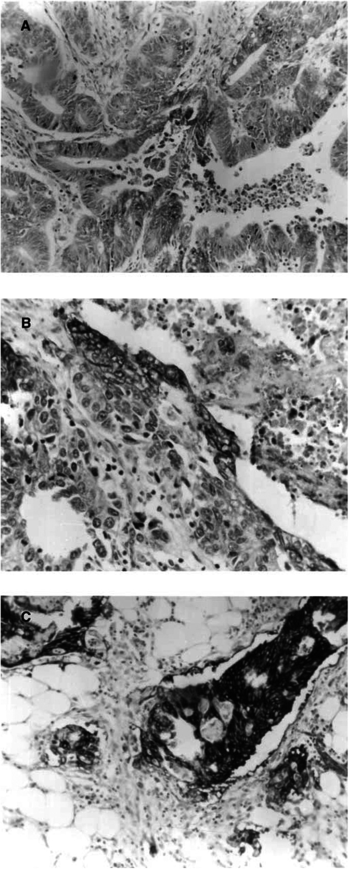
Glucose transporter-1 expression was only observed as membranous staining in tumour cells. Erythrocytes within blood vessels were also seen to stain strongly for Glut-1, which served as an internal control. Perinecrotic and periulcerative areas stained strongly. Two patterns of staining were observed, a focal pattern and a more diffuse staining. Although staining was increased around necrotic areas, the patchy staining pattern was not consistently localised to necrosis. In addition, more cells were positive in the deeper portion of the tumour compared to the superfical luminal part. Normal cells and tumour stroma did not stain for Glut-1. There was a significant correlation between the first and the second score for the general (r=0.87, P<0.0001), superficial (r=0.59 P=0.03) and deep (r=0.82, P<0.0001) parts of the tumour. Figure 1 illustrates sections that were scored 1,2 and 3.
Figure 1.
(A) Score 1. Less than 10% of the cells are stained by anti-Glut-1 (magnification × 10). (B) Score 2. Between 10 and 50% of the cells are stained with Glut-1. Strong membranous staining is seen adjacent to an area of necosis. (magnification × 20). (C) Score 3. More than 50% of cells are stained with Glut-1 at the invasive border. (magnification × 20)
Distribution of patients according to GLUT-1 expression
Of the 43 cases examined, 70% expressed Glut-1. The distribution of scores is shown in Table 2 . There was a strong correlation between the overall tumour score and the scores for the deep (r=0.91, P<0.0005) and superficial (r=0.89, P<0.0005) parts of the tumour.
Table 2. Distribution of scores.
| Score | 0 | 1 | 2 | 3 |
|---|---|---|---|---|
| Patients (number) | 30% (13) | 42% (18) | 21% (9) | 7% (3) |
Correlation of GLUT-1 expression in relation to clinical factors
The distribution of Glut-1 expression according to Duke's stage and nodal status is shown in Table 3 . There was no correlation between the degree of Glut-1 immunostaining and tumour stage (r=−0.047, P=0.77). The patients were also grouped according to the presence or absence of nodal metastasis; however, there was no significant difference in the metastasis status of patients whose tumours showed negative (score 0) or positive (scores 1–3) Glut-1 staining (P=0.52, Fisher's exact test). Likewise, there was no significant difference in the metastasis status of patients whose tumours had weak (score 0–2) or heavy (score 3) Glut-1 staining (P=0.30, Fisher's exact test). Furthermore, there was no significant association between Glut-1 expression and either tumour grade (r=0.10, P=0.58) or pathological size (r=−0.02, P=0.89).
Table 3. Distribution of scores by Duke's stage and nodal status.
| Score | 0 | 1 | 2 | 3 |
|---|---|---|---|---|
| Stage B | 7 | 11 | 8 | 1 |
| Stage C | 6 | 7 | 1 | 2 |
| Node negative | 8 | 10 | 8 | 1 |
| Node positive | 5 | 8 | 1 | 2 |
Relationship between Glut-1 expression and outcome
To examine whether the group of patients studied had similar characteristics to larger groups of patients with rectal cancer, we analysed the relationship between treatment outcome and established clinical prognostic factors (tumour stage, nodal status and tumour size). As expected, advanced stage was a significant adverse prognostic factor (P=0.023). In addition, the 5-year actuarial survival was higher for patients with node negative (89%) vs positive (56%) tumours (P=0.009), and tumours that were pathologically <5 cm (89%) rather than ⩾5 cm (67%) in diameter (P=0.06). Grade was a borderline significant prognostic factor for survival (P=0.06).
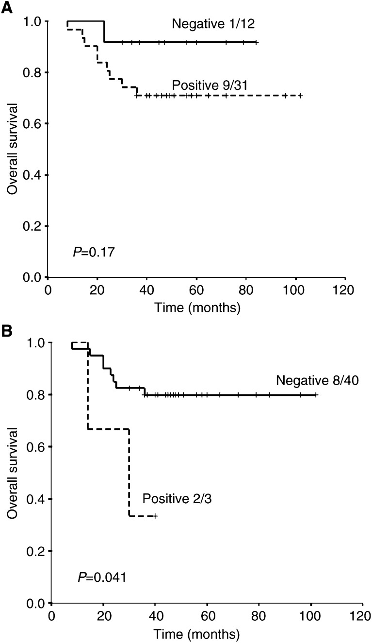
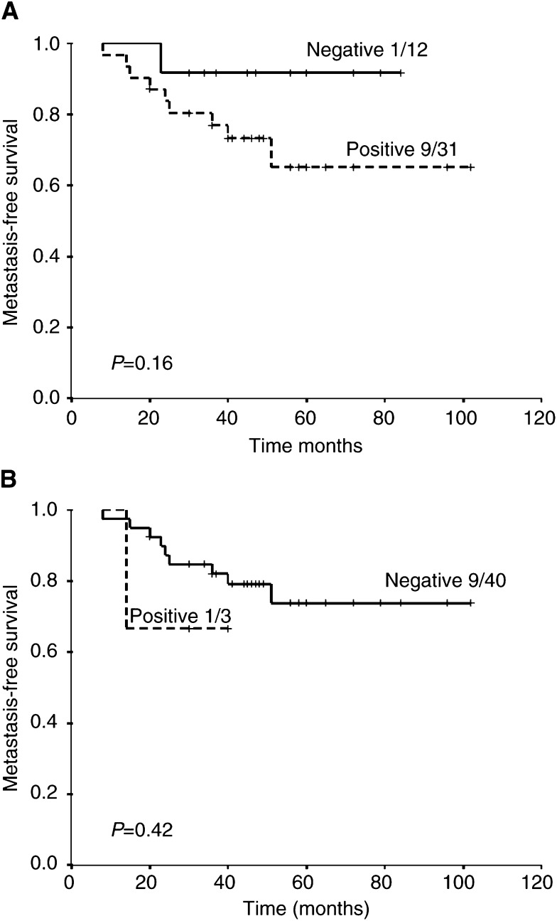
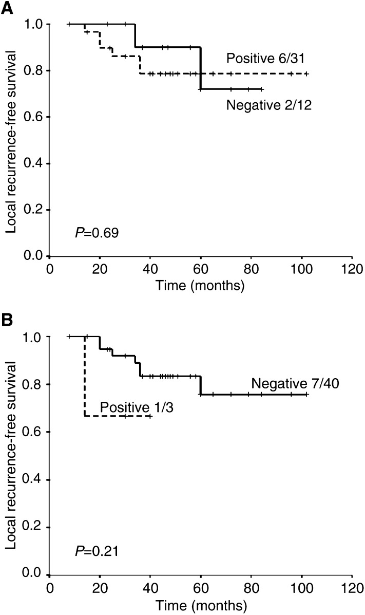
There was no significant difference in the overall survival of patients with Glut-1 positive (score 1–3) vs negative (score 0) tumours (71 vs 92% at 5 years; P=0.17). There was, however, a significant difference in overall survival for patients with strongly positive tumours (score 3) compared to those with a score of 0–2 (33% vs 80%, respectively; P=0.041). Survival curves are shown in Figure 2. Strong Glut-1 expression in tumours appeared to be associated with a poor metastasis-free survival (Figure 3) rather than local control (Figure 4).
Figure 2.
Overall survival for (A) score 0 (negative) vs 1–3 (positive) and (B) score 0–2 (negative) vs 3 (positive).
Figure 3.
Metastasis-free survival for (A) score 0 (negative) vs 1–3 (positive) and (B) score 0–2 (negative) vs 3 (positive).
Figure 4.
Local recurrence-free survival for (A) score 0 (negative) vs 1–3 (positive) and (B) score 0–2 (negative) vs 3 (positive).
Bivariate analyses were performed to test for the independence from tumour stage, nodal status and tumour size. Glucose transporter-1 expression remained a significant prognostic factor for overall survival after allowing for tumour stage (P=0.0052) and tumour size (P=0.05), but not nodal status (P=0.16).
Glut-1 expression in the superficial vs deep part of the tumour
Scores from the deep part of the tumour were also prognostic for overall survival (82 vs 25% actuarial survival at 5 years for score 0–2 vs score 3, respectively; P=0.003). However, the Glut-1 expression of the superficial part of the tumour was not prognostic for survival (actuarial survival at 5 years 78 vs 50%, P=0.46 for score 0–2 vs score 3, respectively).
Independence of Glut-1 staining to known prognostic factors
In order to assess the degree of Glut-1 staining as an independent prognostic marker, all factors that had been significant for overall survival on log-rank univariate analysis (tumour stage, nodal status, strongly positive vs the rest for the whole tumour section and the deep part of the tumour) were included in a multivariate Cox regression analysis. The only factor that was prognostic for survival was the presence of Glut-1 staining in the deep portion of the tumour (P=0.013).
DISCUSSION
The presence of hypoxia in tumours has been shown to correlate with resistance to therapy and reduced survival. Therefore, pretreatment assessment of tumour hypoxia might enable prediction of patients likely to have a poor outcome, and who would be suitable for hypoxia-modifying treatment. Polarographic needle electrodes have been used to give a quantitative, pretreatment measurement of tumour hypoxia that correlates with outcome for patients with carcinoma of the cervix (Hockel et al, 1996; Fyles et al, 1998), head and neck (Nordsmark et al, 1996; Brizel et al, 1997) and soft tissue sarcoma (Brizel et al, 1996). However, the method is invasive and limited to accessible tumours greater than 2 cm in diameter. Furthermore, any measurement will also include areas of necrosis that can bias the results to falsely low values. An alternative method is the immunohistochemical assessment of bound nitroimadazoles, such as pimonidazole, injected prior to biopsy (Kennedy et al, 1997; Nordsmark et al, 2001). This requires an added intervention and, as yet, there are only limited data on their relationship with treatment outcome. This has prompted a number of groups to examine potential endogenous markers of hypoxia, which include proteins that are upregulated under hypoxia. The transcription factor HIF-1α has been the most widely studied potential endogenous marker of hypoxia (Aebersold et al, 2001; Haugland et al, 2002; Janssen et al, 2002; Koukourakis et al, 2002), along with its downstream proteins such as carbonic anhydrase IX (CAIX) (Giatromanolaki et al, 2001; Koukourakis et al, 2001; Loncaster et al, 2001), vascular endothelial growth factor (Loncaster et al, 2000; Koukourakis et al, 2002) and Glut-1 (Airley et al, 2001, 2003). The advantage of using intrinsic markers of hypoxia is that the approach is simple and quick, and could be potentially applied to a wide variety of solid tumour types.
In the present study, we examined the pattern and extent of Glut-1 expression in adenocarcinoma of the rectum as a potential endogenous marker of hypoxia. A total of 70% of tumours expressed Glut-1 to a varying degree. Glucose transporter-1 expression was seen to localise around areas of necrosis and ulceration, while little staining was seen in stromal or normal tissue. In general, two patterns of staining were observed, diffuse and patchy, the latter showing both colocalisation with necrosis and non-necrotic areas. Other studies have reported similar patterns of staining (Haber et al, 1998; Airley et al, 2001). In addition, two recent studies have shown that the levels of Glut-1 expression in carcinoma of the cervix correlates with the level of tumour hypoxia measured using either polarographic needle electrodes (Airley et al, 2001) or pimonidazole staining (Airley et al, 2002). In the former study, the strongest correlation was seen with the most hypoxic fraction of polarographic needle electrode measurements, leading to the suggestion that Glut-1 scores might represent the level of chronic or diffusion-limited hypoxia, while in the second study, there was a good, but not exact, colocalisation of pimonidazole staining and Glut-1 expression. Taken together, these findings suggest a link between hypoxia and Glut-1 expression in human carcinoma of the cervix. However, several factors could reduce the usefulness of Glut-1 as a hypoxic marker and explain the diffuse pattern of staining observed in some of the tumours. Glucose transporter-1 expression is known to be stimulated by a number of other stimuli including growth factors, thyroid hormone, alkaline pH and oncogenic transformation (Flier et al, 1987; Hakimian et al, 1991; Kuruvilla et al, 1991). Furthermore, it is dually controlled by both hypoxia and reduced oxidative phosphorylation in the absence of hypoxia (Behrooz et al, 1997). Chronic hypoxia leads to increased production of Glut-1 that is mediated via the transcription factor HIF-1. Although it was previously thought that HIF-1 was only stabilised under hypoxia, it is now known to be constitutively expressed in some tumours due to genetic alterations occurring during malignant transformation (Jiang et al, 1997; Feldser et al, 1999; Zhong et al, 2000). This might therefore explain the diffuse pattern of staining and the patchy noncolocalisation with necrosis observed in some of the tumours in the present study.
In order to exploit the measurement of Glut-1 expression as a marker of hypoxia in different types of tumours, there is a need to demonstrate that the marker can provide prognostic information in each tumour type of interest. Previous studies have shown a relationship between Glut-1 expression and overall survival in colon cancer (Haber et al, 1998) and lung cancer (Younes et al, 1997), and disease-free survival in ovarian cancer (Cantuaria et al, 2001). In addition, a significant association between Glut-1 expression and metastasis-free survival, but not disease-free survival or local recurrence, has been reported in patients with cervical carcinoma (Airley et al, 2001). In the present study, we were able to show in rectal cancers that strong expression of Glut-1 (>50% of tumour cells) was a significant prognostic factor for overall survival independent of tumour stage and tumour size, and on multivariate analysis the presence of strong staining in the deep part of the tumour was the only significant factor for overall survival. We also observed a nonsignificant relationship with metastases-free survival, but not local recurrence-free survival, for the presence of staining (1–3) vs non (0). However, larger numbers of patients would be required to confirm this.
It is of interest that in the study of Airley et al (2001), where Glut-1 expression was prognostic for metastasis-free survival, but not overall or local recurrence-free survival, patients with cervical carcinoma were treated with radiotherapy. In our study, patients were predominantly treated with surgery, and Glut-1 was predictive for overall and metastasis-free survival but again not local recurrence-free survival. It might be that Glut-1 expression reflects more severe and longer duration hypoxia, especially as in our study intense staining (score 3) showed the best correlation with outcome and heavy staining is more indicative of de novo Glut-1 synthesis which only occurs after prolonged hypoxia (Shetty et al, 1992; Behrooz et al, 1997). This in turn might be more reflective of the tumours propensity to form distant metastases, associated with hypoxia rather than resistance to radiotherapy and is consistent with previous experimental and clinical data (Fyles et al, 2002).
We found no significant relationship between the expression of Glut-1 in tumours and the accepted clinical prognostic factors of tumour stage, depth of invasion (T stage), nodal status, tumour size and grade of differentiation. In contrast, a previous study in patients with mainly colon cancer showed that extensive staining (>50% of cells) for Glut-1 correlated significantly with the presence of nodal metastases (Haber et al, 1998). Similarly, no consistent picture has emerged for any relationships between clinical prognostic factors and the level of tumour hypoxia measured using polarographic needle electrodes (Hockel et al, 1996; Nordsmark et al, 1996; Pitson et al, 2001). The fact that no correlation was found in our study might reflect the low number of patients included and/or the inhomogeneous treatment given. However, the lack of association and the fact that only strong staining in the deep part of the tumour was a significant factor for overall survival on Cox regression analysis suggests that Glut-1 expression in rectal tumours might give additional information over and above that provided by the established clinical prognostic factors. Studies on a larger group of patients are required to clarify this issue.
Of interest in our study was the finding that more cells were seen to express Glut-1 in the deeper compared to the superficial (corresponding with the luminal surface) parts of tumours. In addition, no relationship was found between patient outcome and Glut-1 expression in the superficial part of the tumours. The latter observation is difficult to interpret because of the lack of published data on the oxygenation status of rectal tumours. However, the data do highlight the potential problem of intratumour heterogeneity and the need to take multiple biopsies from different parts of rectal tumours. Taking a mean of three biopsies per patient, as used in our study, is recommended for other studies in this area.
In the results presented here, Glut-1 was expressed in two-thirds of the rectal tumours examined, and high expression was associated with a poor treatment outcome. In addition to Glut-1, several potential endogenous markers of hypoxia are currently under investigation including HIF-1α, HIF-2α, CAIX and VEGF. Studies suggest that different levels and/or durations of tumour hypoxia are required to upregulate each protein (Giatromanolaki et al, 2001; Olive et al, 2001). It is possible, therefore, that a combination of markers might be required to fully evaluate hypoxia within a given tumour type. Furthermore, several studies have shown that combining protein expression with other factors, such as the level of vascularity, improves the definition of patient groups with differing prognoses (Koukourakis et al, 2001,2002). This is the first study of Glut-1 expression in purely rectal carcinoma. Rectal cancer is an interesting area as preoperative radiotherapy or chemoradiotherapy is being increasingly used as part of the treatment. Furthermore, there is still debate as to which node-negative patients should receive adjuvant chemotherapy. As several different regimens are being used, a better understanding of the individual tumour microenvironment might enable a more rational application of these therapies along with the incorporation of hypoxia-modifying treatments for those patients with hypoxic tumours.
References
- Aebersold DM, Burri P, Beer KT, Laissure J, Djonov V, Greiner R, Semenza GL (2001) Expression of hypoxia-inducible factor-1α: A novel predictive and prognostic parameter in radiotherapy of oropharyngeal cancer. Cancer Res 61: 2911–2916 [PubMed] [Google Scholar]
- Airley R, Loncaster J, Davidson S, Bromley M, Roberts S, Patterson A, Hunter R, Stratford I, West C (2001) Glucose transporter Glut-1 expression correlates with tumour hypoxia and predicts metastasis-free survival in advanced carcinoma of the cervix. Clin Cancer Res 7: 928–934 [PubMed] [Google Scholar]
- Airley RE, Loncaster J, Raleigh JA, Harris AL, West CML, Stratford IJ (2003) Glut-1 and CAIX as intrinsic markers of hypoxia in carcinoma of the cervix: relationship to pimonidazole binding. Int J Cancer 104: 85–91 [DOI] [PubMed] [Google Scholar]
- Behrooz A, Isail-Beigi F (1997) Dual control of glut-1 glucose transporter gene expression by hypoxia and by inhibition of oxidative phosphorylation. J Biol Chem 272: 5555–5562 [DOI] [PubMed] [Google Scholar]
- Brown JM, Giaccia AJ (1998) The unique physiology of solid tumours: opportunities (and problems) for cancer therapy. Cancer Res 58: 1408–1416 [PubMed] [Google Scholar]
- Brizel DM, Sculley SP, Harrelsons JM, Layfield LJ, Bean JM, Prosnitz LR, Dewhirst MW (1996) Tumour oxygenation predicts for the likelihood of distant metastases in human soft tissue sarcoma. Cancer Res 56: 941–943 [PubMed] [Google Scholar]
- Brizel DM, Sibley GS, Prosnitz LR, Scher RL, Dewhirst MW (1997) Tumour hypoxia adversely affects the prognosis of carcinoma of the head and neck. Int J Radiat Oncol Biol Phys 38: 285–289 [DOI] [PubMed] [Google Scholar]
- Cantuaria G, Fagotti A, Ferrandina G, Magalhaes A, Nadji M, Angioli R, Penalver M, Mancuso S, Scambia G (2001) Glut-1 expression in ovarian carcinoma. Association with survival and response to chemotherapy. Cancer 92: 1144–1150 [DOI] [PubMed] [Google Scholar]
- Chen C, Pore N, Behrooz A, Ismail-Beigi F, Maity A (2001) Regulation of glut-1 mRNA by hypoxia-inducible factor-1. J Biol Chem 276: 9519–9525 [DOI] [PubMed] [Google Scholar]
- Dachs GU, Chaplin DJ (1998) Microenvironmental control of gene expression: implications for tumour angiogenesis, progression and metastasis. Semin Radiant Oncol 8: 208–216 [DOI] [PubMed] [Google Scholar]
- Feldser D, Agani F, Iyer NV, Pak B, Ferreira G, Semenza GL (1999) Reciprocal positive regulation of hypoxia-inducible factor 1alpha and insulin-like growth factor 2. Cancer Res 59: 3915–3918 [PubMed] [Google Scholar]
- Flier JS, Mueckler MM, Usher P, Lodish HF (1987) Elevated levels of glucose transport and transporter messenger RNA are induced by ras or src oncogenes. Science 235: 1492–1495 [DOI] [PubMed] [Google Scholar]
- Fyles A, Milosevic M, Hedley D, Pintilie M, Levin W, Manchul L, Hill RP (2002) Tumor hypoxia has independent predictor impact only in patients with node-negative cervix cancer. J Clin Oncol 20: 680–687 [DOI] [PubMed] [Google Scholar]
- Fyles AW, Milosevic M, Wong R, Kavanagh MC, Pintilie M, Sun A, Chapman W, Levin W, Manchul L, Keane TJ, Hill RP (1998) Oxygenation predicts radiation response and survival in patients with cervix cancer. Radiother Oncol 48: 149–156 [DOI] [PubMed] [Google Scholar]
- Giatromanolaki A, Koukourakis MI, Sivridis E, Pastorek J, Wykoff C, Gatter KC, Harris AL (2001) Expression of hypoxia inducible carbonic anhydrase-9 relates to angiogenic pathways and independently to poor outcome in non-small cell lung cancer. Cancer Res 61: 7992–7998 [PubMed] [Google Scholar]
- Haber RS, Rathan A, Weiser KR, Pritsker A, Itzkowitz SH, Bodian C, Slater G, Weiss A, Burstein DE (1998) Glut1 glucose transporter expression in colorectal carcinoma* A marker for poor prognosis. Cancer 83: 34–40 [DOI] [PubMed] [Google Scholar]
- Hakimian J, Ismail-Beigi F (1991) Enhancement of glucose transport in clone 9 cells by exposure to alkaline pH: studies on potential mechanisms. J Membr Biol 120: 29–39 [DOI] [PubMed] [Google Scholar]
- Haugland HK, Vukovic V, Pintilie M, Fyles AW, Milosevic M, Hill RP, Hedley DW (2002) Expression of hypoxia-inducible factor-1alpha in cervical carcinomas: correlation with tumor oxygenation. Int J Radiat Oncol Biol Phys 53: 854–861 [DOI] [PubMed] [Google Scholar]
- Haustermans K, Hofland I, Van de Pavert I, Geboes K, Varia Mahesh, Raleigh J, Begg AC (2000) Diffusion limited hypoxia estimated by vascular image analysis: comparison with pimonidazole staining in human tumors. Radiother Oncol 55: 325–333 [DOI] [PubMed] [Google Scholar]
- Hockel M, Schlenger K, Aral B, Mitze M, Schaffer U, Vaupel P (1996) Association between tumour hypoxia and malignant progression in advanced carcinoma of the uterine cervix. Cancer Res 56: 4509–4515 [PubMed] [Google Scholar]
- Isslbacher KJ (1972) Sugar and amino acid transport by cells in culture: differences between normal and malignant cells. N Engl J Med 286: 929–933 [DOI] [PubMed] [Google Scholar]
- Janssen HL, Haustermans KM, Sprong D, Blommestijin G, Hofland I, Hoebers FJ, Blijweert E, Raleigh JA, Semenza GL, Varia MA, Balm AJ, van Velthuysen ML, Delaere P, Sciot R, Begg AC (2002) HIF-1alpha, pimonidazole, and iododeoxyuridine to estimate hypoxia and perfusion in human head-and-neck tumors. Int J Radiat Oncol Biol Phys 54: 1537–1549 [DOI] [PubMed] [Google Scholar]
- Jiang BH, Agani F, Passaniti A, Semenza GL (1997) V-SRC inuces expression of hypoxia-inducible factor 1 (HIF-1) and transcription of genes encoding vascular endothelial growth factor and enolase 1: involvement of HIF-1 in tumour progression. Cancer Res 57: 5328–5335 [PubMed] [Google Scholar]
- Kennedy AS, Raleigh JA, Perez GM, Calkins DP, Thrall DE, Novotny DB, Varia MA (1997) Proliferation and hypoxia in human squamous cell carcinoma of the cervix: first report of combined immunohistochemical assays. Int J Radiat Oncol Biol Phys 37: 897–905 [DOI] [PubMed] [Google Scholar]
- Koukourakis MI, Giatromanolaki A, Sivridis E, Simopoulos C, Pastorek J, Wykoff CC, Gatter KC, Harris AL (2001) Hypoxia-regulated carbonic anhydrase-9 (CA9) relates to poor vascularisation and resistance of squamous cell head and neck cancer to chemoradiotherapy. Clin Cancer Res 7: 3399–3403 [PubMed] [Google Scholar]
- Koukourakis MI, Giatromanolaki A, Sivridis E, Simopoulos C, Turley H, Talks K, Gatter KC, Harris AL (2002) Hypoxia-inducible factor (HIFIA and HIF1B), angiogenesis, and chemoradiotherapy outcome of squamous cell head and neck cancer. Int J Radiat Oncol Biol Phys 53: 1192–1202 [DOI] [PubMed] [Google Scholar]
- Kuruvilla AK, Perez C, Ismail-Beigi F, Leob JN (1991) Regulation of glucose transport in clone 9 cells by thyroid hormone. Biochem Biophys Acta 1094: 300–308 [DOI] [PubMed] [Google Scholar]
- Loncaster JA, Cooper RA, Logue JP, Davidson SE, Hunter RD, West CML (2000) Vascular endothelial growth factor (VEGF) expression is a prognostic factor for radiotherapy outcome in advanced carcinoma of the cervix. Br J Cancer 83: 620–625 [DOI] [PMC free article] [PubMed] [Google Scholar]
- Loncaster JL, Harris AL, Davidson SE, Logue JP, Hunter RD, Wycoff CC, Pastorek J, Ratcliffe PJ, Stratford IJ, West CML (2001) Carbonic anhydrase (CA IX) expression, a potential new intrinsic marker of hypoxia: correlations with tumour oxygen measurements and prognosis in locally advanced carcinoma of the cervix. Cancer Res 61: 6394–6399 [PubMed] [Google Scholar]
- Maxwell PH, Dachs GU, Gleadle JM, Nicholls LG, Harris AL, Stratford IJ, Hankinson O, Pugh CW, Rarcliffe PJ (1997) Proc Natl Acad Sci USA 94: 8104–8109 [DOI] [PMC free article] [PubMed]
- Mueckler M, Caruso C, Baldwin SA, Pancio M, Blench I, Morris H, Allard Wj, Lienhard GE, Lodis HF (1985) Sequence and structure of a human glucose transporter. Science 229: 941–945 [DOI] [PubMed] [Google Scholar]
- Nordsmark M, Loncaster J, Chou SC, Havsteen H, Lindegaard JC, Davidson SE, Varia M, West C, Hunter R, Overgaard J, Raleigh JA (2001) Invasive oxygen measurements and pimonidazole labeling in human cervix carcinoma. Int J Radiat Oncol Biol Phys 49: 581–586 [DOI] [PubMed] [Google Scholar]
- Nordsmark M, Overgaard M, Overgaard J (1996) Pretreatment oxygenation predicts radiation response in advanced squamous cell carcinoma of the head and neck. Radiother Oncol 41: 31–39 [DOI] [PubMed] [Google Scholar]
- Olive PL, Aquino-Parsons C, MacPhail SH, Liao SY, Raleigh JA, Lerman MI, Stanbridge EJ (2001) Carbonic anhydrase 9 as an endogenous marker for hypoxia cells in cervical cancer. Cancer Res 61: 8924–8929 [PubMed] [Google Scholar]
- Pitson G, Fyles A, Milosevic M, Wylie J, Pintilie M, Hill R (2001) Tumor size and oxygenation are independent predictors of nodal diseases in patients with cervix cancer. Int J Radiat Oncol Biol Phys 51: 699–703 [DOI] [PubMed] [Google Scholar]
- Semenza GL (2000) Hypoxia clonal selection, and the role of HIF-1 in tumour progression. Crit Rev Biochem Mol Biol 35: 71–103 [DOI] [PubMed] [Google Scholar]
- Shetty M, Loeb JN, Ismail-Beigi F (1992) Enhancement of glucose transport in response to inhibition of oxidative phosphorylation: pre- and posttranslational mechanisms. Am J Physiol 262: C527–C532 [DOI] [PubMed] [Google Scholar]
- Wendling P, Manz R, Thews G, Vaupel P (1984) Heterogenous oxygenation of rectal carcinomas in humans: critical parameter for preoperative irradiation. Adv Exp Med Biol 180: 293–300 [DOI] [PubMed] [Google Scholar]
- Younes M, Brown RS, Stephenson M, Gondo M, Cagle PT (1997) Overexpression of Glut1 and Glut3 in stage I nonsmall cell lung carcinoma is associated with poor survival. Cancer 80: 1046–1051 [DOI] [PubMed] [Google Scholar]
- Younes M, Lechago LV, Somoana JR, Mosharaaf M, Lechago J (1996) Wide expression of the human erythrocyte glucose transporter Glut1 in human cancers. Cancer Res 56: 1164–1167 [PubMed] [Google Scholar]
- Zhang JZ, Behrooz A, Ismail-Beigi F (1999) Regulaion of glucose transport by hypoxia. Am J Kidney Dis 34: 189–202 [DOI] [PubMed] [Google Scholar]
- Zhong H, Chiles K, Feldser D, Laughner E, Hanrahan C, Georgescu MM, Simons JW, Semenza GL (2000) Modulation of hypoxia-inducible factor 1α exspression by the epidermal growth factor/phosphatidy-linositol 3-kinase/PTEN/AKT/FRAP pathway in human prostate cancer cells: implications for tumour angiogenesis. Cancer Res 60: 1541–1545 [PubMed] [Google Scholar]


