Abstract
A role for glycoprotein (GP)V in platelet function has been proposed on the basis of observations that GP V is the major thrombin substrate on intact platelets cleaved during thrombin-induced platelet aggregation, and that GP V promotes GP Ib-IX surface expression in heterologous cells. We tested the hypotheses that GP V is involved in thrombin-induced platelet activation, in GP Ib-IX expression, and in other platelet responses by generating GP V null mice. Contrary to expectations, GP V −/− platelets were normal in size and expressed normal amounts of GP Ib-IX that was functional in von Willebrand factor binding, explaining why defects in GP V have not been observed in Bernard–Soulier syndrome, a bleeding disorder caused by a lack of functional GP Ib-IX-V. Moreover, in vitro analysis demonstrated that GP V −/− platelets were hyperresponsive to thrombin, resulting in increased fibrinogen binding and an increased aggregation response. Consistent with these findings, GP V −/− mice had a shorter bleeding time. These data support a role for GP V as a negative modulator of platelet activation. Furthermore, they suggest a new mechanism by which thrombin enhances platelet responsiveness independent of activation of the classical G-protein-coupled thrombin receptors.
Platelet thrombosis and hemostasis are complex reactions that depend on adhesive interactions mediated by specific receptors. A major platelet complex is glycoprotein (GP) Ib-IX-V. The initial adhesion of platelets is primarily mediated by binding of platelet membrane GP Ib-IX-V to von Willebrand factor (vWf) found on damaged vessel walls (1). After adhesion, binding of other agonists such as thrombin, ADP, and collagen induce signaling events that ultimately activate the receptor function of αΙΙbβ3 for soluble fibrinogen, leading to platelet aggregation (2). Although platelet aggregates are required for normal hemostasis, they can in addition cause arterial thrombosis in atherosclerotic arteries, e.g., acute myocardial infarction and stroke, inducing ischemic complications of cardiovascular disease (3, 4).
The importance of the GP Ib-IX-V complex in normal platelet function is underscored by the study of Bernard–Soulier syndrome (BSS), an inherited bleeding disorder characterized by large platelets that are defective in adhesion to damaged vessel walls (1). This genetic disorder is caused by a lack of functional GP Ib-IX-V and has been linked to defects in either GP Ib or GP IX (5). The activities mapped to the GP Ib subunit of the GP Ib-IX-V complex include vWf (6) and thrombin binding (7, 8) on the extracellular domain and actin-binding protein (9–11) and 14–3-3 (12–15) binding on the cytoplasmic domain.
Several studies indicate functional activities for the GP V subunit of the GP Ib-IX-V complex. In one example, GP V has been shown to be cleaved by thrombin from the platelet surface during thrombin-mediated platelet stimulation (16), but the role of GP V cleavage in this thrombin-induced platelet response is unresolved (17). In another example, the signaling protein 14–3-3 has been shown to bind to the cytoplasmic domain of GP V (14, 15), raising the possibility that GP V may be involved in platelet signaling during adhesion to vWf. Finally, GP V has been shown to promote the expression of GP Ib-IX in heterologous cells (18–20), suggesting that the synthesis of all subunits of GP Ib-IX-V is required for optimal expression of the complex. In the present study, we determined the role of GP V in platelet biology by evaluating the consequences of GP V gene deletion on thrombin-induced platelet activation, vWf-platelet interactions, GP Ib-IX expression, and the maintenance of normal hemostasis.
Methods
Proteins and Antibodies.
Rabbit anti-mouse GP V antibodies include Ab810 (residues C472-A490) and Ab808 (against residues L432-R450). Anti-GP Ib-IX (rabbit polyclonal 3584) was kindly provided by Beat Steiner, Hoffman–LaRoche (21). Anti-αΙΙbβ3 (Ab41) was described previously (22). Human fibrinogen (Enzyme Research Laboratories, South Bend, IN) or human vWf (Haematologic Technologies, Burlington, VT) were FITC-labeled in 0.1 M NaHCO3 (1 mg/ml) using FITC celite (Molecular Probes) with labeling conditions designed to obtain fluorescein-to-protein ratios between 1 and 4. Snake venom peptide botrocetin was purified as described (23) and kindly provided by J. Rose (COR Therapeutics, Inc.). Human glycocalicin was purified from outdated platelets (24) by affinity chromatography using mouse anti-human glycocalicin [MAb 5A12; kindly provided by Burt Adelman (Biogen)].
Generation of the GP V −/− Mouse.
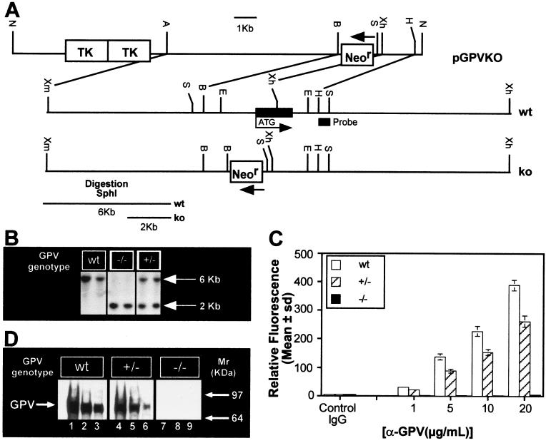
The GP V coding region (700 bp) obtained from rat platelet RNA by reverse transcription–PCR by using degenerate primers based on human GP V sequence (25) was cloned into pCR2.1 (Invitrogen) and sequenced. Genomic DNA was isolated from two positive clones obtained from screening a mouse 129 BAC library with the rat GP V insert, and ≈22 kilobases (kb) was mapped (Fig. 1A). The 5′ XmaI fragment (11 kb) and the EcoRI fragment (4 kb, containing the GP V gene) were subcloned into BlueScript (Stratagene) and sequenced. The ≈8 kb XmaI(blunt)–BamHI fragment from the XmaI plasmid was subcloned into pPN2T (26, 27) to generate the 5′ homology region (HR) downstream of the Neor cassette, followed by the 1.4-kb XhoI–HindIII fragment from the EcoRI plasmid generating the 3′ HR. Thus, the GP V coding region (including the putative initiator Met to Leu389) was replaced by a reverse-oriented Neor cassette (Fig. 1A). pGPVKO was electroporated in RW4 embryonic stem cells (28), and Neor clones were selected in G418 media and microinjected into C57BL/6J embryos (R. Wesselschmidt, Genome Systems). Southern blotting was used to confirm recombination and germline transmission by using a probe designed to show linkage (Fig. 1B), and heterozygote (+/−) animals were bred to homozygosity.
Figure 1.
(A) Genomic organization of the mouse GPV gene and generation of the targeting vector. The targeting vector pGPVKO contained a 5′ HR [8 kb; XmaI(blunt)–BamHI fragment of GP V] and a 3′HR (1.4 kb, XhoI–HindIII fragment of GP V) around the Neor cassette, as shown (B = BamHI, E = EcoRI, A = Acc651, H = HindIII, N = NotI, S = SphI, Xh = XhoI, Xm = XmaI). (B) Southern blot analysis of mouse-tail genomic DNA digested with SphI. The probe used is indicated in A. (C) FACS analysis of wt, +/− and −/− platelets. PRP from wt +/− and −/− mice was incubated with the indicated concentrations of Ab #810 and analyzed by flow cytometry. (D) Western analysis of GP V wt, +/− and −/− platelets. WP lysates were electrophoresed, transferred, and incubated with Ab #808 (5 μg/ml) followed by ECL. Lanes 1–3, wt, lanes 4–6, +/−, and lanes 7–9, −/−. Lanes 1, 4, 7: 2 × 10⁷ platelets; lanes 2, 5, 8: 1 × 10⁷ platelets; lanes 3, 6, 9: 5 × 10⁶ platelets. Mouse GP V has a Mr ≈ 89,000 kDa.
Mouse Platelet Preparation.
Blood from cardiac puncture was diluted into saline containing 1/10 vol of either (i) TSC buffer [3.8% trisodium citrate/0.111 M glucose, pH 7/0.4 μM prostaglandin E1 (PGE1)] for platelet-rich plasma (PRP) or (ii) acid-citrate-dextrose (ACD) (85 mM sodium citrate/0.111M glucose/714 mM citric acid/0.4 μM PGE1), for washed platelets (WP). After centrifugation at 82 × g for 10 min, the supernatant from i normalized to 2 × 10⁸ platelets/ml, and 1 mM Mg2+ (final) was PRP. For WP, supernatant from ii was pooled with a second obtained by centrifugation after the repeat addition of 137 mM NaCl and centrifuged at 325 × g for 10 min. Platelets, washed twice in CGS buffer (12.9 mM sodium citrate/33.33 mM glucose/123.2 mM NaCl, pH7) were resuspended in calcium-free Tyrodes–Hepes buffer (CFTH) (10 mM Hepes/5.56 mM glucose/137 mM NaCl/12 mM NaHCO3/2.7 mM KCl/0.36 mM NaH2PO4/1 mM MgCl2, pH7.4), and normalized to 2 × 10⁸/ml. PRP or WP was incubated at room temperature for 30 min before use.
GP Expression.
For flow cytometry, PRP (10 μl) was incubated with primary rabbit antibody in CFTH/0.1% BSA for 1 hr at 4°C, followed by phycoerythrin (PE)-conjugated donkey anti-rabbit IgG (H + L) F(ab′)2 (1:200, Jackson ImmunoResearch) for 30 min at 4°C in the dark. Samples were diluted to 400 μl in PBS containing 0.1% BSA and analyzed on a facsort (Becton–Dickinson). For Western analysis, WP (5 × 10⁷), solubilized in reducing sample buffer (29), were electrophoresed, transferred to membranes, and probed with antibody overnight at 4°C, followed by incubation with peroxidase-conjugated mouse anti-rabbit secondary antibody (1:5,000; Jackson ImmunoResearch) (1 hr) and developed by ECL (Amersham Pharmacia).
FITC–Ligand-Binding Assay.
For solution-phase vWf binding, pooled PRP was incubated with FITC–vWf and botrocetin (4 to 40 μM) for 10 min and diluted in CFTH before analysis. For FITC–fibrinogen binding, WP, isolated from individual mice, were incubated in duplicate (1 × 10⁶) with α-thrombin for 10 min. The reaction was terminated with PPACK (phenylalaninylprolylargininyl-chloromethylketone; 50 μM final). The platelets were incubated with FITC-labeled fibrinogen (100 μgs/ml) for 30 min, fixed with p-formaldehyde (10%) for 20 min, diluted into 1% p-formaldehyde in CFTH, and analyzed by flow cytometry.
Binding Assays.
Ninety-six-well plates were coated with various concentrations (25–500 ng/well) of human vWf overnight at 4°C. WP in Mg2+-free CFTH buffer (1.2 × 10⁸/ml) containing botrocetin (4 μM) were added to wells for 1 hr at room temperature. After two PBS washes, bound platelets were lysed, and intracellular acid phosphatase activity was quantitated colorimetrically by using the substrate pNPP (Sigma).
Platelet Aggregation.
WP pooled from 6–10 littermate mice and aggregation initiated by thrombin was measured in a lumi-aggregometer (Chrono-Log) with stirring (1,000–1,200 rpm) at 37°C.
Determination of Bleeding Time.
Bleeding time measurements were obtained by using the tail-cut model (30) on littermate mice generated from heterozygous breeding. Because complete litters were not used, the numbers of wt to +/− and −/− do not reflect Mendelian ratios. All experiments were blinded. Briefly, anesthetized mice were transected at the 5-mm mark from the tip of the tail and incubated in warm (37°C) saline. Time for cessation of bleeding was noted as the primary endpoint. If bleeding did not stop in 15 min, the tail was cauterized and 900 sec noted as the bleeding time. Data are presented as mean ± SEM, and statistical significance was assessed by using Student’s t test analysis and Mann–Whitney nonparametric analysis.
Results
GP V −/− Mouse Construction.
To evaluate the specific role of GP V in both platelet function and the GP Ib-IX-V complex expression, we generated a mouse strain that lacked the GP V gene by using homologous recombination techniques (31) (Fig. 1). Because rat GP V would have greater homology to mouse GP V than the cloned human gene, and RNA was easier to isolate from rat platelets than mouse platelets, a cDNA probe for GP V was generated by reverse transcription–PCR from rat-platelet RNA, which was used to isolate, map, and clone mouse GPV from a BAC 129/Sv library. Complete sequencing of three separate clones showed the mouse 129/Sv GP V gene to have 99.9% homology in the coding region to the published mouse C57BL/6J mouse sequence (32) at both the DNA and protein levels§ (not shown). Sequencing also proved that the mouse and rat coding sequences had greater identity (DNA = 92%, protein = 87%) than human and mouse GP V (DNA = 78%, protein = 70%). Recombinants were used to generate the founder chimeras. One founder male (>85% chimeric) was mated with C57BL/6J females to produce +/− offspring, which were bred to generate homozygotes (Fig. 1B). Deficiency in the GP V gene has not affected viability at birth, as evidenced by findings that the litters have expected Mendelian ratios of −/− offspring (1:4) and that the GP V−/− animals are fertile with no gross observable defects.
Analysis of whole blood from GP V-deficient animals showed the platelets were normal both in number and size (as determined by mean platelet volume). Platelet counts in whole blood were within the normal range. However, preparation of PRP from whole blood results in a significant decrease in recovery of platelets from −/− blood compared with wt blood (Table 1), suggesting that −/− platelets may be activated on centrifugation. Platelets were isolated from the −/− animals to confirm that gene deletion resulted in absence of GP V protein expression and were analyzed for GP V expression by using GP V antibodies. No GP V protein was detectable on the intact platelet surface by using FACS analysis (Fig. 1C) or in total platelet lysates as determined by Western blotting (Fig. 1D).
Table 1.
Platelet recovery from whole blood
| Genotype, Sex (n) | Average platelet counts × 108/ml whole blood | % Recovery PRP (mean ± SD) |
|---|---|---|
| wt, ♂ (17) | 6.52 | 74 ± 22* |
| wt, ♀ (8) | 5.6 | 82.4 ± 13† |
| −/−, ♂ (21) | 7.02 | 62 ± 19.5* |
| −/−, ♀ (9) | 5.36 | 67 ± 14† |
*P = 0.05.
†P = 0.03.
Effect of GP V Gene Deletion on GP Ib-IX Expression and Function.
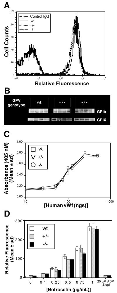
GP V is usually expressed in platelets as a complex with GP Ib-IX (20, 33). We used two techniques to determine whether absence of GP V from the platelet surface affected the expression of the other subunits of the GP Ib-IX-V complex. FACS analysis using an antibody specific for GP Ib-IX showed similar levels of GP Ib-IX in all three genotypes [mean relative fluorescence units (RFU) ± SD for wild type (wt) = 547 ± 106; +/− = 391 ± 65; −/− = 483 ± 56 with p(wt to −/−) = 0.42, p(wt to +/−) = 0.11 and p (+/− to −/−) = 0.14]. Western blot analysis confirmed that similar levels of the GP Ib and GP IX were also present in platelet lysates from all three genotypes (Fig. 2B).
Figure 2.
(A) Surface expression of GP Ib-IX on GP V wt, +/− and −/− platelets. WP were incubated with Ab #3584 (1:1,000 dilution) and analyzed by flow cytometry. The data are representative of six animals of each genotype. Control IgG is on the left. (B) Western analysis of GP Ib-IX expression on GP V wt, +/− and −/− platelets. Lysed WP (5 × 10⁷) were electrophoresed, transferred, and incubated with Ab#3584 (1:1,000) followed by ECL. Four individual mice of each genotype are shown (C) Binding of GP V wt, +/− and −/− platelets to immobilized human vWf. Pooled WP were incubated as described in Methods. The data shown are the average of duplicates and are representative of three such experiments. (D) Binding of FITC–vWf to GP V wt, +/− and −/− platelets. Pooled PRP was incubated with FITC–vWf and botrocetin and analyzed by flow cytometry. The data are representative of three experiments done in duplicate.
Two assays were used to determine whether the GP Ib expressed on GP V −/− platelets was functional. One assay measured the adhesion of platelets to immobilized vWf that was activated by botrocetin to bind GP Ib. Fig. 2C shows that GP V −/− platelets bound to immobilized botrocetin-activated human vWf in a manner indistinguishable from wt platelets. Under these conditions, the binding of vWf to platelets is mediated entirely by GP Ib, because purified human glycocalicin (a soluble extracellular fragment of GP Ibα that contains the vWf binding domain) inhibited botrocetin-induced binding of platelets to vWf in a concentration-dependent manner (not shown). We also found that soluble activated vWf bound identically to platelets from all three genotypes in PRP (Fig. 2D). Again, botrocetin-induced vWf binding could be completely inhibited by purified human glycocalicin (not shown). Furthermore, stimulation of αΙΙbβ3 on platelets by ADP and epinephrine did not induce soluble vWf binding (Fig. 2D). Thus, GP Ib-IX expressed in the GP V−/− platelets was functional.
Effect of GP V Gene Deletion on Thrombin-Induced Platelet Function.
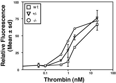
As shown in Fig. 3, thrombin at low concentrations (0.5 nM) induced significantly increased binding of FITC–fibrinogen in GP V −/− platelets compared with wt (mean RFU ± SD wt = 7 ± 1.2, +/− = 13.2 ± 0.9 and −/− = 22 ± 0.8). This difference persisted at 1 nM thrombin (mean RFU ± SD wt = 11.5 ± 6.8, +/− = 27.9 ± 9.45, and −/− = 42 ± 1.8). However, platelets from all genotypes were able to bind FITC–fibrinogen equivalently at high (20 nM) thrombin concentrations (mean RFU ± SD wt = 66.5 ± 8.6, +/− = 76.1 ± 11.3, and −/− = 72 ± 0.2). The apparent EC50 values for thrombin were approximately 2 nM for wt platelets and 0.7 nM for the −/− platelets. P-Selectin expression was also greater in the GP V−/− platelets at low thrombin concentrations, compared with wt (not shown).
Figure 3.
Thrombin-induced FITC–fibrinogen binding in GP V wt, +/− and −/− platelets. WP from individual mice were stimulated with the indicated amounts of thrombin, incubated with FITC-labeled fibrinogen for 30 min, and fixed and analyzed by flow cytometry. The data are representative of three experiments done in duplicate.
Consistent with the FITC–fibrinogen results, platelets lacking GP V exhibited an increased aggregation response to thrombin compared with wt platelets. Indeed, platelets from GP V −/− mice aggregated when treated with subthreshold concentrations of thrombin (0.5 nM) that did not induce a significant response in wt platelets (Fig. 4). As observed in the fibrinogen-binding studies, platelets from GP V+/− heterozygous animals gave an intermediate response in the aggregation assays (Figs. 3 and 4). We determined whether this increased responsiveness was related to increased expression of αΙΙbβ3 by using an antibody specific for the mouse fibrinogen receptor. The levels of αΙΙbβ3 were comparable on platelets from animals of all three genotypes by flow cytometry (Fig. 4B, mean RFU wt = 1,554 ± 386; +/− = 1,246 ± 202; and −/− = 1,435 ± 77; p(wt to −/−) = 0.65, p(wt to +/−) = 0.31 and p (+/− to −/−) = 0.24), and were also similar by Western blotting (Fig. 4C).
Figure 4.
(A) Thrombin-induced aggregation in GP V wt +/− and −/− platelets. WP were prepared from six to ten mice of each genotype, which were littermate controls. Platelet aggregation was determined in a lumi-aggregometer. Five experiments were carried out, and four worked in the manner shown. (B) FACS analysis of surface αΙΙbβ3 levels in GP V wt, +/− and −/− platelets. PRP was incubated with Ab #41 (10 μg/ml), and samples were analyzed by flow cytometry. The data are representative of six individual mice per genotype. Control IgG is on the left. (C) Western analysis of total αΙΙbβ3 levels in GP V wt, +/− and −/− platelets. WP lysates were electrophoresed, transferred to nitrocellulose, and incubated with Ab #41 (10 μg/ml) followed by ECL. Four individual mice of each genotype are shown.
Determination of Bleeding Time.
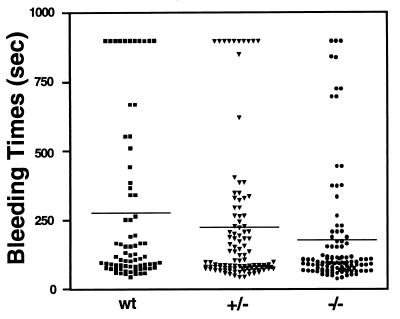
To determine the consequences of enhanced platelet function in GP V −/− mice, bleeding time measurements were performed using a tail-cut model, which was previously shown to be platelet dependent (30, 34). Consistent with the in vitro data, GP V −/− mice had a statistically shorter bleeding time (mean ± SEM = 178 ± 21 sec) than wt littermate control mice (276 ± 35 sec, Student’s t test P = 0.016; Fig. 5). The bleeding time in the +/− animals was intermediate (224 ± 25 sec) but not statistically different from either wt or −/− mice. Furthermore, 70% of the −/− mice had bleeding times less than 120 sec, compared with 50% of the wt and +/− mice. Conversely, 21.6% of the wt mice had bleeding times greater than 500 sec, compared with 9.5% in the +/− mice and 8.5% in the −/− mice. The difference in bleeding time is also statistically significant using nonparametric analysis (Mann–Whitney test, P = 0.046). Thus, the increased aggregability of the platelets from GP V−/− mice observed in in vitro assays translates into a shorter bleeding time in vivo.
Figure 5.
Determination of bleeding time. Bleeding time measurements were obtained by using the tail-cut model on littermate mice (wt n = 74; +/− n = 105, −/− n = 106) generated from heterozygous breeding, as described in Methods. Each symbol represents a single animal.
Discussion
Two conclusions on the role of GP V in platelet function can be made from the present study. First, GP V is a negative regulator of platelet function. Gene targeting of GP V resulted in hyperresponsive platelets, as detected by enhanced fibrinogen binding and enhanced aggregation, resulting in enhanced hemostatic activity in the mice harboring this deletion. The data suggest a role for GP V in decreasing thrombin responsiveness of platelets, with removal of GP V from the platelet surface contributing to platelet stimulation by thrombin. Second, although GP V is a subunit of the GP Ib-IX-V complex, GP V is not required for the expression or function of GP Ib-IX. GP V−/− platelets had normal amounts of GP Ib-IX, and the vWf binding function of GP V−/− platelets was normal. These results are consistent with the fact that mutations in GP Ib and GP IX only and not in GP V have been observed in BSS.
Thrombin is the most potent platelet agonist and is known to function by a proteolytic mechanism (35). The elegant work of Coughlin and coworkers established that thrombin initiates platelet stimulation by cleaving one or more of the protease activated receptor (PAR) family of G-protein-coupled receptors (36–38). Gene targeting of PARs reduces responsiveness of platelets to thrombin (37, 38). The GP Ib subunit of the GP Ib-IX-V complex is a second thrombin-binding site on platelets, providing a high-affinity binding site and thus regulating surface-bound thrombin in human platelets (39, 40). Additionally, the GP V subunit is a thrombin substrate and is cleaved during thrombin-induced platelet aggregation. In considering why GP V−/− platelets are hyperresponsive to thrombin, one possibility is that the presence of intact GP Ib-IX-V serves as a high-affinity reservoir for thrombin. Absence of GP V in platelets as in the GP V null mice or loss of GP V because of proteolysis could decrease the concentration of thrombin required for a stimulatory response. A second possibility is that cleavage of GP V may induce platelet signaling. GP Ib and GP V are both known to bind the adaptor protein 14–3-3 (14, 15). The absence of GP V or GP V cleavage could allow GP Ib to function as a platelet costimulus. However, because we found that GP V −/− platelets adhered normally to immobilized botrocetin-activated vWf, a process dependent on GP Ib signaling (41), we favor the intriguing possibility that GP V acts as a negative modulator of thrombin action on platelets.
Three observations have supported a role for GP V in GP Ib-IX-V complex expression. First, subunits of a complex usually signify a mutual requirement for surface expression (33). Second, all three subunits are missing in the platelets of patients with BSS, an autosomal recessive bleeding disorder (5) linked to defects in GP Ib or GP IX. Third, cotransfection of the GP V gene into heterologous cells expressing GP Ib- IX resulted in increased expression (18, 20, 42). These data taken together have led to the hypothesis that GP V is important for the expression of the vWf receptor (18). Surprisingly, examination of platelets from the GP V-deficient mice by flow cytometry and Western analysis using a GP Ib-IX- specific antisera revealed that lack of GP V did not compromise the expression of GP Ib-IX on the platelet surface. Thus, unlike observations in heterologous cells, GP V is not required for optimal expression of the GP Ib-IX complex on the platelet surface, but that GP Ib-IX is required for GPV expression. Platelets from GP V−/− mice were normal in size, in contrast to the giant size and low numbers of platelets observed in both BSS patients that lack the GP Ib-IX-V complex (5) and in mice deficient in GP Ib (43). The loss of the actin-binding protein anchoring is believed to cause the large platelets in BSS. The present data suggest that GP V is not required for this interaction.
These observations suggest that GP V does not regulate expression of GP Ib-IX, but that thrombin cleavage of GP V may be involved in vascular pathologies. It is of interest to note that we performed bleeding times on a large number of animals (see Fig. 5), to confirm that the phenotype was consistent with the in vitro data. Furthermore, the absence of GP V may occur and yet be undetected in the human population. Recently, it has been shown that platelets from the 12-lipoxygenase-deficient mouse have increased sensitivity to ADP (44), which resulted in a prothombotic phenotype. Thus modifiers of platelet function that up-regulate responses to agonists provide additional means of modifying platelet function. Arterial thrombosis depends on platelet aggregation and is typically associated with a systemic increase in the generation of thrombin, as reflected by increases in thrombin–antithrombin complex (TAT), fibrin degradation product (FDP), and markers indicative of thrombin-induced platelet activation such as P-selectin and PGF2α. Measurement of the loss of GP V from circulating platelets or the appearance of its hydrolytic product GP Vf1 not only may be a retrospective marker of platelet activation but also predict thrombotic events because of enhanced thrombin responsiveness. The contributing roles of the PAR and GP V opposing pathways provides new insight for the development of strategies for pharmacological intervention involving GP V as a negative modulator of thrombin-induced activation.
Acknowledgments
The authors thank Drs. D. A. Law and S. J. Shattil for their critique of the manuscript, and D. DeGuzman and R. Wong for the preparation of this manuscript.
Abbreviations
- BSS
Bernard-Soulier Syndrome
- GP
glycoprotein
- HR
homology region
- PE
phycoerythrin
- PPACK
phenylalaninylprolylargininylchloromethylketone
- PRP
platelet rich plasma
- RFU
relative fluorescence units, vWf, von Willebrand factor
- WP
washed platelets
- wt
wild type
Footnotes
This paper was submitted directly (Track II) to the PNAS office.
The sequence reported in this paper has been deposited in the GenBank database (accession no. AF163101).
Four differences were noted in the protein sequence (P12A, Q88L, G345A, and R431P).
References
- 1.Baumgartner H R, Tschopp T B, Weiss H J. Thromb Haemostasis. 1978;39:782–783. [PubMed] [Google Scholar]
- 2.Clemetson K J, Clemetson J M. Semin Thromb Hemostasis. 1995;21:130–136. doi: 10.1055/s-2007-1000387. [DOI] [PubMed] [Google Scholar]
- 3.Davies M J. Br Med Bull. 1994;50:789–802. doi: 10.1093/oxfordjournals.bmb.a072926. [DOI] [PubMed] [Google Scholar]
- 4.Jang Y, Lincoff A M, Plow E F, Topol E J. J Am Coll Cardiol. 1994;24:1591–1601. doi: 10.1016/0735-1097(94)90162-7. [DOI] [PubMed] [Google Scholar]
- 5.Lopez J A, Andrews R K, Afshar-Kharghan V, Berndt M C. Blood. 1998;91:4397–4418. [PubMed] [Google Scholar]
- 6.Marchese P, Murata M, Mazzucato M, Pradella P, De Marco L, Ware J, Ruggeri Z M. J Biol Chem. 1995;270:9571–9578. doi: 10.1074/jbc.270.16.9571. [DOI] [PubMed] [Google Scholar]
- 7.Handa M, Titani K, Holland L Z, Roberts J R, Ruggeri Z M. J Biol Chem. 1986;261:12579–12585. [PubMed] [Google Scholar]
- 8.De Marco L, Mazzucato M, Masotti A, Ruggeri Z M. J Biol Chem. 1994;269:6478–6484. [PubMed] [Google Scholar]
- 9.Andrews R K, Fox J E. J Biol Chem. 1991;266:7144–7147. [PubMed] [Google Scholar]
- 10.Andrews R K, Fox J E. J Biol Chem. 1992;267:18605–18611. [PubMed] [Google Scholar]
- 11.Xu W, Xie Z, Chung D W, Davie E W. Blood. 1998;92:1268–1276. [PubMed] [Google Scholar]
- 12.Du X, Harris S J, Tetaz T J, Ginsberg M H, Berndt M C. J Biol Chem. 1994;269:18287–18290. [PubMed] [Google Scholar]
- 13.Du X, Fox J E, Pei S. J Biol Chem. 1996;271:7362–7367. doi: 10.1074/jbc.271.13.7362. [DOI] [PubMed] [Google Scholar]
- 14.Calverley D C, Kavanagh T J, Roth G J. Blood. 1998;91:1295–1303. [PubMed] [Google Scholar]
- 15.Andrews R K, Harris S J, McNally T, Berndt M C. Biochemistry. 1998;37:638–647. doi: 10.1021/bi970893g. [DOI] [PubMed] [Google Scholar]
- 16.Berndt M C, Phillips D R. J Biol Chem. 1981;256:59–65. [PubMed] [Google Scholar]
- 17.Bienz D, Schnippering W, Clemetson K J. Blood. 1986;68:720–725. [PubMed] [Google Scholar]
- 18.Calverley D C, Yagi M, Stray S M, Roth G J. Blood. 1995;86:1361–1367. [PubMed] [Google Scholar]
- 19.Li C Q, Dong J F, Lanza F, Sanan D A, Sae-Tung G, Lopez J A. J Biol Chem. 1995;270:16302–16307. doi: 10.1074/jbc.270.27.16302. [DOI] [PubMed] [Google Scholar]
- 20.Meyer S C, Fox J E. J Biol Chem. 1995;270:14693–14699. doi: 10.1074/jbc.270.24.14693. [DOI] [PubMed] [Google Scholar]
- 21.Poujol C, Tronik-Le Roux D, Tropel P, Roullot V, Nurden A, Marguerie G, Nurden P. Blood. 1998;92:2012–2023. [PubMed] [Google Scholar]
- 22.Law D A, Nannizzi-Alaimo L, Ministri K, Hughes P E, Forsyth J, Turner M, Shattil S J, Ginsberg M H, Tybulewicz V L, Phillips D R. Blood. 1999;93:2645–2652. [PubMed] [Google Scholar]
- 23.Andrews R K, Booth W J, Gorman J J, Castaldi P A, Berndt M C. Biochemistry. 1989;28:8317–8326. doi: 10.1021/bi00447a009. [DOI] [PubMed] [Google Scholar]
- 24.Vicente V, Kostel P J, Ruggeri Z M. J Biol Chem. 1988;263:18473–18479. [PubMed] [Google Scholar]
- 25.Lanza F, Morales M, de La Salle C, Cazenave J P, Clemetson K J, Shimomura T, Phillips D R. J Biol Chem. 1993;268:20801–20807. [PubMed] [Google Scholar]
- 26.Tybulewicz V L, Crawford C E, Jackson P K, Bronson R T, Mulligan R C. Cell. 1991;65:1153–1163. doi: 10.1016/0092-8674(91)90011-m. [DOI] [PubMed] [Google Scholar]
- 27.Morrison J R, Paszty C, Stevens M E, Hughes S D, Forte T, Scott J, Rubin E M. Proc Natl Acad Sci USA. 1996;93:7154–7159. doi: 10.1073/pnas.93.14.7154. [DOI] [PMC free article] [PubMed] [Google Scholar]
- 28.Hug B A, Wesselschmidt R L, Fiering S, Bender M A, Epner E, Groudine M, Ley T J. Mol Cell Biol. 1996;16:2906–2912. doi: 10.1128/mcb.16.6.2906. [DOI] [PMC free article] [PubMed] [Google Scholar]
- 29.Laemmli U K. Nature (London) 1970;227:680–685. doi: 10.1038/227680a0. [DOI] [PubMed] [Google Scholar]
- 30.Hodivala-Dilke K M, McHugh K P, Tsakiris D A, Rayburn H, Crowley D, Ullman-Cullere M, Ross F P, Coller B S, Teitelbaum S, Hynes R O. J Clin Invest. 1999;103:229–238. doi: 10.1172/JCI5487. [DOI] [PMC free article] [PubMed] [Google Scholar]
- 31.Koller B H, Smithies O. Annu Rev Immunol. 1992;10:705–730. doi: 10.1146/annurev.iy.10.040192.003421. [DOI] [PubMed] [Google Scholar]
- 32.Ravanat C, Morales M, Azorsa D O, Moog S, Schuhler S, Grunert P, Loew D, Van Dorsselaer A, Cazenave J P, Lanza F. Blood. 1997;89:3253–3262. [PubMed] [Google Scholar]
- 33.Modderman P W, Admiraal L G, Sonnenberg A, von dem Borne A E. J Biol Chem. 1992;267:364–369. [PubMed] [Google Scholar]
- 34.Tsakiris D A, Scudder L, Hodivala-Dilke K, Hynes R O, Coller B S. Thromb Haemostasis. 1999;81:177–188. [PubMed] [Google Scholar]
- 35.Davey M G, Luscher E F. Nature (London) 1967;216:857–858. doi: 10.1038/216857a0. [DOI] [PubMed] [Google Scholar]
- 36.Coughlin S R, Scarborough R M, Vu T K, Hung D T. Cold Spring Harbor Symp Quant Biol. 1992;57:149–154. doi: 10.1101/sqb.1992.057.01.019. [DOI] [PubMed] [Google Scholar]
- 37.Kahn M L, Zheng Y W, Huang W, Bigornia V, Zeng D, Moff S, Farese R V, Jr, Tam C, Coughlin S R. Nature (London) 1998;394:690–694. doi: 10.1038/29325. [DOI] [PubMed] [Google Scholar]
- 38.Kahn M L, Nakanishi-Matsui M, Shapiro M J, Ishihara H, Coughlin S R. J Clin Invest. 1999;103:879–887. doi: 10.1172/JCI6042. [DOI] [PMC free article] [PubMed] [Google Scholar]
- 39.Hayes K L, Tracy P B. J Biol Chem. 1999;274:972–980. doi: 10.1074/jbc.274.2.972. [DOI] [PubMed] [Google Scholar]
- 40.Mazzucato M, Marco L D, Masotti A, Pradella P, Bahou W F, Ruggeri Z M. J Biol Chem. 1998;273:1880–1887. doi: 10.1074/jbc.273.4.1880. [DOI] [PubMed] [Google Scholar]
- 41.Asazuma N, Ozaki Y, Satoh K, Yatomi Y, Handa M, Fujimura Y, Miura S, Kume S. Blood. 1997;90:4789–4798. [PubMed] [Google Scholar]
- 42.Lopez J A, Leung B, Reynolds C C, Li C Q, Fox J E. J Biol Chem. 1992;267:12851–12859. [PubMed] [Google Scholar]
- 43.Ware J, Russell S, Ruggeri Z M. Blood. 1998;92:703a. [Google Scholar]
- 44.Johnson E N, Brass L F, Funk C D. Proc Natl Acad Sci USA. 1998;95:3100–3105. doi: 10.1073/pnas.95.6.3100. [DOI] [PMC free article] [PubMed] [Google Scholar]


