Abstract
The broad distribution and high colonization rates of plant roots by a variety of endophytic fungi suggest that these symbionts have an important role in the function of ecosystems. Semiarid and arid lands cover more than one-third of the terrestrial ecosystems on Earth. However, a limited number of studies have been conducted to characterize root-associated fungal communities in semiarid grasslands. We conducted a study of the fungal community associated with the roots of a dominant grass, Bouteloua gracilis, at the Sevilleta National Wildlife Refuge in New Mexico. Internal transcribed spacer ribosomal DNA sequences from roots collected in May 2005, October 2005, and January 2006 were amplified using fungal-specific primers, and a total of 630 sequences were obtained, 69% of which were novel (less than 97% similarity with respect to sequences in the NCBI database). B. gracilis roots were colonized by at least 10 different orders, including endophytic, coprophilous, mycorrhizal, saprophytic, and plant pathogenic fungi. A total of 51 operational taxonomic units (OTUs) were found, and diversity estimators did not show saturation. Despite the high diversity found within B. gracilis roots, the root-associated fungal community is dominated by a novel group of dark septate fungi (DSF) within the order Pleosporales. Microscopic analysis confirmed that B. gracilis roots are highly colonized by DSF. Other common orders colonizing the roots included Sordariales, Xylariales, and Agaricales. By contributing to drought tolerance and nutrient acquisition, DSF may be integral to the function of arid ecosystems.
Symbiotic associations of fungi and plants are ancient and phylogenetically diverse (4, 9, 11, 42, 43, 60, 64, 69). Some of these fungi, notably mycorrhizal fungi, have received extensive study, but accumulating evidence indicates that many of the fungi associated with plant roots are dark septate fungi (DSF). These fungi are usually described as endophytes, whose functions have only recently been studied (43, 44, 76). DSF comprise a taxonomically diverse group (34, 37) characterized by melanized septate hyphae. Endophytes with hyaline hyphae are also common (e.g., see references 48, 53, and 76), but are less well characterized, because they are more difficult to detect in microscopic analysis and are sometimes considered contaminants in mycorrhizal fungal studies. Growing evidence showing the broad distribution and high root colonization rates of DSF in different ecosystems suggests that their functional importance may rival that of arbuscular mycorrhizal fungi (AMF) (1, 43), particularly for plants growing in stressed environments, such as alpine habitats and arid grasslands (8, 45a, 54, 56, 75). In these habitats, endophytic DSF and other root-associated fungi allow some plants to increase their resistance to drought and heat and facilitate the acquisition of nutrients (23, 43, 45a, 50, 57, 76, 81).
Desert environments are one of the most challenging ecosystems for plants and microorganisms (18, 51, 87). Biological activity, diversity, and distribution are highly restricted by temperature and the heterogeneous availability of moisture and nutrients (7, 82, 86). Desert plants, including grasses (8, 54) and cacti (73), harbor a diversity of fungal endophytes. However, few studies have characterized these communities. Analyses of these fungal communities may yield important information about the ecological roles of root-associated fungi in the maintenance of plant community structure and ecosystem production in arid ecosystems (86).
In a previous study conducted at the Sevilleta National Wildlife Refuge (SNWR) in central New Mexico, we showed that the roots of Bouteloua gracilis (Willd. ex Kunth) Lag. ex Griffiths (blue grama), a dominant C4 perennial grass, harbored few AMF but an abundance of DSF (54). As a result of this assessment, we conducted a more extensive survey of the fungi associated with B. gracilis roots. B. gracilis is one of the most dominant perennial grasses in North America, in part, because it is highly resistant to drought (see the PLANTS database at http://plants.usda.gov) (12, 47). Our hypothesis is that the wide distribution of B. gracilis may be related to its relationship with fungal endophytes. Few previous studies have characterized the root-associated fungal community of this important forage grass (8, 54).
MATERIALS AND METHODS
Methodology.
Bouteloua gracilis roots were collected at the SNWR (New Mexico) in May and October 2005 and January 2006. The samples were collected at the long-term N-amended and control plots (5 × 10 m; 34o24′N, 106o40′W). Nitrogen has been applied twice a year on these plots since 1995 as granular NH4NO3 (100 kg N ha−1 y−1) (33, 72). We randomly collected a minimum of six plants per collection date and at least three plants for each treatment in different plots. The roots were cleaned with sterile water until all soil particles attached to the roots were removed. A subset of the roots was microscopically analyzed to visually evaluate endophytic colonization, and a subset of the roots was stored without additional buffers in Eppendorf tubes at −20°C for DNA extraction for less than a week. We also isolated pure cultures from roots by following the methods of Porras-Alfaro and Bayman (55). Roots were selected if they were (1) connected to green leaves, (2) had root hairs, and (3) did not have obvious lesions. The molecular and microscopy methods used in this paper have been described in detail in a previous paper (54). Plant identification was confirmed with internal transcribed spacer (ITS) sequences (plant GenBank accession numbers, EU144371 to EU144388).
Microscopy.
In preparation for visual assessment of plant-associated fungi, the roots were stained using a modification of the Vierheilig and Piche protocol (80) as described in a previous paper (54).
A total of 93 images from 39 roots from 18 plants were examined. Briefly, roots were surface sterilized with a solution containing 1% sodium hypochlorite and 1% Triton X-100 (a surfactant), placed at 65°C for 1 h in a 1.0 M KOH solution, and kept in the solution at room temperature overnight. During the next day, the KOH was removed and the tissue was washed with water until the solution was clear. A solution of 1% HCl with lactophenol cotton blue (1:50, vol/vol) was added for 12 to 15 h. The roots were subsequently kept in acidified glycerol (1% HCl, vol/vol) until they were assessed for DSF (within 2 weeks).
Because it was difficult to take high-resolution images of longitudinal sections of root tissue that were in focus throughout the images, we opted to strip the cortex from all roots and take digital images of the margins of the vascular cylinder (on the outside margin of the darker-staining endodermis). This method also assured that the fungal structures imaged would be within the root and not superficial fungi resistant to our serial washing. The longitudinal area within the root-harboring fungi was assessed by taking high-resolution images of at least one section of the length of every root using a Sony 3-chip RGB camera (Sony Medical Systems, Montvale, NJ).
Molecular methods.
DNA was extracted with the DNeasy plant mini kit (Qiagen, Chatsworth, CA) from root segments that were cleaned with sterile water (approximately three to five segments, each 3 cm long) before storage at −20°C. Fungal-specific primers ITS1-F and ITS4 were used for amplification by following a method described previously (26), PCR products were cloned by using the TOPO-TA cloning kit (Invitrogen, Carlsbad, CA), and plasmid sequences were amplified by using rolling-circle amplification (TempliPhi; Amersham, Buckinghamshire, England) and sequenced with the BigDye Terminator sequencing kit (version 1.1; Applied Biosystems, Foster City, CA) (54). Negative controls were included in all amplifications and clonings (without DNA template). The sequences were assembled and edited with Sequencher 4.0 (Gene Codes, Ann Arbor, MI). For this study, 18 clone libraries (from 18 different plants; a total of 630 sequences) were generated, representing six samples for each collection (three nitrogen and three control plots). Totals of 358, 124, and 148 sequences were obtained from all the samples collected on May 2005, October 2005, and January 2006, respectively. A preliminary analysis of May samples revealed that B. gracilis roots are mainly colonized by a dominant group of endophytes that in some cases constituted 100% of the clone libraries (66). Therefore, we opted to obtain fewer sequences from plants collected in October and January. In this article, we present a general description of the fungal community colonizing B. gracilis roots. A detailed analysis of the seasonal variation and the effect of nitrogen enrichment on fungal communities will be presented in a subsequent paper.
Chimeric sequences were determined by following a method described by O'Brien et al. (52); ITS1 and ITS2 regions of the same sequence were queried independently in GenBank, and if the ITS1 and ITS2 sequences matched unrelated sequences in the NCBI database, they were considered chimeric, especially in those cases where sequences were obtained only once or in only one sample. Based on the chimeric analysis, seven sequences were removed from the data set.
Complete sequences from ITS regions and the 5.8S gene were used for taxonomic identification and the determination of operational taxonomic units (OTUs) using the Sequencher program (Mountain View, CA). A database created by George Rosenberg, Molecular Biology Facility of the University of New Mexico, was used to obtain taxonomic information and similarity values from the core nucleotide database in GenBank. Sequences were classified at the levels of phylum, class, and order following taxonomy described previously (20, 45, 67, 88). We did not intend to assign sequences at the level of family or genus using BLASTN, because most of our sequences matched other uncultured fungi.
We analyzed the pooled data of all the collection dates and treatments by using the approach reported previously by Arnold et al. (6) and Higgins et al. (31). Contigs for defined OTUs were created with the Sequencher program (Mountain View, CA) at 95% similarity and 40% sequence overlap and rarefaction curves, and Chao estimator curves were constructed using the EstimateS program.
For the phylogenetic analysis, trees were constructed using parsimony analysis with PAUP (version 4.0b10) (74). Only 5.8S ribosomal DNA and small partial regions of ITS1 and ITS2 were included in the phylogenetic analysis, because ITS regions are highly variable. A full heuristic search using tree bisection reconnection as the branch-swapping algorithm was conducted, and all characters were equally weighted and unordered. A total of 100 trees were retained during the analysis. Bootstrap values were obtained based on 1,000 replicates, with 50% majority rule.
Accession numbers.
Sequences were deposited in GenBank under accession numbers EU144362 to EU144370 and EU144389 to EU145018. Sequence alignments from phylogenetic analysis were deposited in the Webin-Align database (ftp://ftp.ebi.ac.uk/pub/databases/embl/align/) under the numbers ALIGN_001182 (all sequences, 184 bp), ALIGN_001183 (Agaricoid clade alignment, 500 bp), ALIGN_001184 (Sordariomycetes alignment, 306 bp), and ALIGN_001185 (Pleosporales alignment, 363 bp).
RESULTS
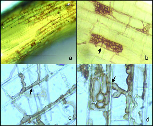
Microscopic analysis showed that DSF were observed in 36 of the 39 (92%) roots examined and in all 18 plants collected from May 2005 to January 2006. Only two roots exhibited structures or hyaline hyphae characteristic of AMF. DSF were observed mainly within the root cortex but seldom penetrated into the vascular cylinder (inside the endodermis) (Fig. 1a). Although fungal hyphae commonly grew within the intercellular grooves created by adjacent endodermis cells (Fig. 1b and c), it was common for many DSF hyphae to traverse the cortex and extend outside the root into the adjacent soil and on occasion to penetrate through the cell walls of adjacent root cortex cells (Fig. 1d).
FIG. 1.
DSF endophytes colonizing B. gracilis roots. (a) Tertiary root of B. gracilis (blue grama) showing the endodermis torn to expose the vascular bundle within. DSF are commonly observed on the external surfaces of the endodermis cells (brown hyphae) but rarely within the vascular cylinder (magnification, ×400). (b and c) DSF grow along intercellular grooves and form clusters of hyphae known as microsclerotia (magnification, ×200). (d) In some cases, hyphae penetrate through cell walls to inhabit adjacent cells (magnification, ×1,000).
The analyses of the pooled molecular data revealed that B. gracilis roots are colonized by a diverse fungal community (the Shannon-Weiner diversity index for the entire data set was 3.1). A total of 51 (95% confidence interval, 39 to 63) OTUs and a Chao diversity estimator of 91 OTUs with a confidence interval of 63 to 179 were obtained from B. gracilis roots.
Most of the sequences obtained exhibited low similarity with sequences previously known. A comparison of the sequences obtained from B. gracilis roots with those in the NCBI database using BLASTN revealed that the fungal community colonizing B. gracilis roots was related largely to uncultured endophytes and coprophilic fungi (together, both groups represent 92.7% of the entire data set); 7.2% represented other known saprobes and plant pathogens, and 0.2% corresponded to AMF. In the data set, 435 (or 69%) of the 630 sequences and 34 (or 67%) of the 51 OTUs were novel (less than 97% similarity with respect to other sequences in the core nucleotide NCBI database). Ascomycota and Basidiomycota represented 83.2% and 16.5% of the sequences in the entire data set (630 sequences obtained from healthy roots), respectively. In a previous study, we reported that no AMF sequences were obtained from our ITS clone libraries (300 sequences at that time) (54). After additional clones were obtained with the ITS primers, only one sequence corresponded to Glomeromycota. These results confirm the low level of AMF colonization observed in the microscopic analysis. Based upon the molecular data, the most common classes colonizing B. gracilis roots are Dothidiomycetes (58.7% of 630 sequences, obtained from all 18 plants), Sordariomycetes (22.7%, obtained from 10 plants), and Agaricomycetes (16.5%, obtained from 10 plants).
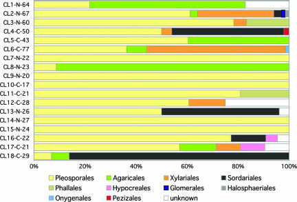
Pleosporales was the most abundantly represented order within the clone libraries, accounting for 58.9% (370 sequences) of all sequences in the database (Fig. 2). Members of the Pleosporales were obtained from all plants, and in some samples, they constituted up to 100% of the clone libraries. Other frequent orders represented in the clone libraries included Sordariales, Agaricales, and Xylariales, accounting for 21.7%, 14.3%, and 11.6% of the fungal community, respectively (Fig. 2).
FIG. 2.
Distribution of fungal orders found in the different ITS clone libraries (CL) of B. gracilis plants. Clonal libraries, nitrogen (N) plots, control (C) plots, and number of sequences obtained per plant are indicated on the left side of the graph. CL1 to CL6, CL7 to CL12, and CL13 to CL18 were collected in May 2005, October 2005, and January 2006, respectively.
Analysis of the fungal orders colonizing B. gracilis showed that Agaricales sequences were more common in May (of a total of 90 sequences of Agaricales, 71.3% were obtained from May samples [four of the six plants]) than in January (5.6% [two of six plants]) and October (23.3% [only one plant]). In contrast, members of the Xylariales were almost absent in October (of a total of 138 sequences of Xylariales, 2.9% were obtained in October [one plant]) but were common in samples collected in May (66.7% [four plants]) and January (30.4% [four samples]) (Fig. 2).
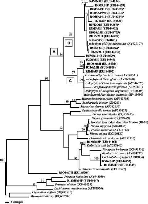
Although B. gracilis roots were colonized by a highly diverse group of endophytic fungi, this fungal community was dominated by a small number of OTUs. The most common OTUs (clade A with 94% bootstrap support and two terminal subclades [B and C with 86% and 77% bootstrap support, respectively]) (Fig. 3) belonged within the order Pleosporales. The number of sequences within clade A alone represented 50% of the entire database (315 out of 630 sequences). Furthermore, with the exception of samples collected from N plots in October 2005, the sequences in clade A were obtained in all samples collected from both N and C plots. DSF within subclade B also were obtained in pure culture. The GenBank sequence most closely related to subclade B is from a root endophyte isolated from a grass (Stipa hymenoides) inhabiting a semiarid grassland in Utah (29). Subclade C is related to endophytes commonly found in coniferous trees, such as Juniperus, Picea, and Pinus (32, 71), and to a DSF in the genus Paraphaeosphaeria (Fig. 3).
FIG. 3.
Phylogenetic analysis of the most common endophytic group within the order Pleosporales (clade A and subclades B and C) (see Results). Sequences from this study are shown in bold, and GenBank numbers are shown in parentheses after the name or sequence code. Isolates in pure culture are indicated with asterisks. One of the most parsimonious trees is shown, and bootstrap values (1,000 replicates [more than 70%]) are indicated above the branches. Capnodium coffeae (DQ461515) and Mycosphaerella sp. (DQ632689) were used as outgroups (67), and the sequence alignment was deposited in Webin-Align under accession number ALIGN-001185.
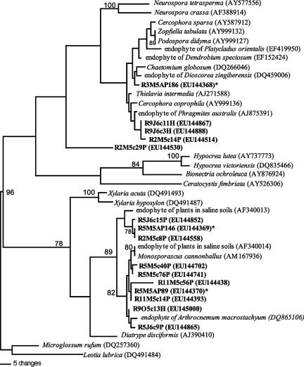
Other common endophytes colonizing B. gracilis included Cercophora (9.7% of the entire data set) and the genus Monosporascus (11% of the entire data set) (Fig. 4). The phylogenetic analysis of the order Sordariales supports the assertion that Monosporascus is more closely related to Xylariales than to Sordariales (17). Most of the sequences obtained from B. gracilis are related to other endophytes that colonize grasses or plants growing in saline soils (14, 17, 29, 49, 58).
FIG. 4.
Common Sordariomycetes colonizing B. gracilis roots. One of the most parsimonious trees is shown, and bootstrap values (1,000 replicates [more than 70%]) are indicated above the branches. Sequences from this study are shown in bold, and GenBank numbers are shown in parentheses after the name or sequence code. Isolates in pure culture are indicated with asterisks. Microglossum rufum (DQ257360) and Leotia lubrica (DQ491484) were used as outgroups (88). Sequence alignment was deposited in Webin-Align under number ALIGN-001184.
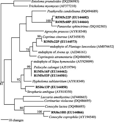
Within the class Agaricomycetes, we included only members of the Agaricoid clade in the phylogenetic analysis, since the ITS regions for the other sequences within the Agaricales are difficult to align (45). All basidiomycete sequences obtained are closely related to previously described species, including several common coprophilic fungi (Fig. 5).
FIG. 5.
Phylogenetic analysis of common endophytes in the Agaricoid clade. One of the most parsimonious trees is shown, and bootstrap values (1,000 replicates [more than 70%]) are indicated above the branches. Sequences from this study are shown in bold, and GenBank numbers are shown in parentheses after the name or sequence code. Ambiguous regions in the alignment (characters 101 to 250 and 441 to 469 [alignment accession number ALIGN-001183]) were excluded from the analysis. Entoloma prunuloides (DQ206983) and Tricholoma myomyces (AF377210) were used as outgroups (45).
DISCUSSION
Root-associated fungal diversity in arid grasslands.
Considering the difficult conditions plants endure in arid environments—high temperatures, low precipitation, variable rainfall patterns, and low soil moisture—we found high diversity of root-associated fungi in B. gracilis (Fig. 2). The low similarity levels between many of our sequences and existing sequences in the NCBI core nucleotide database suggest that a high proportion of the fungal sequences associated with B. gracilis roots are novel to science. Although the ITS is the most widely used region for fungal identification, with more than 77,000 fungal ITS sequences in NCBI, it is possible that some of the sequences obtained here represent known fungi for which ITS sequences are not available at NCBI. Moreover, further analyses need to be conducted using pure cultures to obtain sequences of regions, such as the large-subunit ribosomal DNA, to get a better phylogenetic placement for the species represented by our novel ITS sequences (e.g., see reference 31). The fungal species richness associated with B. gracilis roots is similar to that of other grasses from more mesic environments, such as the perennial grass Arrhenatherum elatius, in which 49 different phylotypes were found associated with roots (77), and the wetland grass Phragmites australis (85).
Most studies of root-associated fungi in arid environments have focused on AMF (e.g., see references 19, 33, and 41). To our knowledge, our study is the first to show that the roots of an arid grass species are associated with a diverse consortium of Ascomycota and Basidiomycota. A study of bacterial diversity found in soil samples at the SNWR by Fierer and Jackson (24) showed that bacteria are also highly diverse in arid grasslands in comparison with other temperate and tropical ecosystems. Why are microbial communities so diverse in desert environments? Chesson et al. (15), Herrera et al. (30), and Zak (86) proposed that the heterogeneous conditions of desert ecosystems (e.g., rainfall variability that controls carbon and nitrogen cycles, disturbance events, soil heterogeneity, and the patchy distribution of plant communities and cyanobacteria crust soils) may be responsible for the greater fungal and enzymatic diversity present in these extreme environments. The high colonization rates of healthy plant roots by root-associated fungi (8, 41, 54, 77), along with evidence showing that endophytes improve resistance to drought conditions and contribute to increasing the efficiency of nutrient transfer processes (1, 23, 28, 37, 38, 45a, 76), suggests that the association of plants with a variety of fungi might be fundamental for plant survival in arid ecosystems. Our knowledge about root-associated fungal diversity is very limited in most ecosystems, and cross-site comparisons are necessary to understand the biogeography and ecological role of these fungal communities. In addition, how these diverse groups of root-associated fungi interact with mycorrhizal fungi, the host, and environmental conditions is for the most part unknown (36).
DSF within the order Pleosporales are common inhabitants of B. gracilis roots.
Although we detected high diversity of fungi associated with B. gracilis roots, microscopic analysis showed that roots are highly colonized by DSF (Fig. 1). These results are supported by the molecular data that showed that the dominant fungal clade colonizing B. gracilis roots (50% of the sequences in the entire data set, found in almost all plants) is closely related to other DSF within the order Pleosporales (Fig. 3). Neubert et al. (49) reported similar results for Phragmites australis, a wetland grass that is colonized by a diverse fungal community, but with only a few dominant species. Although we obtained sequences and observed fungi known to have hyaline hyphae, they represented less than 5% of the total surface area of the endodermis in our roots. It is possible that some of the OTUs related to species known to produce light-colored hyphae possibly inhabit areas in the cortex nearer the root epidermis (indeed, microscopic analysis of the surface of the endodermis showed DSF almost exclusively), or perhaps these fungi are hidden by the high colonization rates of DSF. PCR bias is a recognized problem (35), but our observations under the microscope suggest that the molecular data used to describe the composition and structure of the fungal communities colonizing B. gracilis roots are a good proxy to describe the dominant root-associated fungi in B. gracilis.
Johnson et al. (33) and Allen et al. (2) reported that B. gracilis plants collected from the same SNWR sites that we sampled were highly colonized by AMF. However, the level of colonization evaluated with molecular and microscopy methods in this and a previous study (54) showed high levels of DSF instead of AMF (see reference 8). Different environmental conditions and a long drought period that followed previous studies may account for the low AMF colonization rates observed for B. gracilis roots (54), suggesting that the fungal community of grama roots is highly sensitive to climate.
The abundance and diversity of DSF within B. gracilis roots had no obvious detrimental effects on the plants. Microscopic observations supported molecular data in which only 7.2% of the entire data set corresponds to known pathogens and saprobes. The repeated isolation and identification of DSF from healthy B. gracilis plants by molecular methods and careful visual inspection of the plants roots suggest that the symbiosis with this novel group of DSF within the order Pleosporales (Fig. 3) could be mutualistic (1, 62). Subsequent experiments of reinoculation of the endophyte in the host plant are necessary to further demonstrate that these root-associated fungi are endophytic. It is notable that despite their ubiquity throughout the cortex and epidermis cells of nearly all roots of all plants examined, DSF were never found within any of the vascular bundles. Similar results have previously been described by O'Dell et al. (53), who reported colonization of cortical cells by the DSF Phialocephala fortinii, and by Gao and Mendgen (25), who reported that the fungus Stagonospora sp. inhabits the external areas of the cortex but not the vascular cylinder of Phragmites australis. In our study, however, much of the growth was observed immediately outside the external interface of the vascular cylinder, on the endodermis. Consequently, we suspect that the dominant DSF in B. gracilis (and not AMF) are more intimately involved with important physiological functions that involve the movement of nutrients and water in and out of the vascular cylinder (Fig. 1), as suggested by several studies of DSF (28, 34, 37, 76, 89). Well-described cases of symbiotic endophytic fungi in above-ground tissues of grasses (63-65) have already demonstrated that endophytic fungi can contribute to the survival and adaptation of grasses.
One fundamental characteristic of DSF that may be important in symbiotic associations with plants is the production of wall-bound and extracellular melanins. Melanins provide protection to the fungus and very likely to the plant against desiccation and extreme temperatures (10, 37). In addition, they appear to be important for resistance against microbial attack, providing protection against hydrolytic enzymes and exerting an antibiotic effect (10, 43). The ubiquitous distribution of DSF associated with roots of other dominant grasses related to B. gracilis, such as Bouteloua eriopoda and Bouteloua curtipendula, (8) and the grass Deschampsia flexuosa (89), suggest that DSF could have an important function in the adaptation, survival, and distribution of these plants.
Coprophilous fungi associated with plant roots.
To our knowledge, few other instances of known coprophilous fungi associated with plant roots have been reported (e.g., see references 58 and 84). The fact that 20% of the sequences in our data set have high similarity with those from known coprophilous fungi (Fig. 2, 4, and 5) suggests a previously undisclosed ecological role for these fungal species. Studies by Wicklow and Zak (83) and Wicklow et al. (84) showed that herbivory might have a fundamental role in B. gracilis seed dispersal and establishment. Variable rainfall patterns and moisture in semiarid grasslands complicate the effective establishment of seedlings (68, 86), and herbivore dung may provide a more suitable environment than soil in possessing enough water for seed germination. Wicklow and Zak (83) found a significant increase in the germination of B. gracilis and Sporobolus cryptandrus (sand drop) seeds when they were in contact with herbivore dung in semiarid grasslands. Wicklow et al. (84) also suggest that dung-burying beetles may have a significant role in the establishment of B. gracilis seedlings. A significant percentage of seeds, leaves, and roots are consumed by herbivores in grasslands (70), and B. gracilis plants are highly palatable even during dry periods (27, 46). Thus, it is possible that during the passage of B. gracilis seeds through the digestive tract of an herbivore, those seeds become inoculated with coprophilic fungi. The establishment of a symbiotic association with coprophilous fungi (Fig. 4 and 5) may facilitate access to different nutrients during the decomposition and mineralization of herbivores feces. For example, Angel and Wicklow (3) suggested that coprophilous fungi have an important role in long-term nitrogen immobilization. This nitrogen might contribute to the survival of plants associated with coprophilous fungi when this nutrient is limiting plant growth. The presence of coprophilous fungi associated with plant roots therefore may have a significant role in the survival and establishment of seedlings in semiarid grasslands. In the future, published descriptions of the role of coprophilous fungi in arid grasslands may need to include the possibility that some members of this group are also endophytic as part of their life cycles.
Consortium of root-associated fungi: interactions and implications for ecosystem functioning.
Our study demonstrates that B. gracilis forms symbiotic associations with a broad range of fungi, including coprophilous fungi, DSF, and AMF (also see references 8 and 54). Diverse communities of root-associated fungi have been reported for many plant families and within several species of grasses (22, 37, 56, 75). These findings counter the traditional view that endophytic fungal symbioses in grasses mostly occur within above-ground tissues (60, 63, 65). Although it has been suggested that these fungal communities have important roles in plant survival and ecosystem functioning (1, 13, 37), most studies examining the effects of fungal communities on plant diversity and structure have focused only on mycorrhizae (e.g., see references 39, 78, and 79). Current models of plant-fungi interactions in the roots oversimplify this complex system.
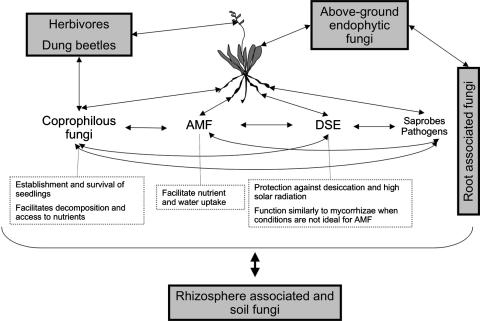
We define root-associated fungi as the consortium of fungal species that symbiotically interact among themselves, with the plant host, and with other ecosystem components with the common effect of enhancing survival and fitness of the symbionts (Fig. 6). In this consortium, roles vary; for example, some fungi will contribute to the uptake of nutrients, while others will improve resistance to drought or pathogens or enhance seed germination and survival of seedlings. As in any consortium, interactions will range from positive to neutral to negative, depending on conditions (5, 55). The composition, activities, and interactions of these fungi will be regulated by the plant host and by the fungal members of the consortium in response to biotic and abiotic factors, such as nitrogen amendment, temperature, rainfall patterns, and the presence of pathogens and herbivores (21, 40, 55, 61, 60).
FIG. 6.
Interactions of root-associated fungi in B. gracilis roots, a dominant grass in semiarid grasslands. The model shows possible functions of root-associated fungi in B. gracilis and the interaction among the fungal endophytic community within the roots, other fungal communities in soil, and aerial plant tissues, primary producers, and consumers.
Such fungal consortia can have ecosystem-level effects. Endophytic colonization of one dominant grass can alter the abundance of other plant species (16), and AMF can alter the diversity and structure of a plant community (59, 78, 79). Rudgers and Clay (60) suggest that the differential competitive ability of plants colonized by endophytic fungi in comparison with plants that are not colonized may be important in defining plant community structure in stressful conditions. At present, we know very little about the variation in fungal consortia among plants, or their responses to environmental changes, or their interactions with rhizosphere and soil fungal communities and consumers in the ecosystem. The complex responses to small-scale spatial heterogeneity and changes in environmental conditions of fungal consortia associated with plant roots and other tissues may act to ensure survival and maximize the benefits of the fungal symbiotic associations for the plant host.
Acknowledgments
We thank Dean DeCock for statistical help.
The Mycological Society of America is acknowledged for the E. Butler Mentor Travel Award and the MSA graduate student fellowship. This research was funded by the National Science Foundation Long-Term Ecological Research Program at the Sevilleta National Wildlife Refuge (DEB-027774 and DEB-0217774) and a grant from the National Science Foundation Ecosystems Sciences Program (DEB-0516113).
Footnotes
Published ahead of print on 14 March 2008.
REFERENCES
- 1.Addy, H. D., M. M. Piercey, and R. S. Currah. 2005. Microfungal endophytes in roots. Can. J. Bot. 83:1-13. [Google Scholar]
- 2.Allen, M. F., W. K. Smith, T. S. Moore, and M. Christensen. 1981. Comparative water relation and photosynthesis of mycorrhizal and non-mycorrhizal Bouteloua gracilis H.B.K. Lag Ex Steud. New Phytol. 86:683-693. [Google Scholar]
- 3.Angel, K., and D. T. Wicklow. 1983. Coprophilous fungal communities in semiarid to mesic grasslands. Can. J. Bot. 61:594-602. [Google Scholar]
- 4.Arnold, A. E., Z. Maynard, G. S. Gilbert, P. D. Coley, and T. A. Kursar. 2000. Are tropical fungal endophytes hyperdiverse? Ecol. Lett. 3:267-274. [Google Scholar]
- 5.Arnold, A. E., L. C. Mejia, D. Kyllo, E. I. Rojas, Z. Maynard, N. Robbins, and E. A. Herre. 2003. Fungal endophytes limit pathogen damage in a tropical tree. Proc. Natl. Acad. Sci. USA 100:15649-15654. [DOI] [PMC free article] [PubMed] [Google Scholar]
- 6.Arnold, A. E., D. A. Henk, R. L. Eells, F. Lutzoni, and R. Vilgalys. 2007. Diversity and phylogenetic affinities of foliar fungal endophytes in loblolly pine inferred by culturing and environmental PCR. Mycologia 99:185-206. [DOI] [PubMed] [Google Scholar]
- 7.Austin, A. T., L. Yahdjian, J. M. Stark, J. Belnap, A. Porporato, U. Norton, D. A, Ravetta, and S. M. Schaeffer. 2004. Water pulses and biogeochemical cycles in arid and semiarid ecosystems. Oecologia 141:221-235. [DOI] [PubMed] [Google Scholar]
- 8.Barrow, J. R. 2003. Atypical morphology of dark septate fungal root endophytes of Bouteloua in arid southwestern USA rangelands. Mycorrhiza 13:239-247. [DOI] [PubMed] [Google Scholar]
- 9.Bayman, P., L. L. Lebron, R. L. Tremblay, and D. J. Lodge. 1997. Variation in endophytic fungi from roots and leaves of Lepanthes (Orchidaceae). New Phytol. 135:143-149. [DOI] [PubMed] [Google Scholar]
- 10.Bell, A. A., and M. H. Wheeler. 1986. Biosynthesis and functions of fungal melanins. Annu. Rev. Phytopathol. 24:411-451. [Google Scholar]
- 11.Brundrett, M. C. 2002. Coevolution of roots and mycorrhizas of land plants. New Phytol. 154:275-304. [DOI] [PubMed] [Google Scholar]
- 12.Buxbaum, C. A. Z., and K. Vanderbilt. 2007. Soil heterogeneity and the distribution of desert and steppe plant species across a desert-grassland ecotone. J. Arid Environ. 69:617-632. [Google Scholar]
- 13.Cairney, J. W. G., and R. M. Burke. 1998. Extracellular enzyme activities of the ericoid mycorrhizal endophyte Hymenoscyphus ericae (Read) Korf & Kernan: their likely roles in decomposition of dead plant tissue in soil. Plant Soil 2:181-192. [Google Scholar]
- 14.Carter, J. P., J. Spink, P. F. Cannon, M. J. Daniels, and A. E. Osbourn. 1999. Isolation, characterization, and avenacin sensitivity of a diverse collection of cereal-root-colonizing fungi. Appl. Environ. Microbiol. 65:3364-3372. [DOI] [PMC free article] [PubMed] [Google Scholar]
- 15.Chesson, P., R. L. E. Gebauer, S. Schwinning, N. Huntly, K. Wiegand, M. S. K. Ernest, A. Sher, A. Novoplansky, and J. F. Weltzin. 2004. Resource pulses, species interactions, and diversity maintenance in arid and semi-arid environments. Oecologia 141:236-253. [DOI] [PubMed] [Google Scholar]
- 16.Clay, K., and J. Holah. 1999. Fungal endophyte symbiosis and plant diversity in succesional fields. Science 285:1742-1744. [DOI] [PubMed] [Google Scholar]
- 17.Collado, J., A. González, G. Platas, A. M. Stchigel, J. Guarro, and F. Peláez. 2002. Monosporascus ibericus sp. nov., an endophytic ascomycete from plants on saline soils, with observation on the position of the genus based on sequence analysis of the 18S DNA. Mycol. Res. 106:118-127. [Google Scholar]
- 18.Collins, S. L., R. L. Sinsabaugh, C. Crenshaw, L. Green, A. Porras-Alfaro, M. Stursova, and L. H. Zeglin. Pulse dynamics and microbial processes in arid land ecosystems. J. Ecol., in press.
- 19.Corkidi, L., D. L. Rowland, N. C. Johnson, and E. B. Allen. 2002. Nitrogen fertilization alters the functioning of arbuscular mycorrhizas at two semiarid grasslands. Plant Soil 240:299-310. [Google Scholar]
- 20.Eriksson, O. E. 2006. Outline of Ascomycota—2006. Myconet 12:1-82. [Google Scholar]
- 21.Escudero, V., and R. Mendoza. 2005. Seasonal variation of arbuscular mycorrhizal fungi in temperate grasslands along a wide hydrologic gradient. Mycorrhiza 15:291-299. [DOI] [PubMed] [Google Scholar]
- 22.Gollotte, A., D. van Tuinen, and D. Atkinson. 2004. Diversity of arbuscular mycorrhizal fungi colonizing roots of the grass species Agrotis capillaries and Lolium perenne in a field experiment. Mycorrhiza 14:111-117. [DOI] [PubMed] [Google Scholar]
- 23.Fernando, A. A., and R. S. Currah. 1996. A comparative study of the effect of Phialocephala fortinii (Fungi Imperfecti) on the growth of some subalpine plants in culture. Can. J. Bot. 74:1071-1078. [Google Scholar]
- 24.Fierer, N., and R. B. Jackson. 2006. The diversity and biogeography of soil bacterial communities. Proc. Natl. Acad. Sci. USA 103:626-631. [DOI] [PMC free article] [PubMed] [Google Scholar]
- 25.Gao, K., and K. Mendgen. 2006. Seed-transmitted beneficial endophytic Stagonospora sp. can penetrate the walls of the root epidermis, but does not proliferate in the cortex, of Phragmites australis. Can. J. Bot. 84:981-988. [Google Scholar]
- 26.Gardes, M., and T. D. Bruns. 1993. ITS primers with enhanced specificity of Basidiomycetes: application to the identification of mycorrhizae and rusts. Mol. Ecol. 2:113-118. [DOI] [PubMed] [Google Scholar]
- 27.Gosz, R. J., and J. R. Gosz. 1996. Species interactions on the biome transition zone in New Mexico: response of blue grama (Bouteloua gracilis) and black grama (Bouteloua eriopoda) to fire and herbivory. J. Arid Environ. 34:101-114. [Google Scholar]
- 28.Haselwandter, K., and R. J. Read. 1982. The significance of root-fungus association in two Carex species of high-alpine plant communities. Oecologia 53:352-354. [DOI] [PubMed] [Google Scholar]
- 29.Hawkes, C. V., J. Belnap, C. D'Antonio, and M. K. Firestone. 2006. Arbuscular mycorrhizal assemblages in native plant roots change in the presence of invasive exotic grasses. Plant Soil 281:369-380. [Google Scholar]
- 30.Herrera, J., C. L. Kramer, and O. J. Reichman. 1997. Patterns of fungal communities that inhabit rodent foot stores: effect of substrate and infection time. Mycologia 86:846-857. [Google Scholar]
- 31.Higgins, K. L., A. E. Arnold, J. Miadlikowska, S. D. Sarvate, and F. Lutzoni. 2007. Phylogenetic relationships, host affinity, and geographic structure of boreal and arctic endophytes from three major plant lineages. Mol. Phylogenet. Evol. 42:543-555. [DOI] [PubMed] [Google Scholar]
- 32.Hoffman, M. T., and A. E. Arnold. 2008. Geographic locality and host identity shape fungal endophyte communities in cupressaceous trees. Mycol. Res. 112:331-344. [DOI] [PubMed] [Google Scholar]
- 33.Johnson, N. C., D. L. Rowland, L. Corkidi, L. M. Egerton-Warburton, and E. B. Allen. 2003. Nitrogen enrichment alters mycorrhizal allocation at five mesic to semiarid grasslands. Ecology 84:1895-1908. [Google Scholar]
- 34.Jumpponen, A. 2001. Dark septate endophytes. Are they mycorrhizal? Mycorrhiza 11:207-211. [Google Scholar]
- 35.Jumpponen, A. 2007. Soil fungal communities underneath willow canopies on a primary successional glacier forefront: rDNA sequence results can be affected by primer selection and chimeric data. Microb. Ecol. 53:233-246. [DOI] [PubMed] [Google Scholar]
- 36.Jumpponen, A., and L. Johnson. 2005. Can rDNA analyses of diverse fungal communities in soil and roots detect effects of environmental manipulation—a case study from tallgrass prairie. Mycologia 97:1177-1194. [DOI] [PubMed] [Google Scholar]
- 37.Jumpponen, A., and J. M. Trappe. 1998. Dark septate endophytes: a review of facultative biotrophic root-colonizing fungi. New Phytol. 140:295-310. [DOI] [PubMed] [Google Scholar]
- 38.Jumpponen, A., K. G. Mattson, and J. M. Trappe. 1998. Mycorrhizal functioning of Phialocephala fortinii: interactions with soil nitrogen and organic matter. Mycorrhiza 7:261-265. [DOI] [PubMed] [Google Scholar]
- 39.Leake, J. R. 2001. Is diversity of ectomycorrhizal fungi important for ecosystem function? New Phytol. 152:1-3. [DOI] [PubMed] [Google Scholar]
- 40.Li, L., A. Yang, and Z. Zhao. 2005. Seasonality of arbuscular mycorrhizal symbiosis and dark septate endophytes in a grassland site in southwest China. FEMS Microb. Ecol. 54:367-373. [DOI] [PubMed] [Google Scholar]
- 41.Li, T., and Z. W. Zhao. 2005. Arbuscular mycorrhizas in a hot and arid ecosystem in southwest China. Appl. Soil Ecol. 29:135-141. [Google Scholar]
- 42.Lin, X., C. H. Lu, Y. J. Huang, Z. H. Zheng, W. J. Su, and Y. M. Shen. 2007. Endophytic fungi from a pharmaceutical plant, Camptotheca acuminata: isolation, identification, and bioactivity. World J. Microbiol. Biotechnol. 23:1037-1040. [Google Scholar]
- 43.Mandyam, K., and A. Jumpponen. 2005. Seeking the elusive function of the root-colonising dark septate endophytic fungi. Studies Mycol. 53:173-189. [Google Scholar]
- 44.Márquez, L. M., R. S. Redman, R. J. Rodriguez, and M. J. Roossinck. 2007. A virus in a fungus in a plant: three-way symbiosis required for thermal tolerance. Science 315:513-515. [DOI] [PubMed] [Google Scholar]
- 45.Matheny, P. B., J. M. Curtis, V. Hofstetter, M. C. Aime, J. Moncalvo, Z. Zai-Wei Ge, Z. Yang, J. C. Slot, J. F. Ammirati, T. J. Baroni, N. L. Bougher, K. W. Hughes, D. J. Lodge, R. W. Kerrigan, M. T. Seidl, D. K. Aanen, M. DeNitis, G. M. Daniele, D. E. Desjardin, B. R. Kropp, L. L. Norvell, A. Parker, E. C. Vellinga, R. Vilgalys, and D. S. Hibbett. 2006. Major clades of Agaricales: a multilocus phylogenetic overview. Mycologia 98:982-995. [DOI] [PubMed] [Google Scholar]
- 45a.McLellan, C. A., T. J. Turbyville, E. M. K. Wijeratne, A. Kerschen, E. Vierling, C. Queitsch, L. Whitesell, and A. A. L. Gunatilaka. 2007. A rhizosphere fungus enhances Arabidopsis thermotolerance through production of an HSP90 inhibitor. Plant Physiol. 145:174-182. [DOI] [PMC free article] [PubMed] [Google Scholar]
- 46.Mellado, M., A. Olvera, A. Quero, and G. Mendoza. 2005. Dietary overlap between prairie dog (Cynomys mexicanus) and beef cattle in a desert rangeland of northern Mexico. J. Arid Environ. 62:449-458. [Google Scholar]
- 47.Minnick, T. J., and D. P. Coffin. 1999. Geographic patterns of simulated establishment of two Bouteloua species: implications for distributions of dominants and ecotones. J. Veg. Sci. 10:343-356. [Google Scholar]
- 48.Narisawa, K., M. Chen, T. Hashiba, and A. Tsuneda. 2003. Ultrastructural study on interaction between a sterile, white endophytic fungus and eggplant roots. J. Gen. Plant Pathol. 69:292-296. [Google Scholar]
- 49.Neubert, K., K. Mendgen, H. Brinkmann, and S. G. R. Wirsel. 2006. Only a few fungal species dominate highly diverse mycofloras associated with common reed. Appl. Environ. Microbiol. 72:1118-1128. [DOI] [PMC free article] [PubMed] [Google Scholar]
- 50.Newsham, K. K. 1999. Phialophora graminicola, a dark septate fungus, is a beneficial associate of the grass Vulpia ciliata ssp. ambigua. New Phytol. 144:517-524. [DOI] [PubMed] [Google Scholar]
- 51.Noy-Meir, I. 1973. Desert ecosystems: environment and producers. Annu. Rev. Ecol. Syst. 4:25-51. [Google Scholar]
- 52.O'Brien, H. E., J. L. Parrent, J. A. Jackson, J. Moncalvo, and R. Vilgalys. 2005. Fungal community analysis by large-scale sequencing of environmental samples. Appl. Environ. Microbiol. 71:5544-5550. [DOI] [PMC free article] [PubMed] [Google Scholar]
- 53.O'Dell, T. E., H. B. Massicotte, and J. M. Trappe. 1993. Root colonization of Lupinus latifolius Agardh. and Pinus contorta Dougl. by Phialocephala fortini Wang & Wilcox. New Phytol. 124:93-100. [Google Scholar]
- 54.Porras-Alfaro, A., J. Herrera, R. L. Sinsabaugh, and D. O. Natvig. 2007. Effect of long-term nitrogen fertilization on mycorrhizal fungi associated with a dominant grass in a semiarid grassland. Plant Soil 296:65-75. [Google Scholar]
- 55.Porras-Alfaro, A., and P. Bayman. 2007. Mycorrhizal fungi of Vanilla: diversity, specificity and effects on seed germination and plant growth. Mycologia 99:510-525. [DOI] [PubMed] [Google Scholar]
- 56.Read, D., and K. Haselwandter. 1981. Observations on the mycorrhizal status of some alpine plant communities. New Phytol. 88:341-352. [Google Scholar]
- 57.Redman, R., K. B. Sheehan, R. G. Stout, R. J. Rodriguez, and J. M. Henson. 2002. Thermo tolerance generated by plant/fungal symbiosis. Science 298:1581. [DOI] [PubMed] [Google Scholar]
- 58.Renker, C., K. Weisshuhn, H. Kellner, and F. Buscot. 2006. Rationalizing molecular analysis of field-collected roots for assessing diversity of arbuscular mycorrhizal fungi: to pool, or not to pool, that is the question. Mycorrhiza 16:525-531. [DOI] [PubMed] [Google Scholar]
- 59.Rudgers, J. A., J. M. Koslow, and K. Clay. 2004. Endophytic fungi alter relationships between diversity and ecosystem processes. Ecol. Lett. 7:42-51. [Google Scholar]
- 60.Rudgers, J. A., and K. Clay. 2005. Fungal endophytes in terrestrial communities and ecosystems, p. 423-442. In J. Dighton, J. F. White, and P. Oudemans (ed.), The fungal community: its organization and role in the ecosystems, 3rd ed. CRC Press, Boca Raton, FL.
- 61.Ruotsalainen, A. L., H. Vare, and M. Vestberg. 2002. Seasonality of root fungal colonization in low alpine herbs. Mycorrhiza 12:29-36. [DOI] [PubMed] [Google Scholar]
- 62.Saikkonen, I., S. H. Faeth, M. Helander, and T. J. Sullivan. 1998. Fungal endophytes: a continuum of interactions with host plants. Annu. Rev. Ecol. Syst. 29:319-343. [Google Scholar]
- 63.Schardl, C. L., J. S. Liu, J. F. White, R. A. Finkel, Z. Q. An, and M. R. Siegel. 1991. Molecular phylogentic relationships of nonpathogenic grass mycosymbionts and clavicipitaceous plant pathogens. Plant Syst. Evol. 178:27-42. [Google Scholar]
- 64.Schardl, C. L., A. Leuchtmann, K. R. Chung, D. Penny, and M. R. Siegel. 1997. Coevolution by common descent of fungal symbionts (Epichloe spp.) and grass hosts. Mol. Biol. Evol. 14:133-143. [Google Scholar]
- 65.Schardl, C. L., A. Leuchtmann, and M. J. Spiering. 2004. Symbioses of grasses with seedborne fungal endophytes. Annu. Rev. Plant Biol. 55:315-340. [DOI] [PubMed] [Google Scholar]
- 66.Schloss, P. D., and J. Handelsman. 2006. Introducing SONS, a tool for OTU-based comparisons of membership and structure between microbial communities. Appl. Environ. Microbiol. 72:6773-6779. [DOI] [PMC free article] [PubMed] [Google Scholar]
- 67.Schoch, C. L., R. A. Shoemaker, K. A. Seifert, S. Hambleton, J. W. Spatafora, and P. W. Crous. 2006. A multigene phylogeny of the Dothideomycetes using four nuclear loci. Mycologia 98:1041-1052. [DOI] [PubMed] [Google Scholar]
- 68.Schwinning, S., and O. E. Sala. 2004. Hierarchy of responses to resource pulses in arid and semi-arid ecosystems. Oecologia 141:211-220. [DOI] [PubMed] [Google Scholar]
- 69.Smith, S. E., and D. J. Read. 1997. Mycorrhizal symbiosis, 2nd ed. Academic Press, New York, NY.
- 70.Stanton, N. L. 1988. The underground in grasslands. Annu. Rev. Ecol. Syst. 19:573-589. [Google Scholar]
- 71.Stefani, F. O. P., and J. Berube. 2006. Biodiversity of foliar fungal endophytes in white spruce (Picea glauca) from southern Quebec. Can. J. Bot. 84:777-790. [Google Scholar]
- 72.Stursova, M., C. L. Crenshaw, and R. L. Sinsabaugh. 2006. Microbial responses to long-term N deposition in a semiarid grassland. Microb. Ecol. 51:90-98. [DOI] [PubMed] [Google Scholar]
- 73.Suryanarayanan, T. S., S. K. Wittlinger, and S. H. Faeth. 2005. Endophytic fungi associated with cacti of Arizona. Mycol. Res. 109:635-639. [DOI] [PubMed] [Google Scholar]
- 74.Swofford, D. 2002. PAUP*. Phylogenetic analysis using parsimony (and other methods), version 4. Sinauer Associates, Sunderland, MA.
- 75.Treu, R., G. A. Laursen, S. L. Stephenson, J. C. Landolt, and R. Densmore. 1996. Mycorrhizae from Denali National Park and Preserve, Alaska. Mycorrhiza 6:21-29. [Google Scholar]
- 76.Usuki, F., and K. Narisawa. 2007. A mutualistic symbiosis between a dark septate endophytic fungus, Heteroconium chaetospira, and a nonmycorrhizal plant, Chinese cabbage. Mycologia 99:175-184. [DOI] [PubMed] [Google Scholar]
- 77.Vandenkoornhuyse, P., S. L. Baldauf, C. Leyval, J. Straczek, and P. W. Young. 2002. Extensive fungal diversity in plant roots. Science 295:2051. [DOI] [PubMed] [Google Scholar]
- 78.van der Heijden, M. G. A., T. Boller, A. Wiemken, and I. R. Sanders. 1998. Different arbuscular mycorrhizal fungal species are potential determinants of plant community structure. Ecology 79:2082-2091. [Google Scholar]
- 79.van der Heijden, M. G. A., J. N. Klironomos, M. Ursic, P. Moutoglis, R. Streitwolf-Engel, T. Boller, A. Wiemken, and I. R. Sanders. 1998. Mycorrhizal fungal diversity determines plant biodiversity, ecosystem variability and productivity. Nature 396:69-72. [Google Scholar]
- 80.Vierheilig, H., and Y. Piche. 1998. A modified procedure for staining arbuscular mycorrhizal fungi in roots. Z. Fur Pflanz. Bodenkunde 161:601-602. [Google Scholar]
- 81.Vohnik, M., J. Albrechtova, and M. Vosatka. 2005. The inoculation with Oidiodendron maius and Phialocephala fortinii alters phosphorus and nitrogen uptake, foliar C:N ratio and root biomass distribution in Rhododendron cv. Azurro. Symbiosis 40:87-96. [Google Scholar]
- 82.Whitford, W. G. 2002. Ecology of desert ecosystems. Academic Press, New York, NY.
- 83.Wicklow, D. T., and J. C. Zak. 1983. Viable grass seeds in herbivore dung from a semi-arid grassland. Grass Forage Sci. 38:25-26. [Google Scholar]
- 84.Wicklow, D. T., R. Kumar, and J. E. Lloyd. 1984. Germination of blue grama seeds buried by dung beetles (Coleoptera: Scarabaeidae). Environ. Entomol. 13:878-881. [Google Scholar]
- 85.Wirsel, S. G. R., W. Leibinger, M. Ernst, and K. Mendgen. 2001. Genetic diversity of fungi closely associated with common reed. New Phytol. 149:589-598. [DOI] [PubMed] [Google Scholar]
- 86.Zak, J. 2005. Fungal communities of desert ecosystems: links to climate change, p. 659-681. In J. Dighton, J. F. White, and P. Oudemans (ed.), The fungal community: its organization and role in the ecosystems, 3rd ed. CRC Press, Boca Raton, FL.
- 87.Zak, J. C., R. Sinsabaugh, and W. P. MacKay. 1995. Windows of opportunity in desert ecosystems: their implications to fungal community development. Can. J. Bot. 73:S1407-S1414. [Google Scholar]
- 88.Zhang, N., L. A. Castlebury, A. N. Miller, S. M. Huhndorf, C. L. Schoch, K. A. Seifert, A. Y. Rossman, J. D. Rogers, J. Kohlmeyer, B. Volkmann-Kohlmeyer, and G. Sung. 2006. An overview of the systematics of the Sordariomycetes based on a four-gene phylogeny. Mycologia 98:1076-1087. [DOI] [PubMed] [Google Scholar]
- 89.Zijlstra, J. D., P. Van't Hof, J. Baar, G. J. M. Verkley, R. C. Summerbell, I. Paradi, W. G. Braakhekke, and F. Berendse. 2005. Diversity of symbiotic root endophytes of the Helotiales in ericaceous plants and the grass, Deschampsia flexuosa. Studies Mycol. 53:147-162. [Google Scholar]


