Abstract
The human 15-lipoxygenase (15-LO) gene was transfected into rat kidneys in vivo via intra-renal arterial injection. Three days later, acute (passive) or accelerated forms of antiglomerular basement membrane antibody-mediated glomerulonephritis were induced in transfected and nontransfected or sham-transfected controls. Studies of glomerular functions (filtration and protein excretion) and ex vivo glomerular leukotriene B4 biosynthesis at 3 hr, and up to 4 days, after induction of nephritis revealed preservation or normalization of these parameters in transfected kidneys that expressed human 15-LO mRNA and mature protein, but not in contralateral control kidneys or sham-transfected animals. The results provide in vivo-derived data supporting a direct anti-inflammatory role for 15-LO during immune-mediated tissue injury.
The enzyme 15-lipoxygenase (15-LO) oxygenates arachidonic acid at carbon 15 to yield 15(S)-hydroxyeicosatetraenoic acid [15(S)-HETE]. 15-LO gene expression is restricted largely to leukocyte cell lines, but also has been detected in reticulocytes and airway epithelial cells (1, 2). Dual lipoxygenation at both the C-5 and C-15 positions in activated neutrophils and macrophages yields a class of LO interaction products, lipoxins (LXs) (3). Macrophages are a particularly rich source of 15-LO and hence of 15(S)-HETE and LXs (4–8). LX synthesis also can occur via transcellular metabolism of the leukocyte-generated intermediate, LTA4, by 12-LO in adjoining cells (9) or platelets (10, 11).
The biological significance of 15-LO has been the focus of intense investigation. In 1990, Brezinski and Serhan (12) reported 15(S)-HETE esterification into inositol-containing lipids (12). Moreover, the esterified phosphoinositol membrane lipid pool containing 15(S)-HETE could serve as a “primed” pool for subsequent generation of LXs and inhibition of LTB4 synthesis from endogenous sources in human neutrophils. This novel membrane priming event was shown to have an impact on signal transduction. Subsequently, our own group (13, 14) and others (15–18) have presented evidence supporting the specific capacity of 15(S)-HETE and LXA4 to effectively paralyze polymorphonuclear cell and macrophage activation, chemotaxis, and trans-endothelial migration. Data from these studies suggest that substitution of 15(S)-HETE/LXA4 for esterified arachidonic acid in the diacylglycerol (DAG) component of phosphatidylinositol bisphosphate leads to remodeling of membrane topology, thereby thwarting recognition of inflammatory agonists and adhesion molecules by leukocyte surface receptors. Legrand et al. (16) have suggested that should cellular activation still occur in 15(S)-HETE preincubated cells, a significant fraction of the “second messenger” DAG will consist of 15(S)-HETE (or LXA4)-substituted [“false” DAG], incapable of activating protein kinases. Although the precise mechanism for potential 15-HETE-DAG/kinase interactions has yet to be elucidatied, this two-step “tripping” of macrophage/polymorphonuclear cell activation potentially endows 15-lipoxygenated eicosanoids with “anti-inflammatory” properties.
Recently, in vivo evidence for the anti-inflammatory impact of the 15-LO pathway has been reported. Stable analogs of LXs have been shown to be topically active and anti-inflammatory in mouse models of inflammation (19, 20). 15-LO transgenic rabbits were developed by Shen et al. (21) and found to be protected against experimental atherosclerosis (21). Finally, IL-13, an anti-inflammatory lymphokine, is known to up-regulate 15-LO gene expression in human monocytes (27) and to induce LXA4 receptor expression in human enterocytes (22).
Recent advances in experimental gene therapy have provided an opportunity to investigate the role of 15-LO in vivo (for a review of gene therapy techniques see refs. 23 and 24). To explore whether overexpression of 15-LO exerts local anti-inflammatory actions, we used a method of gene delivery using high transfection efficiency liposomes fused to the protein coat of hemagglutinating virus of Japan (HVJ) liposomes. This method has the advantage of delivering DNA into the nucleus of postmitotic adult cells (25, 26). With this method, entire functioning genes can be delivered directly to the kidney, localizing almost exclusively in the glomerular mesangial cells (27).
Methods
All of the experiments were performed on adult male Sprague–Dawley rats weighing 220–270 g. Rats were housed at the Veterans Administration Medical Center Animal Care Facility and provided normal rats chow and water ad libitum.
Rat Models of Glomerulonephritis.
Antiglomerular basement membrane nephrotoxic serum (NTS) was produced in sheep by repeated immunization with rat glomerular basement membrane (28, 29). Two models of glomerulonephritis were used.
Passive NTS nephritis (pNTS).
In pNTS, NTS was administered at a dose of 300 μl (i.v.) at time zero, and measurements were performed at 3 and 48 hr. h15-LO-transfected animals were studied at 3 hr (n = 8) and 48 hr (n = 6) post-NTS injection. These animals were compared with animals transfected with green fluorescent protein and NTS in identical protocols (n = 3 each; 3 and 48 hr post-NTS). Each animal’s untransfected, contralateral (right) kidney served as its own control.
Accelerated NTS nephritis (aNTS).
In aNTS, rats were preimmunized with 2 mg sheep IgG mixed 1:1 with complete Freund’s adjuvant (i.p.) and then injected with NTS (200 μl, i.v.) 7 days later. Animals were divided into four groups: group 1 had h15-LO transfection alone; group 2 had h15-LO transfection + aNTS; group 3 had sham transfection only; and group 4 had sham transfection + aNTS. Animals were followed for 4 days post-NTS injection in metabolic cages (see below).
Methods of in Vivo Transfection of h15-LO.
The HVJ-liposome method was used to transfect the kidney as described (25–27). Briefly, the procedure was as follows.
Preparation of HVJ.
HVJ (Sendai virus) was obtained from Enyu Imai (Osaka University Medical School, Japan). The virus was purified by a series of centrifugation steps and used within a week of purification. Activity was estimated from the OD of the purified HVJ by using the following calculation: OD540 of 1.0 = 15,000 hemagglutinating units (HAU)/ml. Approximately 210,000 HAU of HVJ were used per experiment (7–10 rats).
Preparation of lipid mixture.
Phosphatidylserine sodium salt (10 mg), cholesterol (1 g), and phosphatidylcholine (48 mg) were dissolved in tetrahydrofuran buffer and placed into custom-made glass tubes. The tubes were individually processed in a RotavaporR-114 (BUCHI, Flawil, Switzerland) with vacuum pump and water bath (45°C) under nitrogen gas. The tubes were stored at −20°C and used within 1 week.
Preparation of h15-LO plasmid.
h15-LO 2.7-kb gene plasmid was fused to a commercially available vector, pUC-CAGGS, that contains a chicken actin promoter segment and a poly(A) segment. Two hundred micrograms of plasmid was used in each preparation. Sham transfection was performed by using pUC-CAGGS vector alone.
Preparation of HVJ liposome.
h15-LO plasmid was combined with nonhistone nuclear protein high mobility group 1, (Wako Chemical, Richmond, VA) and incubated at 20°C for 1 hr. This mixture then was transferred to the lipid mixture tubes and combined through a series of vortex and sonication steps (Branson 1210 sonicator). Purified-HVJ suspension was then UV light-inactivated and added to the nonhistone nuclear protein high mobility group 1/liposome tubes. This combination was shaken for 1 hr at 37°C, and the final liposomes were purified by ultracentrifugation on a sucrose gradient and used within 24 hr. Just before injection, CaCl2 as 2 mM of final concentration was added to the HVJ liposomes.
Surgical procedure.
We performed two types of transfection. Unilateral transfection was performed in the model of pNTS model. Bilateral transfection was performed in the aNTS model.
Animals were anesthetized with the short-acting barbiturate Brevital (50 mg/kg, i.p.). For unilateral transfection (pNTS), a clamp was placed just above the left kidney; for bilateral transfection (aNTS) a clamp was placed above the right kidney and on the superior mesenteric artery. A flexible canulla was inserted in the aorta and the kidney(s) was (were) perfused with ice-cold saline. The HVJ-liposome mixture was perfused over 1 min, and the kidneys were cooled with sterile ice packs for 10 min. The 10-min cold-ischemia facilitates transfection and prevents ischemic injury. During this time the canulla was removed and the aorta was repaired. After 10 min of transfection, the clamps were removed and the incision was closed. The entire procedure lasted less than 30 min, and the kidneys experienced approximately 11 min of cold ischemia. Transfection was confirmed at the termination of the experiments as described below.
Renal Clearance Studies: Anesthetized Rats.
These studies were performed in the pNTS model. Male Sprague–Dawley rats were prepared for surgery according to protocols described previously (30, 31). In brief, after anesthesia, the femoral artery was cannulated for monitoring mean systemic arterial pressure and sampling of blood. After a tracheostomy, catheters were inserted into both jugular veins for infusion of plasma, and a solution of inulin (70 mg/ml in 0.9% NaCl) and para-aminohippurate (16 mg/ml) at 1.2 ml/hr. The ureters also were cannulated for collection of urine samples. Homologous rat plasma was administered to adequately replace surgically induced plasma losses, thus maintaining euvolemia (32). In all experiments, measurements were started 3 or 48 hr after the administration of either decomplemented rabbit serum or NTS and carried out as follows. Samples of femoral arterial blood were obtained in each period for determination of systemic arterial hematocrit and plasma concentrations of inulin and para-aminohippurate, and samples of urine from the kidneys were collected for the determination of flow rate, protein concentration, inulin concentration, and the calculation of whole kidney glomerular filtration rate and renal plasma flow. The concentration of inulin in plasma and urine samples was determined by colorometric assay (Anthrone method; ref. 29). The concentration of para-aminohippurate in urine and plasma was determined according to the method of Smith et al. (33). Calculation of hemodynamic values was performed by using conventional formulae. Urinary protein concentration was determined by using the Bradford method (34).
Urinary LxA4 Measurements.
Urinary LXA4 was determined by ELISA (Oxford Biomedical Research, Oxford, MI), following the manufacturer’s instructions.
Renal Clearance Studies: Metabolic Cages.
Rats with aNTS were followed for 4 days in rat metabolic cages (Lab Products, Seaford, DE). During this time, the rats had access to standard rat chow and water ad libitum. Urine was collected at 24-hr intervals for measurement of protein, urine flow, and creatinine (Beckman Creatinine II Analyzer). Blood samples were taken daily from the tail vein for measurement of creatinine and blood urea nitrogen (BUN, Beckman BUN analyzer). Kidney tissue was harvested at the end of the experiment for confirmation of transfection.
Expression of h15-LO Protein.
Glomeruli were isolated by mechanical sieving, suspended in lysis buffer (50 mM Hepes/1% Triton X-100/50 mM NaCl/50 mM NaF/10 mM sodium pyrophosphate/5 mM EDTA/1 mM Na3BO4/1 mM PMSF/10 mg/liter aprotinin/10 mg/liter leupeptin), and sonicated for 10 sec. Cell debris was separated from solubilized protein by centrifugation at 1,200 × g for 10 min. Protein in the supernatant was quantitated by the Lowrey method (35). Twenty micrograms of total protein then was separated by SDS/PAGE and transferred to nitrocellulose membrane by using Trans-Blot SD electrophoretic transfer cell (Bio-Rad). The membrane was probed with rabbit anti-human 15-LO antibody (41) for 1 hr after which it was washed with PBS-0.1% Tween. A horseradish peroxidase-labeled anti-rabbit IgG antibody (Amersham Pharmacia) was applied for 1 hr, then washed with PBS-0.1% Tween. The hybridization signal was detected by using Enhanced Chemioluminescence detection reagents (Amersham Pharmacia).
RNA Isolation and Analysis.
h15-LO mRNA expression in rat glomeruli was determined as described (36). Briefly, glomerular were isolated and RNA was extracted by the Chomczynsky method (37), using RNAzol reagent (Biotecx Laboratories, Houston). 15-LO mRNA was amplified by reverse transcriptase–PCR using GeneAmp RNA kits (Perkin–Elmer) and h15-LO-specific oligonucleotide primers as described (38). The DNA band corresponding to the 15-LO segment was identified by its predicted size (952 bp).
Histology and Immunohistochemistry.
Kidneys from anesthetized rats with aNTS were harvested, sliced thinly in cross-section, and immersed in 10% formalin for overnight fixation. Fixed tissues then were dehydrated, cleared, and embedded in paraffin. Tissue sections were cut at 3 μm and stained with hematoxylin and eosin.
For immunohistochemistry, tissue sections were incubated in 3% H2O2 to eliminate endogenous peroxidase activity, and then blocked with 1% BSA in PBS. Polyclonal antibody anti-ED-1 (Chemicon) was used as the primary antibody in a routine indirect immunoperoxidase procedure. Immunostaining was performed according to instructions provided with the kit (Vecstatin ABC kit, Vector Laboratories).
LTB4 Assay.
LTB4 was measured in supernatants from glomeruli harvested from animals with pNTS (n = 4, each time point) as follows. To 250 μl of isolated glomeruli suspension, 250 μl of reaction buffer consisting of Hanks’ balanced salt solution plus 30 mM Hepes, pH 7.5, 0.2% BSA, and 4 μM A23,187 was added and incubated at 37°C for 30 min. The incubation was terminated by adding 2 ml of ice-cold ethanol and vortexed. After being placed on ice for 5 min, samples were centrifuged at 1,500 g for 10 min, and supernatant was transferred and added to 8 ml of 0.1 M phosphate buffer, pH 4.0. LTB4 was extracted by applying samples on C-18 cartridge rinsed with 5 ml of ethanol followed by 5 ml of distilled water. After rinsing 5 ml of distilled water followed by 5 ml of hexane, LTB4 was eluted in 4.5 ml of ethyl acetate plus 1% methanol. The extracted samples were dried up under nitrogen gas and reconstituted in EIA buffer (PBS plus 0.1% BSA, pH 7.4). LTB4 was measured by EIA system (ELISA Technologies, Lexington, KY) and normalized as ng/10,000 isolated glomeruli.
Statistical Analysis.
Mean (±SEM) values within each group and between groups were compared by using ANOVA, with Bonferroni modification for multiple preplanned comparisons. Differences were considered significant if P < 0.05.
Results
pNTS: Confirmation of Transfection.
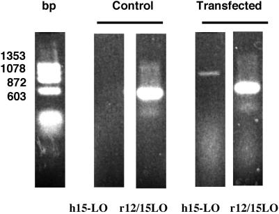
Rats in which h15-LO cDNA was selectively transfected into the left kidney 3 days before NTS injection were studied at 3 hr (n = 8) and 48 hr (n = 6) post-NTS injection. These animals were compared with animals unilaterally transfected with green fluorescent protein cDNA and injected with NTS in identical protocols (n = 3 each; 3 and 24 hr post-NTS). In each group, the untransfected (right) kidney served as an internal control. A representative reverse transcription–PCR experiment is shown in Fig. 1. The right kidney was not transfected and does not show h15-LO expression. The left, transfected kidney shows both h15-LO and the native rat 12/15-LO (see above).
Figure 1.
Representative reverse transcription–PCR experiment. The five lanes from left to right are: DNA size marker, RNA from nontransfected kidney amplified for h15-LO, nontransfected kidney amplified for rat 12/15-LO, transfected kidney amplified for h15-LO, and transfected kidney amplified for rat 12/15-LO.
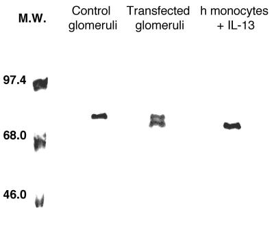
Fig. 2 illustrates Western blot analysis of h15-LO protein from the same experiment. Human peripheral blood monocytes stimulated with IL-13 were used as a positive control for h15-LO expression (36). The rat and hLOs are very close in size and thus the bands run close together. Also, rat glomeruli show a faint positive upper band of unknown nature, whereas the human monocytes do not. Thus, only transfected glomeruli express the h15-LO protein, whereas contralateral kidneys do not.
Figure 2.
Western blot analysis of h15-LO protein. The lanes show from left to right: molecular weight markers, nontransfected kidney, transfected glomeruli, and human peripheral blood monocytes stimulated with IL-13 as a positive control for h15-LO.
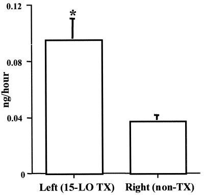
We also measured LXA4 concentration in urine from unilateraly tranfected animals. Urine from left (transfected) kidneys showed a 3-fold increase in LXA4 excretion compared with the right (control) kidney (Fig. 3).
Figure 3.
Urinary LXA4 excretion from unilaterally 15-LO transfected rats. Left kidney: transfected (TX). Right kidney: control. n = 10. *, P < 0.05 vs. control.
pNTS: Renal Clearance Studies.
Induction of pNTS leads to substantial acute reductions in the glomerular filtration rate (GFR) and renal plasma flow rate (RPF) (33, 39, 40). While these parameters were depressed to an equivalent degree in all animals at 3 hr post-NTS, h15-LO-transfected (left) kidneys regained a significantly greater amount of renal function 48 hr after NTS challenge than the nontransfected controls. Data are derived from measurements taken under Inactin anesthesia at 3 and 48 hr post-NTS nephritis (GFR, 0.47 ± 0.07 vs. 0.66 ± 0.07, 3 hr, 0.56 ± 0.14 vs. 0.62 ± 0.10, 48 hrs; RPF, 2.29 ± 0.32 vs. 3.24 ± 0.35, 3 hr, 3.02 ± 0.71 vs. 3.59 ± 0.56, 48 hr; ml/min, n = 14, h15-LO transfected, left kidney vs. nontransfected, right kidney, respectively).
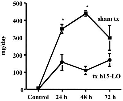
Total urinary protein excretion rates/day are shown in Fig. 4. 15-LO transfected kidneys had substantially less total urinary protein excretion than their contralateral controls.
Figure 4.
Total urinary protein excretion rate/day. ■, sham-transfected (sham tx) rats; ●, values from rats transfected with h15-LO (tx h15-LO) (n = 7); *, P < 0.05 vs. transfected group.
pNTS: Green Fluorescent Protein Transfection.
To assess the impact of the transfection procedure per se, we performed identical experiments in which green fluorescent protein was substituted for h15-LO (n = 6). No significant differences in renal function or histology were noted between transfected and nontransfected kidneys.
From these studies we determined that in vivo renal 15-LO transfection using the HVJ-liposome approach was feasible, therapeutically promising, and without deleterious effects on normal function. We therefore performed additional experiments using aNTS in which both kidneys were transfected and renal parameters were compared with mock-transfected controls. This macrophage-dominated model is a closer representation of the majority of human glomerulonephritides (33, 41, 42).
aNTS: Confirmation of Transfection.
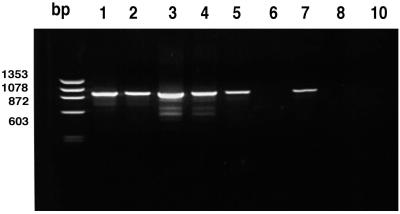
Rats were transfected with h15-LO cDNA into both kidneys 3 days before NTS injection. Fig. 5 shows h15-LO mRNA expression in glomeruli isolated from control rats (numbered 1–3) and nephritic rats (numbered 4–10). 15-LO mRNA is readily apparent in isolated glomeruli from transfected rats. The far left lane in Fig. 5 contains molecular weight markers and lanes 1–10 are isolated glomerular mRNA from individual rats. Note that rat #9 died and is not included in this study. Note also that three rats did not show expression at the end of the experiment (numbers 6, 8, and 10). These animals therefore were removed from the study; however, interestingly these three animals showed higher degrees of proteinuria and BUN than the successfully transfected rats (data not shown). Thus, our percent transfection success (about 60%) is very close to that obtained previously (24). Expression of h15-LO protein was confirmed by Western blot in animals that expressed mRNA for h15-LO (data not shown).
Figure 5.
Glomerular h15-LO mRNA expression from transfected rats with aNTS. The far left lane contains molecular weight markers, and lanes 1–10 are isolated glomerular mRNA from individual rats. Note that rat #9 is not included in this study. Glomeruli from rats #6, 8, and 10 do not show expression.
aNTS: Renal Functional Studies.
Animals were divided into four groups: group 1 underwent h15-LO transfection alone, group 2 underwent h15-LO transfection + accelerated NTS, group 3 underwent sham transfection only, and group 4 underwent sham transfection + aNTS.
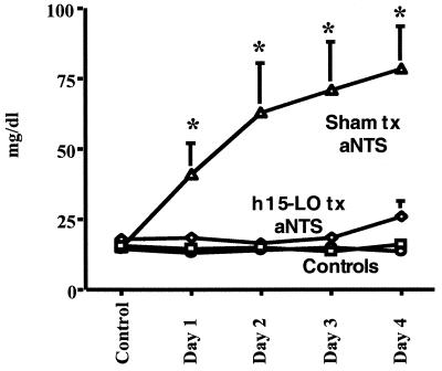
Fig. 6 shows the group mean BUN levels. As can be seen, NTS administration to sham-transfected animals (group 4) resulted in a prompt elevation of BUN, indicating a substantial fall in glomerular filtration rate. In group 2, 15-LO transfection completely abolished the NTS-induced rise in BUN until day 4. Of interest, day 4 post-NTS is day 7 posttransfection, a time at which a significant reduction in gene expression achieved by the HVJ liposome in renal glomeruli has been demonstrated. BUN levels in animals not exposed to NTS but transfected with sham liposomes (group 3) or human 15-LO (group 1) did not change throughout the length of the experiment. Thus, transfection alone did not alter renal function.
Figure 6.
Group mean BUN levels. The upper line (▵) represents sham-transfected (Sham tx) animals; h15-LO transfection (h15-LO tx) is shown as ◊. The lower lines (□ and ○) indicate BUN levels in animals not exposed to NTS, but transfected with sham liposomes (group 3) or h15-LO (group 1) (n = 7). *, P < 0.05 vs. h15-LO transfected + aNTS.
Creatinine clearances also were measured in these groups and were significantly different at days 1 and 2 of disease between sham and h15-LO-transfected animals with aNTS (1.6 ± 0.3 vs. 2.4 ± 0.2*, day 1; and 1.4 ± 0.3 vs. 2.2 ± 0.3*, day 2; ml/min, two-kidney clearance, 15-LO vs. sham tranfection in aNTS; *, P < 0.05).
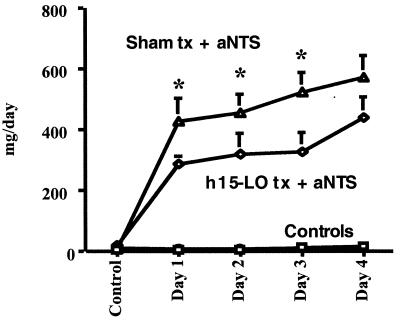
Fig. 7 illustrates, in a similar manner, the urinary protein excretion rates over 4 days. Under normal conditions, urinary protein levels are minimal, as demonstrated by the sham or 15-LO transfected healthy rats (controls). Sham-transfected rats exhibited prompt and sustained increases in urinary protein excretion upon administration of NTS. h15-LO transfection significantly reduced urinary protein excretion rates over 4 days by about 40% compared with sham-transfected controls.
Figure 7.
Urinary protein excretion over 4 days. The upper line (▵) represents sham-transfected (Sham tx) animals (group 4); h15-LO transfection (h15-LO tx) (group 2) is shown as ◊. The lower lines (□ and ○) indicate animals not exposed to NTS, but transfected with sham liposomes (group 3) or h15-LO (group 1) (n = 7). *, P < 0.05 vs. h15-LO transfected + aNTS.
Histology and Immunohistochemistry.
Kidneys from NTS rats showed the typical pathology of this disease, including marked focal segmental or diffuse proliferative glomerulonephritis. Immunostaining for ED-1 revealed only a few immunoreactive cells in scattered glomeruli from non-nephritic rats. However, glomeruli from nephritic rats contained an abundance of ED-1-positive cells. No differences in cell infiltrate or histopathology were detected among transfected and nontransfected nephritic rats. Sham-operated and sham-transfected rats did not show any indication of glomerular infiltrates or any other specific pathological changes.
LTB4 Generation in Isolated Glomeruli.
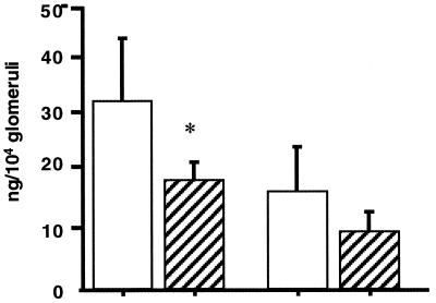
Previous studies have shown that glomerular LTB4 production peaks in the early phase of glomerulonephritis and has pro-inflammatory properties in this model (14, 39). Fig. 8 illustrates LTB4 production in glomeruli isolated from h15-LO-transfected rats during pNTS at 3 and 48 hr after initiation of disease (n = 4 each time point). In pNTS (3 hr), glomerular LTB4 production was elevated in nontransfected kidneys. Glomeruli from transfected kidneys produced significantly less LTB4 at 3 hr (33 ± 10 vs. 19 ± 3 ng/104 glomeruli, P < 0.05, control vs. h15-LO, respectively). As demonstrated previously (41), after 2 days of disease, glomerular LTB4 production falls. Although the temporal pattern of changes in LTB4 production at 48 hr was similar, differences between the transfected and nontransfected kidneys were maintained (left < right), although the mean values were not statistically different.
Figure 8.
Glomerular LTB4 production from h15-LO transfected rats during pNTS at 3 and 48 hr (n = 4 each time point) after initiation of disease. Solid bars represent right, nontransfected kidneys, and crosshatched bars represent h15-LO-transfected left kidneys. *, P < 0.05 vs. nontranfected right kidney.
Discussion
Evidence for a generalized anti-inflammatory role for 15-LO products has been derived from clinical observations and experimental studies in vivo and in vitro (6, 12–15, 42–48). The anti-inflammatory actions of 15-LO-derived eicosanoids during experimental glomerulonephritis demonstrated here are likely mediated through two major pathways: (a) specific antagonism of leukotriene synthesis, receptor interactions, and biologic effects (41–49), and (b) generalized inactivation of polymorphonuclear cell/macrophage functions through remodeling of leukocyte membrane phospholipids (12, 17, 50).
Previous studies on the regulation of rat 12/15-LO gene expression in NTS nephritis (51), as well as studies on cytokine regulation of 15-LO expression in human monocytes (14), provide compelling evidence supporting a specialized role for the induction of 15-LO activity as an “arrest” signal for macrophage-mediated tissue injury. Using quantitative PCR, we demonstrated a gradual increase in glomerular 12/15-LO mRNA over the first 48 hr of immune injury, reaching peak expression at 2–3 days (51). Over an identical time course, glomerular infiltration by macrophages and T lymphocytes is observed (52, 53), and gene expression of TH2-derived cytokines, IL-4 and IL-13, also increases, reaching a maximum at 24–48 hr (54). Because these cytokines are known to exert anti-inflammatory actions (55–57) and are unique inducers of 15-LO mRNA in human monocytes (14, 58), it is tempting to postulate that, at 24–72 hr postonset of acute inflammation, lymphocyte-derived anti-inflammatory effects are activated in the inflammatory micromilieu, resulting in macrophage inactivation and arrest of tissue injury.
In the present studies, we sought to provide direct evidence that overexpression of 15-LO is an effective anti-inflammatory strategy. We took advantage of the differences between human and rodent 15-LO to reliably detect human enzyme expression in rat kidney. The marked differences in LXA4 excretion between transfected versus nontransfected kidneys attests to the functional integrity of the overexpressed enzyme. The clear-cut differences between transfected and nontransfected kidneys of the same rat argue against circulating factors, or other downstream events resulting from presence of 15-LO, as underlying the protection of function and down-regulation of inflammation in transfected kidneys. Furthermore, mesangial cells are known to be the primary site of HVJ-liposome transfection, and thus overexpression of 15-LO in this area of the glomerulus will be expected to influence activation, but not migration, of macrophages. No significant differences were noted in the intensity or pattern of histopathologic expression between 15-LO-transfected and nontransfected nephritic kidneys. This finding is not surprising because 15-LO products have not been shown to influence macrophage migration, and the clear implications from our functional and biochemical analysis is that 15-LO products impacted on cellular activation rather than infiltration.
It has been proposed previously that underexpression (or total absence by gene knockout) of the 15-LO inducers IL-4 and IL-13 predisposes to severe and destructive inflammatory reactions in rats and mice (54, 59, 60). Conversely, administration of IL-4 is associated with amelioration of such injury (61). Susceptibility to auto-immune injury, therefore, might arise, in part, from a genetic or acquired deficiency of endogenous anti-inflammatory stimuli. The unique action of 15(S)-lipoxygenated eicosanoids in abrogating activation of polymorphonuclear cells and macrophages is a particularly effective tissue-preserving strategy because, regardless of inciting mechanisms, it arrests injury at the crucial final common mechanism mediating tissue damage, namely, effector cell activation (12–15). The present studies support this notion.
Acknowledgments
This work was supported by National Institutes of Health Grant DK 43883 and a fellowship grant from Ministerio Español de Educación y Cultura (J.M.V.).
Abbreviations
- LO
lipoxygenase
- 15-LO
15-LO gene
- h15-LO
human 15-LO
- 15(S)-HETE
15(S)-hydroxyeicosatetraenoic acid
- LX
lipoxin
- LTB
leukotriene B
- HVJ
hemagglutinating virus of Japan
- NTS
nephrotoxic serum
- pNTS
passive NTS nephritis
- aNTS
accelerated NTS nephritis
- BUN
blood urea nitrogen
Footnotes
This paper was submitted directly (Track II) to the PNAS office.
References
- 1.Sigal E, Grunberger D, Highland E, Goprsst C, Dixon R A F, Craik C S. J Clin Invest. 1990;92:1572–1579. [Google Scholar]
- 2.Ford-Hutchinson A W. Eicosanoids. 1991;4:65–74. [PubMed] [Google Scholar]
- 3.Samuelsson B, Dahlen S-E, Lindren J A, Rouzer C A, Serhan C N. Science. 1987;237:1171–1176. doi: 10.1126/science.2820055. [DOI] [PubMed] [Google Scholar]
- 4.Feinmark S J, Lingren J-Å, Claesson H-E, Malmsten C, Samuelsson B. FEBS Lett. 1981;136:141–144. doi: 10.1016/0014-5793(81)81233-1. [DOI] [PubMed] [Google Scholar]
- 5.Levy B D, Romano M, Chapman H A, Reillt J J, Drazen J, Serhan C N. J Clin Invest. 1993;92:1572–1579. doi: 10.1172/JCI116738. [DOI] [PMC free article] [PubMed] [Google Scholar]
- 6.Serhan C N. Biochem Biophys Acta. 1994;1212:1–25. doi: 10.1016/0005-2760(94)90185-6. [DOI] [PubMed] [Google Scholar]
- 7.Taylor S M, Liggit D H, Laegreid W W, Siflow R, Leid R W. Inflammation. 1986;10:157–165. doi: 10.1007/BF00915997. [DOI] [PubMed] [Google Scholar]
- 8.Kouzan S, Brady A R, Nettesheim P, Eling T. Am Rev Respir Dis. 1985;131:642–632. doi: 10.1164/arrd.1985.131.4.624. [DOI] [PubMed] [Google Scholar]
- 9.Garrick R, Shen S-Y, Ogunc S, Wong P Y-K. Biochem Biophys Res Commun. 1989;162:626–633. doi: 10.1016/0006-291x(89)92356-5. [DOI] [PubMed] [Google Scholar]
- 10.Romano M, Chen X-S, Takahashi Y, Yamamoto S, Funk C D, Serhan C N. Biochem J. 1993;296:127–133. doi: 10.1042/bj2960127. [DOI] [PMC free article] [PubMed] [Google Scholar]
- 11.Serhan C N, Sheppard K-A. J Clin Invest. 1990;85:772–780. doi: 10.1172/JCI114503. [DOI] [PMC free article] [PubMed] [Google Scholar]
- 12.Brezinski M E, Serhan C N. Proc Natl Acad Sci USA. 1990;87:6248–6252. doi: 10.1073/pnas.87.16.6248. [DOI] [PMC free article] [PubMed] [Google Scholar]
- 13.Badr K F, DeBoer D, Schwartzberg M, Serhan C N. Proc Natl Acad Sci USA. 1989;86:3438–3442. doi: 10.1073/pnas.86.9.3438. [DOI] [PMC free article] [PubMed] [Google Scholar]
- 14.Fisher D B, Christman J W, Badr K F. Kidney Int. 1992;41:1155–1160. doi: 10.1038/ki.1992.176. [DOI] [PubMed] [Google Scholar]
- 15.Brady H R, Lamas S, Papayianni A, Takata S, Matsubara M, Marsden P A. Am J Physiol. 1995;268:F1–F12. doi: 10.1152/ajprenal.1995.268.1.F1. [DOI] [PubMed] [Google Scholar]
- 16.Legrand A B, Lawson J A, Meyrick B O, Blair I A, Oates J A. J Biol Chem. 1991;266:7570–7577. [PubMed] [Google Scholar]
- 17.Takata S, Matsubara M, Allen P G, Janmey P A, Serhan C N, Brady H R. J Clin Invest. 1994;93:499–508. doi: 10.1172/JCI116999. [DOI] [PMC free article] [PubMed] [Google Scholar]
- 18.Takata S, Papayianni A, Matsubara M, Wladimiro J, Pronovost P H, Brady H R. Am J Pathol. 1994;145:541–549. [PMC free article] [PubMed] [Google Scholar]
- 19.Takano T, Fiore S, Maddox J F, Brady H R, Petasis N A, Serhan C N. J Exp Med. 1996;185:1693–1704. doi: 10.1084/jem.185.9.1693. [DOI] [PMC free article] [PubMed] [Google Scholar]
- 20.Takano T, Clish C B, Gronert K, Petasis N, Serhan C N. J Clin Invest. 1998;101:819–826. doi: 10.1172/JCI1578. [DOI] [PMC free article] [PubMed] [Google Scholar]
- 21.Shen J, Herderick E, Cornhill J F, Zsigmond E, Kim H S, Kuhn H, Guevara N V, Chan L. J Clin Invest. 1996;98:2201–2208. doi: 10.1172/JCI119029. [DOI] [PMC free article] [PubMed] [Google Scholar]
- 22.Gronert K, Gewirtz A, Madara J L, Serhan C N. J Exp Med. 1998;187:1285–1294. doi: 10.1084/jem.187.8.1285. [DOI] [PMC free article] [PubMed] [Google Scholar]
- 23.Fine L G. Kidney Int. 1996;49:612–619. doi: 10.1038/ki.1996.88. [DOI] [PubMed] [Google Scholar]
- 24.Isaka Y, Imai E. Semin Nephrol. 1996;16:591–598. [PubMed] [Google Scholar]
- 25.Kanada Y, Iwai K, Uchida T. Science. 1989;243:375–378. doi: 10.1126/science.2911748. [DOI] [PubMed] [Google Scholar]
- 26.Isaka Y, Fujiwara Y, Ueda N, Kaneda Y, Kamada T, Imai E. J Clin Invest. 1993;92:2597–2601. doi: 10.1172/JCI116874. [DOI] [PMC free article] [PubMed] [Google Scholar]
- 27.Akagi Y, Isaka Y, Arai M, Kaneko T, Tukenaka M, Moriyama T, Kaneda Y, Ando A, Orita Y, Kamada T, et al. Kidney Int. 1996;50:148–155. doi: 10.1038/ki.1996.297. [DOI] [PubMed] [Google Scholar]
- 28.Albrightson C R, Short B, Dytko G, Zabko-Potapovich B, Brickson B, Adams J L, Griswold D E. Kidney Int. 1994;45:1301–1310. doi: 10.1038/ki.1994.170. [DOI] [PubMed] [Google Scholar]
- 29.Munger K A, Fogo A, Nassar G, Badr K F. J Am Soc Nephrol. 1994;5:588. doi: 10.1681/ASN.V4111847. (abstr.). [DOI] [PubMed] [Google Scholar]
- 30.Munger K, Baylis C. Am J Physiol. 1988;254:F223–F231. doi: 10.1152/ajprenal.1988.254.2.F223. [DOI] [PubMed] [Google Scholar]
- 31.Badr K F, Schreiner G F, Wasserman M, Ichikawa I. J Clin Invest. 1988;81:1702–1709. doi: 10.1172/JCI113509. [DOI] [PMC free article] [PubMed] [Google Scholar]
- 32.Ichikawa I, Maddox D A, Cogan M G, Brenner B M. Renal Physiol. 1978;1:121–131. [Google Scholar]
- 33.Smith H W, Finkelstein N, Aliminosa L. J Clin Invest. 1945;24:388–391. doi: 10.1172/JCI101618. [DOI] [PMC free article] [PubMed] [Google Scholar]
- 34.Bradford M M. Anal Biochem. 1976;72:248–252. doi: 10.1016/0003-2697(76)90527-3. [DOI] [PubMed] [Google Scholar]
- 35.Lowrey O H, Rosebrough N J, Farr A L, Randall R J. J Biol Chem. 1951;193:265–275. [PubMed] [Google Scholar]
- 36.Nassar G M, Morrow J D, Roberts L J, II, Lakkis F G, Badr K F. J Biol Chem. 1994;269:27631–27634. [PubMed] [Google Scholar]
- 37.Chomczynski P, Sacchi N. Anal Biochem. 1987;162:156–159. doi: 10.1006/abio.1987.9999. [DOI] [PubMed] [Google Scholar]
- 38.Sigal E, Craik C S, Highland E, Grunberger D, Costello L C, Dixon R A, Nadel J A. Biochem Biophys Res Commun. 1988;157:457–464. doi: 10.1016/s0006-291x(88)80271-7. [DOI] [PubMed] [Google Scholar]
- 39.Lianos E A. J Clin Invest. 1988;82:427–435. doi: 10.1172/JCI113615. [DOI] [PMC free article] [PubMed] [Google Scholar]
- 40.Bailey J M, Fiskum G, Makheja A N, Simon T H. Atherosclerosis. 1995;113:247–258. doi: 10.1016/0021-9150(94)05452-o. [DOI] [PubMed] [Google Scholar]
- 41.Badr K F. Adv Nephrol Necker Hosp. 1995;24:19–31. [PubMed] [Google Scholar]
- 42.Badr K F. Kidney Int. 1992;42:S101–S108. [PubMed] [Google Scholar]
- 43.Brady H R, Persson U, Ballerman B J, Brenner B M, Serhan C N. Am J Physiol. 1990;259:F809–F815. doi: 10.1152/ajprenal.1990.259.5.F809. [DOI] [PubMed] [Google Scholar]
- 44.Lee T H, Horton C E, Kyan-Aung U, Haskard D, Crea A E G, Spur B W. Clin Sci. 1989;77:195–203. doi: 10.1042/cs0770195. [DOI] [PubMed] [Google Scholar]
- 45.Hedqvist P, Raud J, Palmertz U, Haeggstrom J, Nicolau K C, Dahlen S-E. Acta Physiol Scand. 1989;137:571–572. doi: 10.1111/j.1748-1716.1989.tb08805.x. [DOI] [PubMed] [Google Scholar]
- 46.Fogh K, Sogaard H, Herlin T, Kragballe K. J Am Acad Dermatol. 1988;18:279–285. doi: 10.1016/s0190-9622(88)70040-7. [DOI] [PubMed] [Google Scholar]
- 47.Fogh K, Hansen S E, Herlin V, Knudsen T, Henriksen T B, Ewald H, Bungerand C, Kraballe K. Prostaglandins. 1989;37:213–228. doi: 10.1016/0090-6980(89)90058-0. [DOI] [PubMed] [Google Scholar]
- 48.Badr K F, Serhan C N, Nicolau K C, Samuelsson B. Biochem Biophys Res Commun. 1987;145:408–414. doi: 10.1016/0006-291x(87)91337-4. [DOI] [PubMed] [Google Scholar]
- 49.Lefer A M, Stahl G L, Lefer D J, Brezinski M E, Nicolau K C, Veale C A, Abe Y, Bryan-Smith J. Proc Natl Acad Sci USA. 1988;85:8340–8344. doi: 10.1073/pnas.85.21.8340. [DOI] [PMC free article] [PubMed] [Google Scholar]
- 50.Badr K F. Curr Opin Nephrol Hypertens. 1997;6:111–118. [PubMed] [Google Scholar]
- 51.Katoh K, Lakkis F G, Makita N, Badr K F. Kidney Int. 1994;46:341–349. doi: 10.1038/ki.1994.280. [DOI] [PubMed] [Google Scholar]
- 52.Rahman M A, Nakazawa M, Emancipator S N, Dunn M J. J Clin Invest. 1988;81:1945–1952. doi: 10.1172/JCI113542. [DOI] [PMC free article] [PubMed] [Google Scholar]
- 53.Schreiner G F, Rovin B, Lefkowith J B. J Immunol. 1989;143:3192–3199. [PubMed] [Google Scholar]
- 54.Coelho S N. Kidney Int. 1997;51:646–652. doi: 10.1038/ki.1997.94. [DOI] [PubMed] [Google Scholar]
- 55.Sozzani P, Cambon C, Vita N, Séguélas M-H, Caput D, Ferrara P, Pipy B. J Biol Chem. 1995;270:5084–5088. doi: 10.1074/jbc.270.10.5084. [DOI] [PubMed] [Google Scholar]
- 56.Lehn M, Weiser W Y, Engelhorn S, Gillis S, Remold H G. J Immunol. 1989;143:3020–3024. [PubMed] [Google Scholar]
- 57.Abramson S L, Gallin J I. J Immunol. 1990;144:625–630. [PubMed] [Google Scholar]
- 58.Conrad D J, Kuhn H, Mulkins M, Highland E, Sigal E. Proc Natl Acad Sci USA. 1992;87:217–221. doi: 10.1073/pnas.89.1.217. [DOI] [PMC free article] [PubMed] [Google Scholar]
- 59.Kitching A R, Tipping P G, Mutch D A, Huang X R, Holdsworth S R. Kidney Int. 1998;53:112–118. doi: 10.1046/j.1523-1755.1998.00733.x. [DOI] [PubMed] [Google Scholar]
- 60.Saleem S, Dai Z, Coelho S N, Konieczny B, Assmann K J M, Baddoura F K, Lakkis F G. J Immunol. 1998;160:979–984. [PubMed] [Google Scholar]
- 61.Holdsworth S R, Kitching A R, Huang X R, Mutch D, Tipping P G. J Am Soc Nephrol. 1996;7:1704. (abstr.). [Google Scholar]


