Abstract
The lack of exposure to helminth infections, as a result of improved living standards and medical conditions, may have contributed to the increased incidence of IBD in the developed world. Epidemiological, experimental, and clinical data sustain the idea that helminths could provide protection against IBD. Studies investigating the underlying mechanisms by which helminths might induce such protection have revealed the importance of regulatory pathways, for example, regulatory T-cells. Further investigation on how helminths influence both innate and adaptive immune reactions will shed more light on the complex pathways used by helminths to regulate the hosts immune system. Although therapy with living helminths appears to be effective in several immunological diseases, the disadvantages of a treatment based on living parasites are explicit. Therefore, the identification and characterization of helminth-derived immunomodulatory molecules that contribute to the protective effect could lead to new therapeutic approaches in IBD and other immune diseases.
1. INFLAMMATORY BOWEL DISEASES AND THE HYGIENE HYPOTHESIS
Inflammatory bowel diseases (IBDs), such as Crohn's disease and ulcerative colitis, are chronic immune diseases of the gastrointestinal tract. Although the aetiologies of these diseases still remain unknown, the current hypothesis indicates that IBD results from an uncontrolled immune response to the normal gut flora [1, 2]. Genetic factors and environmental factors both contribute to the damaging mucosal immune response [3, 4].
The incidence of IBD has steadily increased in the developed world since 1950 [5, 6]. According to the hygiene hypothesis, this is directly related to the higher hygienic standards in these countries [7, 8]. It is suggested that the lack of exposure to infectious agents like helminths, as a result of improved living standards and medical conditions, modulates the development of the immune system and thereby increases the risk of immune diseases [9, 10].
The hygiene hypothesis was initially proposed by Strachan in 1989 for hay fever [11] and additional epidemiological studies were performed to further investigate the link between this hygiene concept and the incidence of other immunological diseases. As a consequence, the hygiene hypothesis is now proposed for several immunological disorders such as asthma and allergic diseases [12], cardiovascular diseases [13], Type 1 diabetes mellitus [14], multiple sclerosis [15], and IBD [16].
The hygiene hypothesis for IBD is clearly supported by the geographical distribution of the disease. There is a well described north-south gradient for the incidence of IBD. Northern Europe and North America have the highest IBD incidence rates whereas Crohn's disease and ulcerative colitis remain scarce in South America, Africa, and Asia [6, 17]. However, the gap between high- and low-incidence areas in northern versus southern regions is narrowing. In Asia, for example, incidence rates still remain low as compared to Europe, but they are rapidly increasing [18]. Changing lifestyle is thought to be the major cause of the disease increase in low-incidence areas [18]. The most important factor to explain these geographical differences is the socioeconomic level [16]. IBD is more frequently seen among patients with a higher socioeconomic status [19, 20]. Higher socioeconomic levels can be associated with better sanitation conditions, high-quality water, and better medical standards [2].
Another factor supporting the hygiene hypothesis is the inverse relationship between infant mortality rates and the incidence of IBD. Infant mortality might be linked to worse hygiene and medical conditions. Countries with high infant mortality rates consequently have lower reported incidence of IBD [21].
As mentioned previously, better hygienic circumstances translate into diminished exposure to infectious agents like helminths. The absence of such parasitic infections during childhood renders the immune system more prone to allergic and immune diseases. Thus infections seem to activate an important protective factor against these disorders [7]. Identifying the nature of this protective effect and implementing this notion in therapeutic strategies against IBD and other immune diseases is now the challenge for basic research.
2. IMMUNOLOGY OF THE GASTROINTESTINAL TRACT
2.1. Initiating innate and adaptive immune responses to enteric antigens in the gut
The gastrointestinal tract is continuously exposed to a wide range of dietary and environmental antigens, both harmless and pathogenic. Mounting protective immune responses against harmful pathogens whilst also preventing excessive responses to harmless antigens from food and bacterial flora is one of the major dichotomous functions of the mucosal immune system [22].
There are different levels of host defence that pathogens have to trespass to induce inflammation. Numerous mechanisms are acting to form a physical barrier to prevent micro-organisms from gaining access to the underlying tissues. Production of saliva and mucus, gastric and pancreatic juices, intestinal peristalsis, and epithelial cells all contribute to the elimination of pathogens from the gut lumen [23, 24]. Tight junctions between epithelial cells form a barrier to prevent bacterial pathogens from invading the gut tissue [25–27]. Once a pathogen breaks through this physical barrier, innate and adaptive immune responses work closely together to eliminate the intruder [28].
Antigens in the gut lumen can be taken up via different transport routes [29]. The innate immune system will respond to pathogen associated molecular patterns (PAMPs). As a part of the innate immune system, phagocytes like monocytes, macrophages and dendritic cells, and cytotoxic cells like natural killer cells rapidly control the invasion [30]. The adaptive immune system responds to antigens which have been presented by cells of the innate immune system [30]. Once antigens are taken up by antigen presenting cells, such as dendritic cells, fragments of the antigen are presented to T-cells locally or in mesenteric lymph nodes (MLNs) after migration of the antigen presenting cells [31, 32]. Adaptive immune responses are initiated by stimulation of lymphocytes. T-cells will help B lymphocytes to secrete immunoglobulins, the antigen-specific antibodies that are responsible for eliminating extracellular pathogens. On the other hand, T lymphocytes eradicate intracellular pathogens and mediate, for example, antihelminth and allergic responses [23]. Adaptive immune responses improve on repeated exposure to a given antigen by the formation of B and T memory cells [28].
2.2. T cell subsets and the immunological basis of helminth therapy in IBD
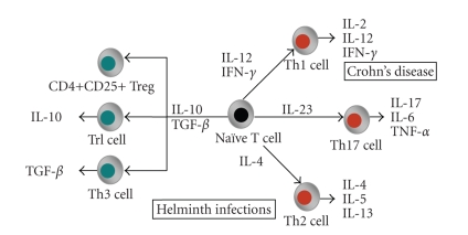
T lymphocytes are characterized by their cell-surface antigens called CD (cluster of differentiation) antigens. A common CD antigen found on all T-cells is the CD3 molecule which forms an essential part of the T-cell receptor and is important in the recognition of antigens presented by antigen presenting cells [33]. Within this pool of T lymphocytes, a difference is made between cytotoxic T-cells (CD8+) and helper T-cells (CD4+). CD4+ T-cells can orchestrate the functional activity of both innate and adaptive immune systems by “helping” macrophages, NK cells, CD8+ T cells, and B cells. These T helper (Th) cells can be divided into several subsets of CD4+ cells and each subset is suited for coordinating the effector activities that best combat the invading pathogen [34]. CD4+ T lymphocytes can be classified into distinct populations based on the cytokines they produce (Figure 1) [35–37].
Figure 1.
T-cell subsets. Naïve CD4+ T cells are stimulated by antigen presenting cells and the cytokine environment to proliferate into a certain subset. There are three distinct effector T-cell subsets (red): Th1, Th2, and Th17. CD4+ regulatory T-cells (green) can be subdivided in CD4+CD25+ Treg, Tr1, and Th3 cells. Crohn's disease is characterized by Th1, Th17 inflammation, whereas helminths induce Th2 and regulatory T-cells (modified from [36, 37]).
Effector CD4+ T cells are divided into three distinct lineages. T helper 1 (Th1) cells are engaged in the eradication of intracellular pathogens (e.g., intracellular bacteria and viruses) and are characterized by the production of IL-2, IL-12, and IFN-γ [38]. Gastrointestinal inflammation during Crohn's disease is Th1 mediated [23]. T helper 2 (Th2) cells stimulate B-cell antibody production, eosinophil recruitment and mucosal expulsion mechanisms and are characterized by the secretion of IL-4, IL-5, and IL-13 [38]. Th2 cells enhance elimination of parasitic helminth infections and support allergic responses. During helminth infection, the host evokes a strong Th2 immune response to provide protection against worm colonization [39]. The cytokines produced by Th1 and Th2 cells crossregulate each other's development and activity, for example, IFN-γ produced by Th1 cells amplifies Th1 development and inhibits proliferation of Th2 cells [35]. In this way, helminths can evoke an immune response that might be able to attenuate the Th1 response found during Crohn's disease.
A third lineage of effector CD4+ cells has been recently discovered and is characterized by the production of IL-17, the Th17 cell. IL-17 induces expression of many innate inflammatory mediators such as IL-6, acute phase proteins, granulocyte-colony stimulating factor, and prostaglandin E2. Th1 and Th2 cytokines can inhibit Th17 development, while Th1 and Th2 effector cells seem resistant to IL17 expression [40]. It is now clear that the Th17 pathway is critical for the development of inflammation. IL17 is elevated in a variety of inflammatory conditions as shown for rheumatoid arthritis, asthma, and recently IBD [41]. Furthermore, it has been shown that IL-23 supports the proliferation of Th17 cells. IL-23 is mainly produced by activated myeloid cells such as macrophages and dendritic cells. The discovery of this new IL-23/IL-17 pathway was a major breakthrough in the immunopathogenesis of IBD and the exact role of this axis needs to be further defined [41, 42]. Investigation of the effect of helminth infections on the IL-23/IL-17 pathway may uncover additional immunological pathways by which helminths can provide protection against immune disorders.
Aside from these effector T-cells, another population of CD3+ cells called regulatory T (Treg) cells have been described. Treg cells have immunosuppressive function and cytokine profiles distinct from either Th1, Th2, or Th17 T-cells [43]. By suppressing excessive Th1, Th2, or Th17 immune responses, Treg cells play an important role in the maintenance of self-tolerance, thus preventing autoimmune diseases, as well as inhibiting harmful inflammatory diseases such as asthma and inflammatory bowel disease [44]. There is emerging evidence that distinct subgroups of CD4+, CD8+, and natural killer T cells mediate immune regulatory mechanisms [45]. The most attention is being paid to the CD4+ Treg cells which can be subdivided into different subsets. These include the natural CD4+CD25+ Treg cells, which inhibit immune responses through cell-cell contact and through the production of immunosuppressive cytokines, type 1 Tr (Tr1) cells which secrete high levels of IL-10 and type 3 T (Th3) cells which primarily secrete TGF-β [43]. Treg lymphocytes suppress the differentiation of both Th1 and Th2 lymphocytes and are considered real gatekeepers of the mucosal immune response [2].
The balance between Th1, Th2, and Treg cells is of special interest in the gastrointestinal immune system. The gut provides a unique microenvironment prone to Treg cell differentiation. This microenvironment is characterized by the constant exposure to commensal flora and food antigens and by the presence of immunomodulatory factors and cytokines that participate in the differentiation of the mucosal immune system [22]. Defects of the regulatory mechanisms may lead to development of specific Th1- or Th2- mediated diseases.
Given that helminths induce a distinct immunological mechanism compared to IBD, worms can be used as immunomodulators to downregulate the immune response in IBD. Helminths induce Th2 and Treg cells which are capable of suppressing Th1 effector cells, the cells responsible for maintenance of inflammation in IBD patients.
3. HELMINTHS AS THERAPEUTIC AGENTS IN IBD
3.1. Experimental and clinical studies supporting helminth-based therapy
Helminths colonize more than one third of the world population [46]. In developed countries, these parasites have been largely eradicated as a public health concern due to the availability of efficacious drugs and better sanitation conditions [47]. In developing countries, however, helminth colonization is still common [48]. As shown by epidemiological studies, there is an inverse relation between the frequency of worm colonization and the prevalence of IBD [46]. It was Elliott et al. who first proposed the hypothesis that the loss of exposure to parasitic worms increased the risk of IBD [49, 50].
Preliminary data of Elliott et al. illustrating a protective response of Schistosoma mansoni infection on trinitrobenzene sulphate (TNBS)-induced colitis in mice [49] have led to several experimental animal studies investigating the effect of helminth infections on IBD [2]. The first full study on helminth modulation of experimentally induced colitis was published by Reardon et al. in 2001. They showed that infection of mice with the tapeworm Hymenolepis diminuta ameliorated dextran sodium sulphate (DSS)-induced colitis [51]. Khan et al. subsequently showed that infection with the nematode, Trichinella spiralis, protected mice from colitis induced by intrarectal challenge with dinitrobenzene sulphate (DNBS) [52]. Elliott et al. demonstrated that schistosome eggs had a protective effect on TNBS-induced colitis in mice [53] and that Heligmosomoides polygyrus could reduce established colitis in IL-10 deficient mice [54]. We previously demonstrated a protective effect of infection with the blood fluke, Schistosoma mansoni, on trinitrobenzene sulfonic acid (TNBS)-induced colitis in rats [55]. Taken together, different helminth parasites (nematode, cestode, and trematode) can ameliorate colitis in different experimental animal models [56]. Furthermore, helminths also protect against other immunological diseases as shown in rodent models for asthma [57], type 1 diabetes mellitus [14], and experimental autoimmune encephalomyelitis [17, 58].
Based on the promising findings of helminth infections on experimental colitis, clinical studies were initiated. Treatment of patients with the porcine whipworm, Trichuris suis, resulted in clinical amelioration of both Crohn's disease and ulcerative colitis [59, 60]. In the same line, a proof of concept study showed clinical efficacy of experimental infection with the human hookworm Necator americanus on Crohn's disease [61]. Clinical trials of Necator americanus in asthma are being organized [62]. An international multicentre clinical trial is in preparation (awaiting FDA approval) to further investigate the clinical efficacy of helminth-based therapy in IBD [2].
3.2. The use of helminth-derived molecules as therapeutic agents
Although helminth infections appear to be effective against IBD, treatment of patients with living helminths may envision drawbacks. Persistent infection and/or invasion of the parasite (particularly zoonotic ones) to other tissues in the human host, where they might cause pathology, should be considered [63, 64]. In 2006, Kradin et al. reported that treatment of a pediatric Crohn's disease patient with five oral doses of Trichuris suis ova caused infection with living sexually immature worms in the ileocecal region and a sexually mature male worm within the cecum [64]. Although helminths may be beneficial in the treatment of IBD, using living helminth ova can lead to infection, therefore, therapeutic human helminth colonization needs to be closely examined for potential adverse side effects. Furthermore, intestinal helminths influence gastrointestinal physiology. Infection with certain nematodes may induce enhanced intestinal propulsive activity, goblet cell hyperplasia, and increased mucus secretion [65]. As a consequence, intestinal helminths may alter gastrointestinal motility, possibly resulting in intestinal symptoms like diarrhoea and abdominal cramps [65]. Moreover, the idea of being infected with a living parasite could be psychologically hard to accept for some patients. Therefore, treatment with immunologically active helminth molecules might overcome the possible disadvantages of a therapy with living parasites.
Identification and characterization of helminth-derived immunomodulatory molecules that contribute to the anticolitis effect could lead to new therapeutic approaches in IBD without the need for helminth infection [56, 66]. Using parasite extracts or synthetic drugs designed to mimic the disease-modulating effect of helminth molecules also allows greater flexibility in dosing routes and therapeutic applications [67].
Helminths possess evolved mechanisms to turn off proinflammatory cascades by secreting and expressing certain molecules [37]. Multiple studies have characterized a broad spectrum of helminth-derived immunomodulatory products. A detailed review of these products is beyond the scope of this paper so we will bring only some molecules of interest into focus. Maizels et al. showed that the filarial nematode Brugia malayi produces homologues of the mammalian cytokine TGF-β. Bm-tgh-2 is secreted by adult worms and binds to mammalian TGF-β receptors thus performing an immunomodulatory function in the host [39]. Helminths secrete cysteine protease inhibitors which interfere with antigen presentation and increase IL-10 secretion from macrophages [68]. Helminth-derived carbohydrates contribute to the induction of Th2 immune responses [69]. Lacto-N-fucopentaose III is the predominant carbohydrate component of Schistosma mansoni egg antigens and this glycan stimulates the secretion of Th2 cytokines [70]. Harnett et al. recently showed that the phosphorylcholine part of the glycoconjugate ES-62, secreted by filarial nematodes, is responsible for its anti-inflammatory action in arthritis [71]. Research focusing on the development of vaccines against helminth infections also showed the effectiveness of helminth antigens as immunomodulators. Vaccination studies against Schistosomiasis are focusing on the protective effect of several Schistosoma antigens [72]. Vaccination studies against human hookworm infections tested recombinant excretory/secretory (ES) products from L3 larval stages of Ancylostoma caninum and promising results were observed [73–75]. Furthermore, vaccination studies against hookworm infection revealed that administration of a cocktail of recombinant antigens has an improved protective effect compared to the protection achieved with separate antigens [76].
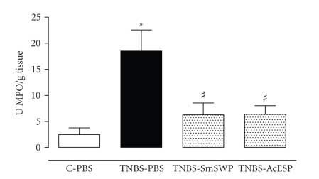
In respect to IBD, there is need for in-depth experimental studies on the effect of helminth antigens on colitis. We are currently investigating the therapeutic potential of protein mixtures of Schistosoma mansoni and Ancylostoma caninum on TNBS-induced colitis in mice. As shown in Figure 2, preliminary experiments showed that both S. mansoni soluble worm proteins and A. caninum ES products attenuated TNBS-induced inflammation of the murine colon [77]. These results indicate that the beneficial effect of treatment with living worms on experimental colitis may be reproduced with soluble extracts of helminths. Yang et al. showed that Schistosoma japonicum egg antigens inhibited the development of asthma in a murine model [57]. S. mansoni antigens are also able to modulate innate immune responses and prevent onset of type 1 diabetes [78]. These studies indicate that treatment with helminth extracts may be as effective as treatment with living helminths and that the achieved protection is not specific for just one helminth species. Isolated helminth proteins may provide a more readily acceptable form of therapy for patients than living worms.
Figure 2.
Effect of Schistosoma mansoni soluble worm proteins (SmSWPs) and Ancylostoma caninum excretory/secretory products (AcESPs) on myeloperoxidase (MPO) activity. MPO activity was measured to monitor the degree of myeloid cell infiltration in the colon. Data are presented as units MPO per gram of colon tissue and 1 unit equals the amount of MPO necessary to degrade 1 μmol of H2O2 to H2O per minute at 25°C. TNBS-induced colitis caused a significant increase in MPO activity compared to control mice treated with phosphate-buffered saline (PBS). Intraperitoneal injection of helminth-derived products significantly ameliorated inflammation as shown by the significant decrease in MPO activity. Treatment of control mice with SmSWP or AcESP had no effect (data not shown). ∗ p ≤ .05, significantly different from contol PBS; # p ≤ .05, significantly different from TNBS-PBS; two way ANOVA, n = 7 − 10 [77].
4. CONCLUDING REMARKS
The hygiene hypothesis suggests an inverse relationship between parasitic infections and the incidence of IBD. Epidemiological, experimental, and clinical data sustain the idea that helminths could provide protection against IBD. The importance of regulatory pathways such as regulatory T-cells, by which helminths induce such protection have been described. However, the complex pathways helminths activate to regulate the host's immune system need further investigation. Helminths influence innate as well as adaptive immune responses and this knowledge can contribute to new therapeutic approaches of helminth-induced protection. Therapy with living helminths appears to be effective in several immunological diseases. A logical next step, to avoid the possible disadvantages of a treatment with living parasites, is the identification and characterization of helminth-derived immunosuppressive molecules that contribute to the protective effect.
References
- 1.Podolsky DK. Inflammatory bowel disease. The New England Journal of Medicine. 2002;347(6):417–429. doi: 10.1056/NEJMra020831. [DOI] [PubMed] [Google Scholar]
- 2.Moreels TG, Pelckmans PA. The hygiene hypothesis and inflammatory bowel diseases: role of helminths. Acta Gastro-Enterologica Belgica. 2006;69(4):413–417. [PubMed] [Google Scholar]
- 3.Fiocchi C. Inflammatory bowel disease: etiology and pathogenesis. Gastroenterology. 1998;115(1):182–205. doi: 10.1016/s0016-5085(98)70381-6. [DOI] [PubMed] [Google Scholar]
- 4.Fiocchi C. The multiple components of inflammatory bowel disease pathogenesis: should we invest in all of them or should we pick and choose? Current Opinion in Gastroenterology. 2005;21(4):399–400. [Google Scholar]
- 5.Russel MGVM. Changes in the incidence of inflammatory bowel disease: what does it mean? European Journal of Internal Medicine. 2000;11(4):191–196. doi: 10.1016/s0953-6205(00)00090-x. [DOI] [PubMed] [Google Scholar]
- 6.Lakatos PL. Recent trends in the epidemiology of inflammatory bowel diseases: up or down? World Journal of Gastroenterology. 2006;12(38):6102–6108. doi: 10.3748/wjg.v12.i38.6102. [DOI] [PMC free article] [PubMed] [Google Scholar]
- 7.Strachan DP. Family site, infection and atopy: the first decade of the ‘hygiene hypothesis’. Thorax. 2000;55(supplement 1):S2–S10. doi: 10.1136/thorax.55.suppl_1.s2. [DOI] [PMC free article] [PubMed] [Google Scholar]
- 8.Bresciani M, Parisi C, Manghi G, Bonini S. The hygiene hypothesis: does it function worldwide? Current Opinion in Allergy and Clinical Immunology. 2005;5(2):147–151. doi: 10.1097/01.all.0000162307.89857.88. [DOI] [PubMed] [Google Scholar]
- 9.Vercelli D. Mechanisms of the hygiene hypothesis—molecular and otherwise. Current Opinion in Immunology. 2006;18(6):733–737. doi: 10.1016/j.coi.2006.09.002. [DOI] [PubMed] [Google Scholar]
- 10.Garn H, Renz H. Epidemiological and immunological evidence for the hygiene hypothesis. Immunobiology. 2007;212(6):441–452. doi: 10.1016/j.imbio.2007.03.006. [DOI] [PubMed] [Google Scholar]
- 11.Strachan DP. Hay fever, hygiene, and household size. British Medical Journal. 1989;299(6710):1259–1260. doi: 10.1136/bmj.299.6710.1259. [DOI] [PMC free article] [PubMed] [Google Scholar]
- 12.Bach J-F. The effect of infections on susceptibility to autoimmune and allergic diseases. The New England Journal of Medicine. 2002;347(12):911–920. doi: 10.1056/NEJMra020100. [DOI] [PubMed] [Google Scholar]
- 13.Magen E, Borkow G, Bentwich Z, Mishal J, Scharf S. Can worms defend our hearts? Chronic helminthic infections may attenuate the development of cardiovascular diseases. Medical Hypotheses. 2005;64(5):904–909. doi: 10.1016/j.mehy.2004.09.028. [DOI] [PubMed] [Google Scholar]
- 14.Cooke A, Zaccone P, Raine T, Phillips JM, Dunne DW. Infection and autoimmunity: are we winning the war, only to lose the peace? Trends in Parasitology. 2004;20(7):316–321. doi: 10.1016/j.pt.2004.04.010. [DOI] [PubMed] [Google Scholar]
- 15.Fleming JO, Cook TD. Multiple sclerosis and the hygiene hypothesis. Neurology. 2006;67(11):2085–2086. doi: 10.1212/01.wnl.0000247663.40297.2d. [DOI] [PubMed] [Google Scholar]
- 16.Feillet H, Bach J-F. Increased incidence of inflammatory bowel disease: the price of the decline of infectious burden? Current Opinion in Gastroenterology. 2004;20(6):560–564. doi: 10.1097/00001574-200411000-00010. [DOI] [PubMed] [Google Scholar]
- 17.Weinstock JV. Helminths and mucosal immune modulation. Annals of the New York Academy of Sciences. 2006;1072:356–364. doi: 10.1196/annals.1326.033. [DOI] [PubMed] [Google Scholar]
- 18.Loftus EV, Jr., Sandborn WJ. Epidemiology of inflammatory bowel disease. Gastroenterology Clinics of North America. 2002;31(1):1–20. doi: 10.1016/s0889-8553(01)00002-4. [DOI] [PubMed] [Google Scholar]
- 19.Alic M. Inflammatory bowel diseases are diseases of higher socioeconomic status: dogma or reality? The American Journal of Gastroenterology. 2000;95(11):3332–3333. doi: 10.1111/j.1572-0241.2000.03330.x. [DOI] [PubMed] [Google Scholar]
- 20.Green C, Elliott L, Beaudoin C, Bernstein CN. A population-based ecologic study of inflammatory bowel disease: searching for etiologic clues. American Journal of Epidemiology. 2006;164(7):615–623. doi: 10.1093/aje/kwj260. [DOI] [PubMed] [Google Scholar]
- 21.Montgomery SM, Pounder RE, Wakefield AJ. Infant mortality and the incidence of inflammatory bowel disease. The Lancet. 1997;349(9050):472–473. doi: 10.1016/S0140-6736(05)61183-X. [DOI] [PubMed] [Google Scholar]
- 22.Dubois B, Goubier A, Joubert G, Kaiserlian D. Oral tolerance and regulation of mucosal immunity. Cellular and Molecular Life Sciences. 2005;62(12):1322–1332. doi: 10.1007/s00018-005-5036-0. [DOI] [PubMed] [Google Scholar]
- 23.Moreels TG, Pelckmans PA. Gastrointestinal parasites: potential therapy for refractory inflammatory bowel diseases. Inflammatory Bowel Diseases. 2005;11(2):178–184. doi: 10.1097/00054725-200502000-00012. [DOI] [PubMed] [Google Scholar]
- 24.Magalhaes JG, Tattoli I, Girardin SE. The intestinal epithelial barrier: how to distinguish between the microbial flora and pathogens. Seminars in Immunology. 2007;19(2):106–115. doi: 10.1016/j.smim.2006.12.006. [DOI] [PubMed] [Google Scholar]
- 25.Berkes J, Viswanathan VK, Savkovic SD, Hecht G. Intestinal epithelial responses to enteric pathogens: effects on the tight junction barrier, ion transport, and inflammation. Gut. 2003;52(3):439–451. doi: 10.1136/gut.52.3.439. [DOI] [PMC free article] [PubMed] [Google Scholar]
- 26.Liu Z, Li N, Neu J. Tight junctions, leaky intestines, and pediatric diseases. Acta Paediatrica. 2005;94(4):386–393. doi: 10.1111/j.1651-2227.2005.tb01904.x. [DOI] [PubMed] [Google Scholar]
- 27.Shen L, Turner JR. Role of epithelial cells in initiation and propagation of intestinal inflammation. Eliminating the static: tight junction dynamics exposed. American Journal of Physiology. 2006;290(4):G577–G582. doi: 10.1152/ajpgi.00439.2005. [DOI] [PubMed] [Google Scholar]
- 28.Delves PJ, Roitt IM. The immune system—first of two parts. The New England Journal of Medicine. 2000;343(1):37–49. doi: 10.1056/NEJM200007063430107. [DOI] [PubMed] [Google Scholar]
- 29.Rumbo M, Anderle P, Didierlaurent A, et al. How the gut links innate and adaptive immunity. Annals of the New York Academy of Sciences. 2004;1029:16–21. doi: 10.1196/annals.1309.003. [DOI] [PubMed] [Google Scholar]
- 30.Medzhitov R, Janeway C., Jr. Innate immunity. The New England Journal of Medicine. 2000;343(5):338–344. doi: 10.1056/NEJM200008033430506. [DOI] [PubMed] [Google Scholar]
- 31.Garside P, Millington O, Smith KM. The anatomy of mucosal immune responses. Annals of the New York Academy of Sciences. 2004;1029:9–15. doi: 10.1196/annals.1309.002. [DOI] [PubMed] [Google Scholar]
- 32.Powrie F. Immune regulation in the intestine: a balancing act between effector and regulatory T cell responses. Annals of the New York Academy of Sciences. 2004;1029:132–141. doi: 10.1196/annals.1309.030. [DOI] [PubMed] [Google Scholar]
- 33.Delves PJ, Roitt IM. The immune system—second of two parts. The New England Journal of Medicine. 2000;343(2):108–117. doi: 10.1056/NEJM200007133430207. [DOI] [PubMed] [Google Scholar]
- 34.Weaver CT, Murphy KM. T-cell subsets: the more the merrier. Current Biology. 2007;17(2):R61–R63. doi: 10.1016/j.cub.2006.12.015. [DOI] [PubMed] [Google Scholar]
- 35.Abbas AK, Murphy KM, Sher A. Functional diversity of helper T lymphocytes. Nature. 1996;383(6603):787–793. doi: 10.1038/383787a0. [DOI] [PubMed] [Google Scholar]
- 36.Ince MN, Elliott DE. Immunologic and molecular mechanisms in inflammatory bowel disease. Surgical Clinics of North America. 2007;87(3):681–696. doi: 10.1016/j.suc.2007.03.005. [DOI] [PubMed] [Google Scholar]
- 37.Zaccone P, Burton OT, Cooke A. Interplay of parasite-driven immune responses and autoimmunity. Trends in Parasitology. 2008;24(1):35–42. doi: 10.1016/j.pt.2007.10.006. [DOI] [PubMed] [Google Scholar]
- 38.Harrington LE, Mangan PR, Weaver CT. Expanding the effector CD4 T-cell repertoire: the Th17 lineage. Current Opinion in Immunology. 2006;18(3):349–356. doi: 10.1016/j.coi.2006.03.017. [DOI] [PubMed] [Google Scholar]
- 39.Maizels RM, Balic A, Gomez-Escobar N, Nair M, Taylor MD, Allen JE. Helminth parasites—masters of regulation. Immunological Reviews. 2004;201(1):89–116. doi: 10.1111/j.0105-2896.2004.00191.x. [DOI] [PubMed] [Google Scholar]
- 40.Bi Y, Liu G, Yang R. Th17 cell induction and immune regulatory effects. Journal of Cellular Physiology. 2007;211(2):273–278. doi: 10.1002/jcp.20973. [DOI] [PubMed] [Google Scholar]
- 41.Zhang Z, Hinrichs DJ, Lu H, Chen H, Zhong W, Kolls JK. After interleukin-12p40, are interleukin-23 and interleukin-17 the next therapeutic targets for inflammatory bowel disease? International Immunopharmacology. 2007;7(4):409–416. doi: 10.1016/j.intimp.2006.09.024. [DOI] [PubMed] [Google Scholar]
- 42.Bamias G, Cominelli F. Immunopathogenesis of inflammatory bowel disease: current concepts. Current Opinion in Gastroenterology. 2007;23(4):365–369. doi: 10.1097/MOG.0b013e3281c55eb2. [DOI] [PubMed] [Google Scholar]
- 43.McGuirk P, Mills KHG. Pathogen-specific regulatory T cells provoke a shift in the Th1/Th2 paradigm in immunity to infectious diseases. Trends in Immunology. 2002;23(9):450–455. doi: 10.1016/s1471-4906(02)02288-3. [DOI] [PubMed] [Google Scholar]
- 44.Thompson C, Powrie F. Regulatory T cells. Current Opinion in Pharmacology. 2004;4(4):408–414. doi: 10.1016/j.coph.2004.05.001. [DOI] [PubMed] [Google Scholar]
- 45.Jiang H, Chess L. Regulation of immune responses by T cells. The New England Journal of Medicine. 2006;354(11):1166–1176. doi: 10.1056/NEJMra055446. [DOI] [PubMed] [Google Scholar]
- 46.Weinstock JV, Summers R, Elliott DE. Helminths and harmony. Gut. 2004;53(1):7–9. doi: 10.1136/gut.53.1.7. [DOI] [PMC free article] [PubMed] [Google Scholar]
- 47.MacDonald AS, Araujo MI, Pearce EJ. Immunology of parasitic helminth infections. Infection and Immunity. 2002;70(2):427–433. doi: 10.1128/iai.70.2.427-433.2002. [DOI] [PMC free article] [PubMed] [Google Scholar]
- 48.Weinstock JV, Summers RW, Elliott DE. Role of helminths in regulating mucosal inflammation. Seminars in Immunopathology. 2005;27(2):249–271. doi: 10.1007/s00281-005-0209-3. [DOI] [PubMed] [Google Scholar]
- 49.Elliott DE, Urban JF, Jr., Argo CK, Weinstock JV. Does the failure to acquire helminthic parasites predispose to Crohn's disease? The FASEB Journal. 2000;14(12):1848–1855. doi: 10.1096/fj.99-0885hyp. [DOI] [PubMed] [Google Scholar]
- 50.Weinstock JV, Summers RW, Elliott DE, Qadir K, Urban JF, Jr., Thompson R. The possible link between de-worming and the emergence of immunological disease. Journal of Laboratory and Clinical Medicine. 2002;139(6):334–338. doi: 10.1067/mlc.2002.124343. [DOI] [PubMed] [Google Scholar]
- 51.Reardon C, Sanchez A, Hogaboam CM, McKay DM. Tapeworm infection reduces epithelial ion transport abnormalities in murine dextran sulfate sodium-induced colitis. Infection and Immunity. 2001;69(7):4417–4423. doi: 10.1128/IAI.69.7.4417-4423.2001. [DOI] [PMC free article] [PubMed] [Google Scholar]
- 52.Khan WI, Blennerhasset PA, Varghese AK, et al. Intestinal nematode infection ameliorates experimental colitis in mice. Infection and Immunity. 2002;70(11):5931–5937. doi: 10.1128/IAI.70.11.5931-5937.2002. [DOI] [PMC free article] [PubMed] [Google Scholar]
- 53.Elliott DE, Li J, Blum A, et al. Exposure to schistosome eggs protects mice from TNBS-induced colitis. American Journal of Physiology. 2003;284(3):G385–G391. doi: 10.1152/ajpgi.00049.2002. [DOI] [PubMed] [Google Scholar]
- 54.Elliott DE, Setiawan T, Metwali A, Blum A, Urban JF, Jr., Weinstock JV. Heligmosomoides polygyrus inhibits established colitis in IL-10-deficient mice. European Journal of Immunology. 2004;34(10):2690–2698. doi: 10.1002/eji.200324833. [DOI] [PubMed] [Google Scholar]
- 55.Moreels TG, Nieuwendijk RJ, De Man JG, et al. Concurrent infection with Schistosoma mansoni attenuates inflammation induced changes in colonic morphology, cytokine levels, and smooth muscle contractility of trinitrobenzene sulphonic acid induced colitis in rats. Gut. 2004;53(1):99–107. doi: 10.1136/gut.53.1.99. [DOI] [PMC free article] [PubMed] [Google Scholar]
- 56.Hunter MM, McKay DM. Review article: helminths as therapeutic agents for inflammatory bowel disease. Alimentary Pharmacology & Therapeutics. 2004;19(2):167–177. doi: 10.1111/j.0269-2813.2004.01803.x. [DOI] [PubMed] [Google Scholar]
- 57.Yang J, Zhao J, Yang Y, et al. Schistosoma japonicum egg antigens stimulate CD4+CD25+ T cells and modulate airway inflammation in a murine model of asthma. Immunology. 2007;120(1):8–18. doi: 10.1111/j.1365-2567.2006.02472.x. [DOI] [PMC free article] [PubMed] [Google Scholar]
- 58.La Flamme AC, Ruddenklau K, Bäckström BT. Schistosomiasis decreases central nervous system inflammation and alters the progression of experimental autoimmune encephalomyelitis. Infection and Immunity. 2003;71(9):4996–5004. doi: 10.1128/IAI.71.9.4996-5004.2003. [DOI] [PMC free article] [PubMed] [Google Scholar]
- 59.Summers RW, Elliot DE, Urban JF, Jr., Thompson R, Weinstock JV. Trichuris suis therapy in Crohn's disease. Gut. 2005;54(1):87–90. doi: 10.1136/gut.2004.041749. [DOI] [PMC free article] [PubMed] [Google Scholar]
- 60.Summers RW, Elliott DE, Urban JF, Jr., Thompson RA, Weinstock JV. Trichuris suis therapy for active ulcerative colitis: a randomized controlled trial. Gastroenterology. 2005;128(4):825–832. doi: 10.1053/j.gastro.2005.01.005. [DOI] [PubMed] [Google Scholar]
- 61.Croese J, O'Neil J, Masson J, et al. A proof of concept study establishing Necator americanus in Crohn's patients and reservoir donors. Gut. 2006;55(1):136–137. doi: 10.1136/gut.2005.079129. [DOI] [PMC free article] [PubMed] [Google Scholar]
- 62.Falcone FH, Pritchard DI. Parasite role reversal: worms on trial. Trends in Parasitology. 2005;21(4):157–160. doi: 10.1016/j.pt.2005.02.002. [DOI] [PubMed] [Google Scholar]
- 63.Van Kruiningen HJ, West AB. Potential danger in the medical use of Trichuris suis for the treatment of inflammatory bowel disease. Inflammatory Bowel Diseases. 2005;11(5):515 pages. doi: 10.1097/01.mib.0000160369.47671.a2. [DOI] [PubMed] [Google Scholar]
- 64.Kradin RL, Badizadegan K, Auluck P, Korzenik J, Lauwers GY. Iatrogenic Trichuris suis infection in a patient with Crohn disease. Archives of Pathology and Laboratory Medicine. 2006;130(5):718–720. doi: 10.5858/2006-130-718-ITSIIA. [DOI] [PubMed] [Google Scholar]
- 65.Khan WI, Collins SM. Immune-mediated alteration in gut physiology and its role in host defence in nematode infection. Parasite Immunology. 2004;26(8-9):319–326. doi: 10.1111/j.0141-9838.2004.00715.x. [DOI] [PubMed] [Google Scholar]
- 66.Zaccone P, Fehervari Z, Phillips JM, Dunne DW, Cooke A. Parasitic worms and inflammatory diseases. Parasite Immunology. 2006;28(10):515–523. doi: 10.1111/j.1365-3024.2006.00879.x. [DOI] [PMC free article] [PubMed] [Google Scholar]
- 67.Raine T, Zaccone P, Dunne DW, Cooke A. Can helminth antigens be exploited therapeutically to downregulate pathological Th1 responses? Current Opinion in Investigational Drugs. 2004;5(11):1184–1191. [PubMed] [Google Scholar]
- 68.Manoury B, Gregory WF, Maizels RM, Watts C. Bm-CPI-2, a cystatin homolog secreted by the filarial parasite Brugia malayi, inhibits class II MHC-restricted antigen processing. Current Biology. 2001;11(6):447–451. doi: 10.1016/s0960-9822(01)00118-x. [DOI] [PubMed] [Google Scholar]
- 69.Fallon PG, Alcami A. Pathogen-derived immunomodulatory molecules: future immunotherapeutics? Trends in Immunology. 2006;27(10):470–476. doi: 10.1016/j.it.2006.08.002. [DOI] [PubMed] [Google Scholar]
- 70.Okano M, Satoskar AR, Nishizaki K, Harn DA., Jr. Lacto-N-fucopentaose III found on Schitosoma mansoni egg antigens functions as adjuvant for proteins by inducing Th2-type response. Journal of Immunology. 2001;167(1):442–450. doi: 10.4049/jimmunol.167.1.442. [DOI] [PubMed] [Google Scholar]
- 71.Harnett MM, Kean DE, Boitelle A, et al. The phosphorycholine moiety of the filarial nematode immunomodulator ES-62 is responsible for its anti-inflammatory action in arthritis. Annals of the Rheumatic Diseases. 2008;67(4):518–523. doi: 10.1136/ard.2007.073502. [DOI] [PubMed] [Google Scholar]
- 72.McManus DP, Loukas A. Current status of vaccines for schistosomiasis. Clinical Microbiology Reviews. 2008;21(1):225–242. doi: 10.1128/CMR.00046-07. [DOI] [PMC free article] [PubMed] [Google Scholar]
- 73.Loukas A, Bethony J, Brooker S, Hotez P. Hookworm vaccines: past, present, and future. The Lancet Infectious Diseases. 2006;6(11):733–741. doi: 10.1016/S1473-3099(06)70630-2. [DOI] [PubMed] [Google Scholar]
- 74.Loukas A, Bethony JM, Mendez S, et al. Vaccination with recombinant aspartic hemoglobinase reduces parasite load and blood loss after hookworm infection in dogs. PLoS Medicine. 2005;2(10):1009–1017. doi: 10.1371/journal.pmed.0020295. [DOI] [PMC free article] [PubMed] [Google Scholar]
- 75.Hotez PJ, Zhan B, Bethony JM, et al. Progress in the development of a recombinant vaccine for human hookworm disease: the Human Hookworm Vaccine Initiative. International Journal for Parasitology. 2003;33(11):1245–1258. doi: 10.1016/s0020-7519(03)00158-9. [DOI] [PubMed] [Google Scholar]
- 76.Mendez S, Zhan B, Goud G, et al. Effect of combining the larval antigens Ancylostoma secreted protein 2 (ASP-2) and metalloprotease 1 (MTP-1) in protecting hamsters against hookworm infection and disease caused by Ancylostoma ceylanicum . Vaccine. 2005;23(24):3123–3130. doi: 10.1016/j.vaccine.2004.12.022. [DOI] [PubMed] [Google Scholar]
- 77.Ruyssers NE, De Man JG, De Winter BY, et al. Therapeutic potential of antigens of Ancylostoma caninum and Schistosoma mansoni in TNBS-induced colitis in mice. Gastroenterology. 2007;132:A229 pages. Abstract. [Google Scholar]
- 78.Zaccone P, Fehérvári Z, Jones FM, et al. Schistosoma mansoni antigens modulate the activity of the innate immune response and prevent onset of type 1 diabetes. European Journal of Immunology. 2003;33(5):1439–1449. doi: 10.1002/eji.200323910. [DOI] [PubMed] [Google Scholar]


