Several well-established MR techniques are routinely used for evaluation of the heart and great vessels. Tl-weighted spin-echo images gated to the cardiac cycle provide black-blood images of the heart and vessels. Spin-echo images provide good anatomic detail, but imaging times are relatively long (approximately 5–7 min, depending on the heart rate). In addition, the images are static: only a single cardiac phase is typically acquired during this interval to avoid excessively long imaging times.
Cine image acquisition often provides essential information about the cardiac chambers and flowing blood—information complementatary to the anatomic information provided on T1-weighted spin-echo images. A cine sequence of the heart consists of multiple static images that are rapidly displayed in a continuous loop. Most often, gradient-recalled echo pulse sequences are used to provide bright-blood cine images of the heart and vessels. Standard gradient-recalled echo sequences require 3–5 min to produce a cine acquisition of one to four image slices using either retrospective on prospective cardiac gating [1]. Segmented K-space cardiac sequences were described by Atkinson and Edelman [2] and allow 12–18 phases of the cardiac cycle (referred to later in this article as movie frames of a cine sequence) to be acquired in a single breath-hold using prospective cardiac gating. Segmented K-space acquisitions have been used extensively in research protocols—for example, in conjunction with MR tagging techniques [3]—although they have been less frequently used in clinical studies [4]. Commercial implementations of the pulse sequence are becoming more widely available. This essay reviews segmented K-space acquisition and illustrates different modes of application of this method for rapidly obtaining cardiac cine sequences.
Pulse Sequence Description
The pulse sequence consists of a fast gradient-recalled echo acquisition with a relatively short TR (e.g., 5–10 msec; a TR of 10 msec is used in our examples for simplicity) combined with a very short TE (approximately 2 msec) and a flip angle of 10–15°. The TR and TE are usually the minimum values achievable by the MR gradient system so that these parameters may be automatically set by the system software (FastCard, General Electric Medical Systems, Milwaukee, WI). These acquisition parameters result in saturation of background and stationary tissues, whereas inflowing blood with unsaturated spins have a higher signal intensity (flow-related enhancement).
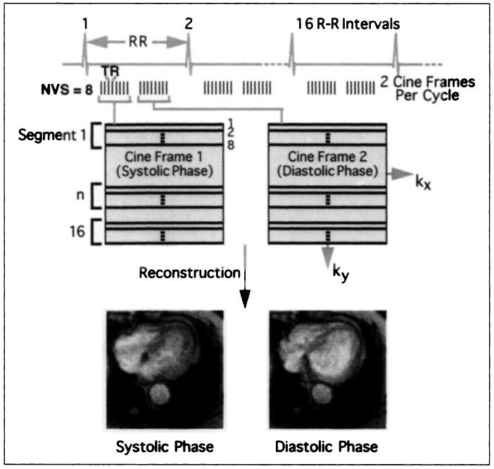
The unique aspect of the pulse sequence is the manner in which K-space is acquired (Fig. 1). During each heartbeat, the R-R interval of the cardiac cycle is divided into multiple cine frames typically of 20- to 80-msec duration. Only a portion, or segment, of K-space is then collected over the duration of each cine frame. The number of lines of K-space acquired over the duration of a cine frame is referred to as the number of views per segment (NVS). NVS multiplied by TR equals the duration of the cine frame. When multiple lines of K-space are collected per R-R interval, the imaging time decreases by a factor of NVS. As an example, to complete 128 phase-encoding steps of K-space with NVS = 8 would require 128 (size of K-space) / 8 (NVS) = 16 R-R intervals (or heartbeats). The total acquisition time is thus determined by the patient's heart rate: for an R-R interval of 1 sec (60 beats per minute), the breath-hold time is 16 R-R intervals × 1 sec = 16 sec. In this example, if TR = 10 msec, the duration of the cine frame is 8 (NVS) × 10 msec (the TR interval) = 80 msec. Because the R-R interval is 1 sec (1000 msec). 920 msec remain (1000 msec − 80 msec) and can be used to collect other lines of K-space for different phases of the cardiac cycle (a single-slice mode with multiple cardiac phases) (Figs. 2-5). Alternatively, if multiple frames of the cardiac cycle are not needed, the remaining portion of the R-R interval can be used to collect lines of K-space for other slice locations (multiple-slice mode with a single cardiac phase).
Fig. 1.
Diagram summarizes segmented K-space gradient-recalled echo acquisition in a multiphase, single-slice mode. Each R-R interval is divided into multiple cardiac phases (cine frames). Cine frame has relatively short duration, so that only portion, or segment, of K-space can be acquired. Multiple RF pulses can be applied during this segment, each corresponding to line of K-space. Number of views per segment (NVS) is number of RF pulses that are applied during each segment, shown as NVS = 8 in this diagram. For TR of 10 msec, duration of each cine frame is 10 msec × 8 = 80 msec; hence, multiple cine frames can be acquired during typical R-R interval (which lasts approximately 1000 msec). Two segments are shown here, one obtained during systole and one obtained during diastole. Note that segments of K-space are usually not contiguous (like contiguous segments shown here) but are interleaved to minimize image artifacts. To complete 128 phase-encoding steps requires 128/8 = 16 R-R intervals.
Fig. 2.
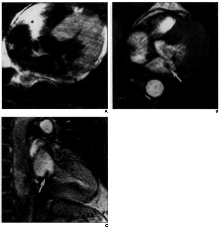
Cardiac valve assessment using segmented K-space acquisition in 67-year-old man with supravalvular mass on echocardiogram. Patient was referred for MR imaging for characterization of relationship between left atrial mass and mitral valve. A-C, Spin-echo T1-weighted MR image (A) and segmented K-space acquisitions in axial (B) and sagittal (C) planes reveal fixed thrombus in left atrium (arrow, B and C). Acquisition time for A was 5 min 43 sec. Each cine sequence, one frame of which is shown in B and C, was acquired in 14 heartbeats and consisted of 15 cine frames using eight views per segment.
Fig. 5.
Cardiac wall motion evaluation using segmented K-space acquisition and cardiac tagging in 45-year-old man with known myocardial infarct of inferior wall. Short-axis cine images of left ventricle (TR/TE, 6.6/2.1; five views per segment; 20 heartbeats) show parallel tags being progressively deformed during ventricular contraction (contraction increasing from left to right). Tags remain parallel in inferior wall at 6- to 7-o'clock position (arrow), indicating lack of contraction as result of myocardial infarction.
Image Acquisition Times and Cardiac Cine Frames
The NVS value is user-defined and is analogous to a turbo factor or echo train length with fast spin-echo imaging; a higher value reduces imaging time in direct proportion to NVS. Similar to using a high echo train length (but for different reasons), higher NVS values potentially result in image blurring because, as NVS is increased, each cine frame captures a longer portion of the cardiac cycle (frame duration = NVS × TR) and cardiovascular structures are moving rapidly. Also, as NVS is increased, the number of movie frames available for cine display correspondingly decreases. For example, for a movie frame duration of 80 msec (NVS × TR = 8 × 10 msec), the maximum number of cardiac cycles is R-R divided by the segment duration, or 1000/80 msec. or approximately 12 frames. If NVS = 16, the number of separate cardiac frames available is reduced to six, so that the cine display will appear less smooth. View sharing is a technique that can interpolate in time between cardiac movie frames and can be used to double the number of frames available for cine display [5].
Multislice Technique
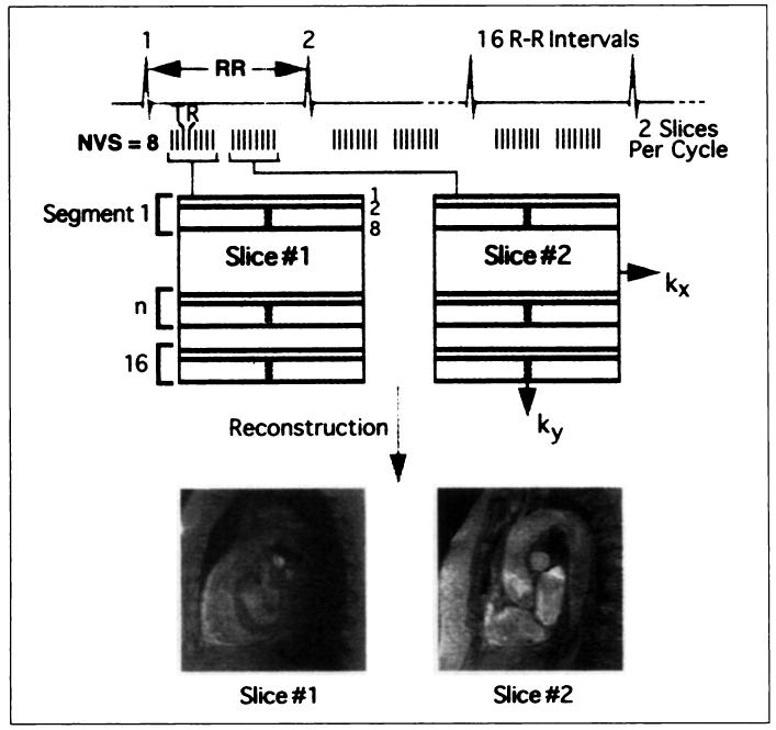
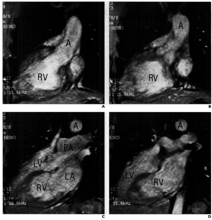
Multiple cardiac cine frames are acquired in one breath-hold at one slice location (sequentially); rapid display of such images can result in a movie loop of a beating heart. In some cases, multiple cardiac cine frames are not necessary, and it is instead desirable to maximize the rate of acquisition of multiple different slice locations (Fig. 6). In a multislice acquisition, similar to multislice spin-echo imaging, additional slice locations are acquired, albeit each at a different point in the cardiac cycle. Thus, instead of 12 cine movie frames in the examples for NVS = 8, 12 different slice locations can be acquired in a breath-hold. This number of locations is helpful if a rapid vascular screening of the entire aorta in one breath-hold is desirable (Fig. 7). Image contrast is affected, however, and for low values of NVS, the effective TR becomes related to the R-R interval. Finally, using a very high NVS value (e.g., 32), cine acquisition can be performed on uncooperative or ill patients with a breath-hold of only about 4–6 sec, at the expense of a limited number of cine movie frames. Typically, we use a 10–15° flip angle for these studies, with an intenslice spacing of 0–20% of the slice thickness.
Fig. 6.
Diagram summarizes segmented K-space gradient-recalled echo acquisition in a multislice, single-phase mode. Each R-R interval is again divided into short intervals, each yielding number of views per segment, except each interval now corresponds to different slice location, each obtained during different portion of cardiac cycle. In this example, one slice location is obtained during systole, and another is obtained during diastole. This technique provides for rapid vascular screening of entire aorta in one breath-hold. Because flow-related enhancement is greater during systole, using only first portion of cardiac cycle increases vascular signal, at expense of slightly increased image acquisition time. NVS = number of views per segment.
Fig. 7.
Cardiac anatomy evaluation using multislice-mode, segmented K-space acquisition in 49-year-old man with corrected transposition of great vessels and dextrocardia. A–D, Segmented K-space acquisition (TR/TE, 6.10/2.5; 32 views per segment) of 12 slice locations obtained in four heartbeats shows progression from most anterior ventricular chamber connected via aorta to most posterior cardiac chamber connected to main pulmonary artery. A = aorta, PA = main pulmonary artery, LV = left ventricle, RV = right ventricle, LA left atrium.
Other Imaging Parameters
The field of view is adjusted for patient size (e.g., 32–36 cm) to avoid wraparound artifacts. Anti-aliasing (no phase wrap) is not used, because this requires two signal averages and doubles the acquisition time. The matrix size is typically 256 in the frequency direction and 128 in the phase direction but is adjusted to compensate for breath-hold capability. Although the body coil can be used as the receiver coil for the aorta using lower bandwidths (±16 kHz) to increase signal-to-noise ratio, a torso phased-array surface coil is preferred. The torso coil allows increased bandwidth (±32 kHz or greater) with adequate signal-to-noise ratio, which, in turn, reduces minimum TR and TE values, allowing more lines of K-space to be collected per unit time. Fractional echo sampling is routinely used to reduce TE. An additional parameter, referred to as the delay time, indicates when imaging should start in relationship to the QRS complex; normally, the delay time is the minimum value accepted by the system (i.e., image during systole), although coronary arteries are more optimally visualized during diastole. View sharing [5] is used if available and results in a smoother cine animation loop. First-order flow compensation can be used to increase vascular signal, but flow artifacts can also be reduced using very short TEs (<2.5 msec).
Limitations
The primary limitation of segmented K-space acquisition is the reduced signal-to-noise ratio that is due to the short TR and low flip angle. However, because images are acquired during a breath-hold with elimination of motion artifacts (both cardiac and respiratory), the image quality in many cases is similar to or even better than that of non-breath-hold sequences. When possible, surface or phased-array coils should be used to improve image quality. Patients with arrhythmias impose an additional limitation, because the method depends on high-quality ECG gating. Patients with slow heart rates, often associated with beta-blocking cardiac medications, will have relatively longer breath-hold times; in these cases NVS should be increased to acquire more lines of K-space during each segment.
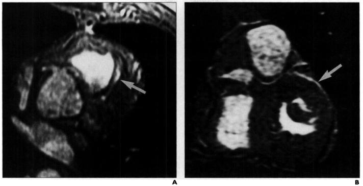
Fig. 3.
Coronary artery evaluation using segmented K-space acquisition in 38-year-old man with abnormal coronary angiogram.
A, Segmented K-space acquisition prescribed along oblique axis shows aberrant origin of left coronary artery (arrow), arising adjacent to right coronary artery. Nine cardiac movie frames were acquired in 20 heartbeats with six views per segment.
B, Coronal oblique acquisition prescribed posterior to pulmonary artery along course of left main coronary artery shows division to left anterior descending (arrow) and circumflex branches.
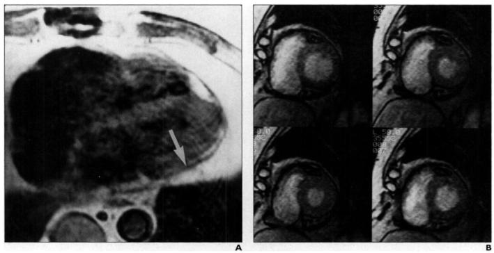
Fig. 4.
Cardiac wall motion evaluation using segmented K-space acquisition in 59-year-old man evaluated for possible constrictive pericarditis after cardiac transplant.
A, T1-weighted spin-echo image shows normal thickness of pericardium (arrow).
B, Short-axis images of left and right ventricles show cine frames at 45, 175, 305, and 435 msec after R wave (displayed left to right, top to bottom) obtained in 16 heartbeats with eight views per segment. Normal function and diastolic filling of both ventricles were noted on cine review. A myocardial biopsy subsequently revealed evidence of rejection.
References
- 1.Didier D, Ratib O, Friedli B, et al. Cine gradient-echo MR imaging in the evaluation of cardiovascular diseases. RadioGraphics. 1993;13:561–573. doi: 10.1148/radiographics.13.3.8316664. [DOI] [PubMed] [Google Scholar]
- 2.Atkinson DJ, Edelman RR. Cineangiography of the heart in a single breath hold with a segmented turbo-FLASH sequence. Radiology. 1991;178:357–360. doi: 10.1148/radiology.178.2.1987592. [DOI] [PubMed] [Google Scholar]
- 3.McVeigh ER, Atalar E. Cardiac tagging with breath-hold cine MRI. Magn Reson Med. 1992;28:3l8–327. doi: 10.1002/mrm.1910280214. [DOI] [PMC free article] [PubMed] [Google Scholar]
- 4.Laissy J-P, Blanc F, Soyer P, et al. Thoracic aortic dissection: diagnosis with transesophageal echocardiography versus MR imaging. Radiology. 1995;194:331–336. doi: 10.1148/radiology.194.2.7824707. [DOI] [PubMed] [Google Scholar]
- 5.Foo TK, Berstein MA, Aisen AM, et al. Improved ejection fraction and flow velocity estimates with use of view sharing and uniform repetition time excitation with fast cardiac techniques. Radiology. 1995;195:471–478. doi: 10.1148/radiology.195.2.7724769. [DOI] [PubMed] [Google Scholar]


