Abstract
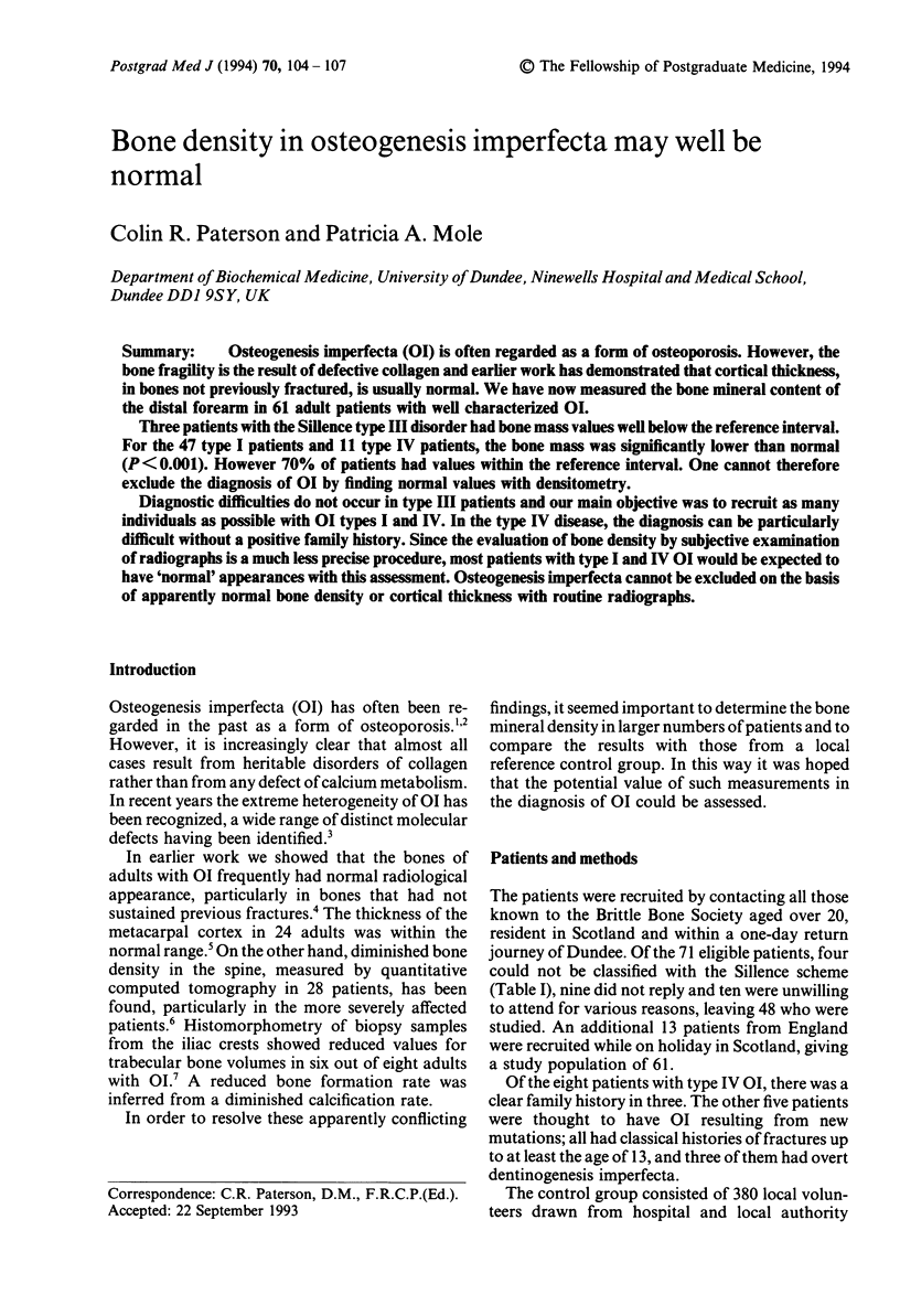
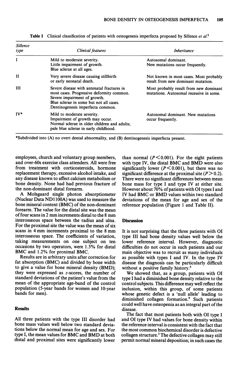
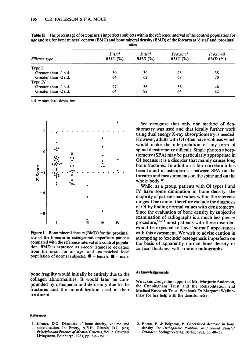
Osteogenesis imperfecta (OI) is often regarded as a form of osteoporosis. However, the bone fragility is the result of defective collagen and earlier work has demonstrated that cortical thickness, in bones not previously fractured, is usually normal. We have now measured the bone mineral content of the distal forearm in 61 adult patients with well characterized OI. Three patients with the Silence type III disorder had bone mass values well below the reference interval. For the 47 type I patients and 11 type IV patients, the bone mass was significantly lower than normal (P < 0.001). However 70% of patients had values within the reference interval. One cannot therefore exclude the diagnosis of OI by finding normal values with densitometry. Diagnostic difficulties do not occur in type III patients and our main objective was to recruit as many individuals as possible with OI types I and IV. In the type IV disease, the diagnosis can be particularly difficult without a positive family history. Since the evaluation of bone density by subjective examination of radiographs is a much less precise procedure, most patients with type I and IV OI would be expected to have 'normal' appearances with this assessment. Osteogenesis imperfecta cannot be excluded on the basis of apparently normal bone density or cortical thickness with routine radiographs.
Full text
PDF


Selected References
These references are in PubMed. This may not be the complete list of references from this article.
- Byers P. H., Wallis G. A., Willing M. C. Osteogenesis imperfecta: translation of mutation to phenotype. J Med Genet. 1991 Jul;28(7):433–442. doi: 10.1136/jmg.28.7.433. [DOI] [PMC free article] [PubMed] [Google Scholar]
- Doyle F. H., Gutteridge D. H., Joplin G. F., Fraser R. An assessment of radiological criteria used in the study of spinal osteoporosis. Br J Radiol. 1967 Apr;40(472):241–250. doi: 10.1259/0007-1285-40-472-241. [DOI] [PubMed] [Google Scholar]
- Kurtz D., Morrish K., Shapiro J. Vertebral bone mineral content in osteogenesis imperfecta. Calcif Tissue Int. 1985 Jan;37(1):14–18. doi: 10.1007/BF02557672. [DOI] [PubMed] [Google Scholar]
- Nilas L., Pødenphant J., Riis B. J., Gotfredsen A., Christiansen C. Usefulness of regional bone measurements in patients with osteoporotic fractures of the spine and distal forearm. J Nucl Med. 1987 Jun;28(6):960–965. [PubMed] [Google Scholar]
- Paterson C. R., McAllion S. J., Shaw J. W. Clinical and radiological features of osteogenesis imperfecta type IVA. Acta Paediatr Scand. 1987 Jul;76(4):548–552. doi: 10.1111/j.1651-2227.1987.tb10519.x. [DOI] [PubMed] [Google Scholar]
- Paterson C. R. Metacarpal morphometry in adults with osteogenesis imperfecta. Br Med J. 1978 Jan 28;1(6107):213–214. doi: 10.1136/bmj.1.6107.213. [DOI] [PMC free article] [PubMed] [Google Scholar]
- Ste-Marie L. G., Charhon S. A., Edouard C., Chapuy M. C., Meunier P. J. Iliac bone histomorphometry in adults and children with osteogenesis imperfecta. J Clin Pathol. 1984 Oct;37(10):1081–1089. doi: 10.1136/jcp.37.10.1081. [DOI] [PMC free article] [PubMed] [Google Scholar]
- Williamson M. R., Boyd C. M., Williamson S. L. Osteoporosis: diagnosis by plain chest film versus dual photon bone densitometry. Skeletal Radiol. 1990;19(1):27–30. doi: 10.1007/BF00197924. [DOI] [PubMed] [Google Scholar]
- Willing M. C., Pruchno C. J., Atkinson M., Byers P. H. Osteogenesis imperfecta type I is commonly due to a COL1A1 null allele of type I collagen. Am J Hum Genet. 1992 Sep;51(3):508–515. [PMC free article] [PubMed] [Google Scholar]


