
Fig. 5.
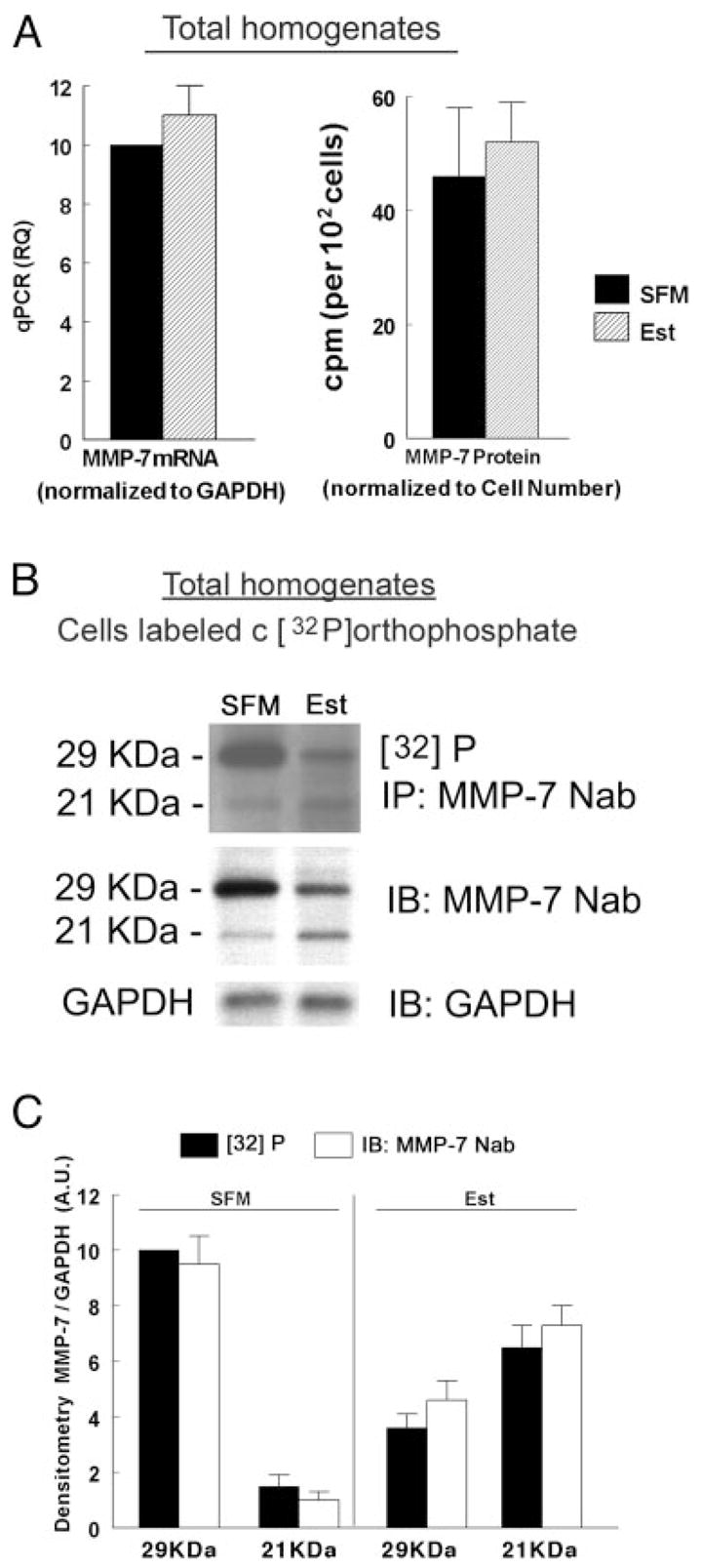
Estrogen effects on MMP-7 expression and phosphorylation. A, Left panel, Quantitative PCR analysis of MMP-7 mRNA in homogenates of estrogen-depleted (SFM) and estrogen-treated cells (Est). Data were normalized to GAPDH mRNA. Right panel, MMP-7 protein analysis. Estrogen-depleted and estrogen-treated cells were labeled with Trans35S-label. Total cells homogenates were immonoprecipitated (IP) with the MMP-7 Nab antibody, and radioactivity was determined in aliquots containing equal amounts of proteins. Data were normalized to cell number per dish. Shown are means (± SD) of three experiments. B and C, Effects on MMP-7 phosphorylation. Estrogen-depleted or estrogen-treated cells were labeled with [32P]orthophosphate. Total homogenates were immonoprecipitated with the MMP-7 Nab antibody and fractionated by gel electrophoresis. After visualization of the radioactive bands (upper panel), gels were immunoblotted (IB) with the MMP-7 Nab antibody, reblotted, and immunoblotted with anti GAPDH antibody (lower panels). The experiment was repeated three times with similar trends. C, Densitometry of the data in B (MMP-7 per GAPDH normalized to an arbitrary unit (A.U.) of 10 for the [32]P-29kDa-SFM category in panel B).