Abstract
We have previously shown that the glypican 3 (GPC3) gene was expressed in neuroblastoma (NB) and Wilms' tumour (WT), two embryonal tumours. GPC3 is an X-linked gene that has its peak expression during development and that is downregulated in all investigated tissues after birth. GPC3 expression could be involved in the aetiology of embryonal tumours such as NB and WT. Methylation is known to play a role in gene silencing, notably in chromosome X inactivation. Southern blot- and PCR-based methylation assays were used to assess the methylation status of the GPC3 promoter on genomic DNA from both normal and embryonal tumour cells. In normal cells, the promoter was not methylated in males and partially methylated in females. Our results suggest that DNA methylation of the promoter region is not essential for the transcriptional repression of the GPC3 gene and that the methylation observed in females is probably linked to the inactive X chromosome. In tumour samples, methylation abnormalities have been found exclusively in female NB samples (loss of methylation) and mainly in male WT samples (gain of methylation). Overall, methylation did not significantly correlate with the expression status of GPC3. Although promoter methylation is likely to affect the expression status of the gene, our results suggest that the deregulation of GPC3 transcriptional expression seen in NB and WT involves other regulatory levels.
Keywords: GPC3, embryonal tumours, transcriptional regulation, methylation, X chromosome
Glypican 3 (GPC3) gene has been shown to be expressed in neuroblastoma (NB) and Wilms' tumour (WT), two embryonal tumours (Saikali and Sinnett, 2000; Toretsky et al, 2001). This gene is expressed in a tissue-specific manner and has its peak expression during development (Filmus et al, 1988; Hsu et al, 1997; Pellegrini et al, 1998). After birth, GPC3 is downregulated in all investigated tissues (Filmus et al, 1988; Hsu et al, 1997; Pellegrini et al, 1998). GPC3 is located at chromosome Xq26.1 and spans more than 500 kb (Pilia et al, 1996; Huber et al, 1997; Shen et al, 1997). The gene product is a heparan sulphate proteoglycan located on the cell surface and attached to the cellular membrane by a glycosyl-phosphatidyl inositol anchor (Pilia et al, 1996). The role of this protein is not yet exactly known. Many studies suggest that GPC3 is a negative cellular growth regulator (Pilia et al, 1996; Weksberg et al, 1996; Cano-Gauci et al, 1999; Lin et al, 1999; Murthy et al, 2000; Xiang et al, 2001), one of the most compelling evidence being that a germline mutation of the gene causes the Simpson–Golabi–Behmel overgrowth syndrome (Pilia et al, 1996) and that Gpc3 knockout mice partly recapitulate the syndrome (Cano-Gauci et al, 1999). On the other hand, GPC3 has been shown to be overexpressed in hepatocellular carcinomas (Hsu et al, 1997; Toretsky et al, 2001; Zhu et al, 2001; Midorikawa et al, 2003) and to be associated with advanced stages as well as with the invasive potential of this cancer (Hsu et al, 1997). Moreover, colorectal carcinoma-associated liver metastases express GPC3 significantly more than primary tumours (Lage et al, 1998). These data suggest that GPC3 is regulating different growth and survival factors in a cell-dependent manner (Filmus, 2001).
The mechanisms regulating the transcription of GPC3 are of particular interest to understand the altered expression of GPC3 in cancer cells. GPC3 has been shown to be overexpressed preferentially in female as compared to male hepatocellular carcinomas (females, 95% and males, 67%) (Hsu et al, 1997). As the gene is located on the X chromosome and DNA methylation is implicated in chromosome X inactivation (Monk, 1986), this observation raises the possibility that a loss of methylation could be implicated in the overexpression of GPC3 in some cancer forms. Hypermethylation of the GPC3 promoter associated with gene silencing has been observed in certain adult cancers (Huber et al, 1999; Lin et al, 1999; Murthy et al, 2000; Xiang et al, 2001). Owing to the potential involvement of GPC3 expression in the aetiology of embryonal tumours, we tested the methylation status of the GPC3 promoter in DNA samples derived from both normal and embryonal tumour cells.
MATERIALS AND METHODS
DNA samples
In this study, we used genomic DNA from the following sources: 14 peripheral blood samples (obtained from volunteer healthy donors at the Sainte-Justine Hospital, Montreal, Canada); 11 placenta samples (DNA was obtained from C Deal); NB cell lines SK-N-AS, SK-N-DZ, SK-N-FI, IMR-32, SK-N-SH (obtained from ATCC, Manassas, VA, USA), NBL-S (obtained from GM Brodeur), SJNB-1, SJNB-7, SJNB-10 (obtained from T Look); primary NB and WT specimens (obtained from patients treated at Sainte-Justine Hospital); and two peripheral blood samples from Turner syndrome patients (DNA and karyotypes were obtained from C Deal). This study was approved by our Institutional Review Board.
Methylation assays
Cytosine methylation assay
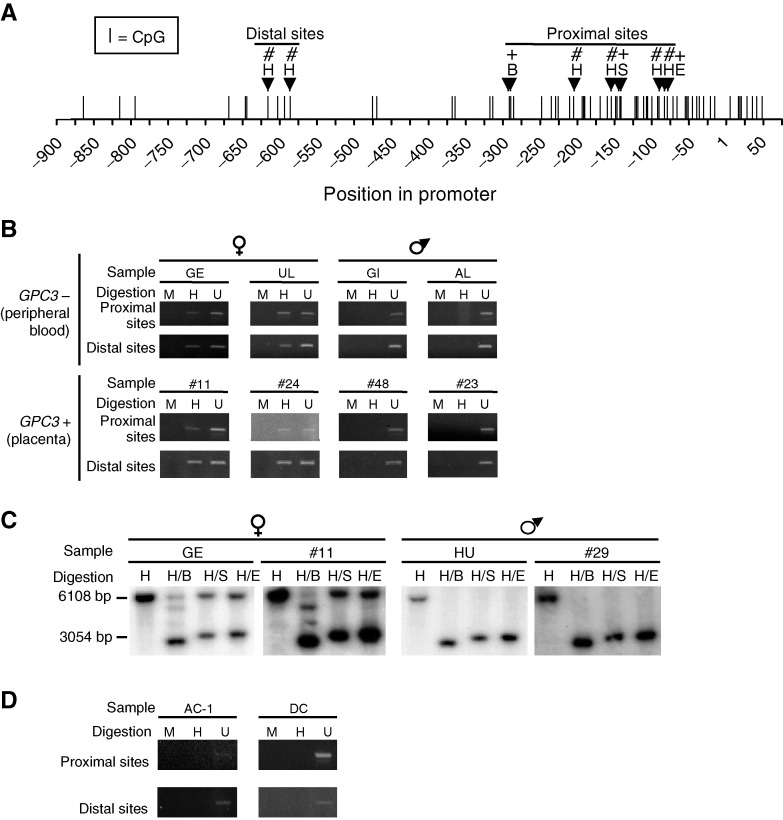
The promoter region of GPC3 contains a CpG island (Figure 1A) (Huber et al, 1997). Several attempts to apply bisulphite protocols (e.g. Cote et al, 1998) in order to assess the methylation status of this promoter region were unsuccessful (data not shown). This apparent resistance to deamination is not unique to GPC3 (e.g. Bearzatto et al, 2000) and could be explained by the high CG content (Harrison et al, 1998). We decided to use two alternative approaches (see below).
Figure 1.
Methylation analysis of the GPC3 promoter in nontumoural samples. (A) CpG dinucleotides positions in the GPC3 promoter region. The methylation status of 11 of these CpG sites was determined either by the PCR-based methylation assay (#) or by the Southern blot-based methylation assay (+) using methyl-sensitive restriction endonucleases HpaII (H), SacII (S), EagI (E), and BssHII (B). HpaII contains one CpG site, whereas SacII, EagI and BssHII contain two CpG sites each. The distal and proximal sites were amplified in distinct PCRs. (B and C) Representative results of the methylation analysis in normal peripheral blood and placental DNA samples obtained by the PCR-based (B) and Southern blot-based (C) methylation assays. See Table 1 for details concerning the samples. (D) PCR-based methylation assay performed on DNA samples from two individuals (AC-1 and DC) affected by the Turner syndrome. Digestions: (B and D) H, HpaII; M, MspI; U, undigested; (C) H, HindIII; B, BssHII; S, SacII and E, EagI. GPC3+, expression of GPC3; GPC3−, no expression of GPC3.
PCR-based methylation assay
For the PCR-based method, 200 ng of genomic DNA and 200 fg of a control plasmidic DNA construct (pBlueScript vector with, as an insert, a 102 bp HPRT gene fragment containing three HpaII/MspI sites within position 1256–1357, Accession number M26434) were digested with 100 U of HpaII or MspI (New England Biolabs, Beverly, MA, USA) for 16 h (see Benachenhou et al, 1998). The cleavage at the six HpaII/MspI sites located within the 700 bp upstream of the GPC3 transcription initiation site was examined by the means of two PCR reactions (one for the two distal sites and one for the four proximal sites; Figure 1A). Polymerase chain reactions were performed in a total volume of 20 μl containing 1 μl of the HpaII or MspI digestion reactions (10 ng of genomic DNA), 1 × of ‘GC Genomic PCR Reaction Buffer’ (Clontech, Palo Alto, CA, USA), 1.1 mM Mg(OAc)2, 200 μM of each of the four dNTPs, 1 M ‘GC-Melt’ (Clontech, Palo Alto, CA, USA), 0.4 μM of each primer (proximal sites: B2 (ACGTGCTGCTACCCAGCCGCTGCA) and L2 (GGAACTTCTCCCAGAGCCAGTCAGAGCG); distal sites: E2 (CCGCTCATTGGCCTACAGCCTGGAGGGC) and J2 (TATTCAAAGGTGAGGCAGGCTGTGAAAAGC)) and 1 × of ‘Advantage GC Genomic Polymerase Mix’ (Clontech, Palo Alto, CA, USA). Polymerase chain reactions for the proximal sites were performed for one cycle of 95°C for 1 min, followed by 38 cycles of 95°C for 45 s, and 74°C for 2 min, followed by one cycle of 74°C for 10 min. PCR reactions for the distal sites were performed for one cycle of 95°C for 1 min, followed by 28 cycles of 95°C for 30 s, and 68°C for 2 min, followed by one cycle of 68°C for 10 min. Complete cleavage was verified by a PCR amplification of the control construct insert with 1 μl of the HpaII or MspI digestion reactions (10 fg plasmid DNA) under standard conditions.
Southern blot-based methylation assay
Genomic DNA was digested with HindIII, either alone or with methyl-sensitive restriction endonucleases EagI, SacII or BssHII. The digestion products were electrophoresed on agarose gels and transferred onto Hybond N+ nylon membranes (Amersham Pharmacia Biotech, Baie d'Urfé, Canada). The membranes were hybridised with a radiolabelled GPC3 promoter-specific PCR product (positions −969 to −346, Figure 1A). In this assay, a 6.1 kb fragment is expected when the investigated sites are fully methylated, whereas a fragment of about 3 kb should be obtained when the sites are not methylated.
Statistical analysis
In order to evaluate whether methylation abnormalities was significantly more frequent in female or male tumour samples, the Fisher's exact test was used. A methylation profile was considered abnormal when it was different from the methylation profile observed in apparently normal samples (peripheral blood and placentas) of the same gender.
RESULTS
In all, 11 CpG sites located in the promoter of the GPC3 gene have been tested for methylation using methyl-sensitive restriction endonuclease assays (Figure 1A). Six of them were located within HpaII sites and were tested by the PCR-based methylation assay along with undigested and methyl-insensitive MspI-digested samples as controls. The five others were located within EagI, SacII and BssHII methyl-sensitive restriction sites and were investigated with the Southern blot-based methylation assay.
These sites were first tested in DNA samples derived from normal cells including 14 peripheral blood samples, known not to express GPC3 (GPC3−) (Hsu et al, 1997), and 11 placenta samples, which strongly express GPC3 (GPC3+) (Hsu et al, 1997). We found that, independent of the expression status, methylation correlated with the gender: females presented a partial methylation, whereas males had no methylation (Figure 1B and C). These results suggest that methylation is not essential for the repression of the GPC3 gene, since GPC3 nonexpressing male samples are not methylated at the studied sites (Table 1 ). Southern blot methylation vs nonmethylation signal intensities presented a ratio of approximately 1 : 1 in females, indicating the presence of methylation in about half of the DNA molecules (Figure 1C). This suggests that the methylation detected in females could be linked to the inactive X chromosome. Male sample #32 GPC3 promoter has been shown to be partially methylated as opposed to other male samples (Table 1). Sex determination assay and X chromosome microsatellite amplification (DXS102, DXS538 and DXS981) showed that this sample has a Y chromosome and only one X chromosome (data not shown). This suggests that the partial methylation seen in sample #32 reflects cell heterogeneity for GPC3 promoter methylation. PCR-based methylation assay on female sample #25 showed that at least one of the proximal sites was not methylated (Table 1). However, the Southern blot-based assay methylation profile of this sample was similar to that of the other female samples, suggesting that the GPC3 promoter is methylated but not at every site.
Table 1. Summary of the GPC3 promoter methylation data of normal cells.
|
Methylation status |
|||||||
|---|---|---|---|---|---|---|---|
|
PCR-based assaya |
Southern blot-based assayb |
||||||
| Sample | Origin | Genderc | Proximal sites | Distal sites | EagI | SacII | BssHII |
| GE | Periph. blood | F | + | + | +/− | +/− | +/− |
| CA | Periph. blood | F | + | + | +/− | +/− | +/− |
| UL | Periph. blood | F | + | + | +/− | +/− | +/− |
| CP41 | Periph. blood | F | + | + | ND | ND | ND |
| CP42 | Periph. blood | F | + | + | ND | ND | ND |
| CP43 | Periph. blood | F | + | + | ND | ND | ND |
| GI | Periph. blood | M | − | − | − | − | − |
| AL | Periph. blood | M | − | − | − | − | − |
| HU | Periph. blood | M | − | − | − | − | − |
| PED 93 | Periph. blood | M | − | − | ND | ND | ND |
| CP111 | Periph. blood | M | − | − | ND | ND | ND |
| CP112 | Periph. blood | M | − | − | ND | ND | ND |
| CP113 | Periph. blood | M | − | − | ND | ND | ND |
| CP114 | Periph. blood | M | − | − | ND | ND | ND |
| #11 | Placenta | F | + | + | +/− | +/− | +/− |
| #24 | Placenta | F | + | + | +/− | +/− | +/− |
| #25 | Placenta | F | − | + | +/− | +/− | +/− |
| #26 | Placenta | F | + | + | ND | ND | ND |
| #27 | Placenta | F | + | + | ND | ND | ND |
| #28 | Placenta | F | + | + | ND | ND | ND |
| #20 | Placenta | M | − | − | ND | ND | ND |
| #23 | Placenta | M | − | − | ND | ND | ND |
| #29 | Placenta | M | − | − | − | − | − |
| #32 | Placenta | M | + | + | ND | +/− | +/− |
| #48 | Placenta | M | − | − | − | − | − |
GPC3=glypican 3; PCR=polymerase chain reaction.
+=methylated; −=not methylated.
+=methylation signal; −=nonmethylation signal; +/−=both methylation and nonmethylation signals; ND=not determined.
F=female; M=male.
In order to test the hypothesis that GPC3 promoter methylation in females is linked to the inactive X chromosome, the PCR-based methylation assay was performed on peripheral blood DNA samples from two Turner syndrome patients with karyotype (45, X), having no inactive X chromosome. No methylation signal was detected (Figure 1D), supporting the hypothesis that the methylation signal detected at the GPC3 promoter is linked to the inactive X chromosome.
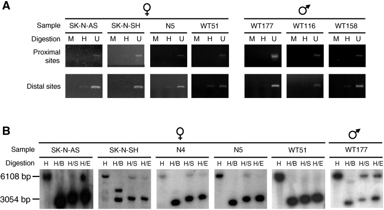
PCR- and Southern blot-based methylation assays were performed on the GPC3 promoter of NB cell lines, primary NBs and primary WTs (Figure 2). Overall in NB samples, four females out of six (67%) showed some loss of methylation, whereas every males had normal methylation status (Figure 2, Table 2 ), suggesting that methylation abnormalities are predominantly found in females (Fisher's test: P=0.011). Methylation analysis of the GPC3 promoter in WT samples also revealed abnormalities when compared to the normal cells. One female out of four (WT51) presented a loss of methylation and three males out of four (75%) showed partial methylation (Figure 2, Table 2). Therefore, in contrast to NB, in WT samples, methylation abnormalities seem to be more frequent in males than in females. However, more samples need to be investigated to confirm this trend (Fisher's test: P=0.243).
Figure 2.
Methylation analysis of the GPC3 promoter in tumour cell DNA samples. PCR- (A) and Southern blot- (B) based methylation assays were performed on tumour cell DNA samples from NB cell lines (SK-N-AS, SK-N-SH), primary NBs (N4, N5) and primary WTs (WT51, WT116, WT158, WT177). Only results for samples with abnormal DNA methylation patterns are shown. Digestions: (A) H: HpaII; M: MspI; U: undigested; (B) H: HindIII; B: BssHII; S: SacII and E: EagI.
Table 2. Summary of the GPC3 promoter methylation data of tumour cells and their GPC3 mRNA expression status.
|
Methylation status |
||||||||
|---|---|---|---|---|---|---|---|---|
|
PCR-based assaya |
Southern blot-based assayb |
|||||||
| Sample | Origin | Genderc | Expressiond | Proximal sites | Distal sites | EagI | SacII | BssHII |
| SK-N-AS | NB cell line | F | +++ | − | tr | − | − | − |
| SK-N-SH | NB cell line | F | ++ | − | tr | tr/− | tr/− | +/− |
| SK-N-DZ | NB cell line | F | − | + | + | +/− | +/− | +/− |
| SJNB-1 | NB cell line | M | ++ | − | − | − | − | − |
| SJNB-7 | NB cell line | M | +++ | − | − | − | − | − |
| SJNB-10 | NB cell line | M | +++ | − | − | ND | ND | ND |
| NBL-S | NB cell line | M | ++ | − | − | ND | ND | ND |
| IMR-32 | NB cell line | M | ++ | − | − | ND | ND | ND |
| SK-N-FI | NB cell line | M | − | − | − | − | − | − |
| NB4 | Primary NB | F | + | + | + | tr/− | tr/− | tr/− |
| NB5 | Primary NB | F | + | − | tr | − | tr/− | tr/− |
| NB8 | Primary NB | F | ND | + | + | ND | ND | ND |
| NB11 | Primary NB | M | − | − | − | − | − | − |
| NB13 | Primary NB | M | + | − | − | − | − | − |
| NB193 | Primary NB | M | ND | − | − | ND | ND | ND |
| WT130 | Primary WT | F | ++ | + | + | +/− | +/− | +/− |
| WT42 | Primary WT | F | + | + | + | +/− | +/− | +/− |
| WT51 | Primary WT | F | ++ | − | + | − | − | − |
| WT106 | Primary WT | F | + | + | ND | ND | ND | ND |
| WT40 | Primary WT | M | +++ | − | − | − | − | − |
| WT177 | Primary WT | M | ++ | − | tr | +/− | +/− | +/− |
| WT116 | Primary WT | M | + | tr | − | ND | ND | ND |
| WT158 | Primary WT | M | ND | tr | + | ND | ND | ND |
GPC3=glypican 3; PCR=polymerase chain reaction; NB=neuroblastoma; WT=Wilms' tumour.
+=methylated; −=not methylated.
+=methylation signal; −=nonmethylation signal; tr=traces; +/−=both methylation and nonmethylation signals; tr/−=both traces of methylation signal and nonmethylation signal; ND=not determined.
F=female; M=male.
Transcriptional expression: −=no expression; +=weak expression; ++=moderate expression; +++=strong expression. The analysis of the expression levels was reported in Saikali et al (2000) and was based on the visual inspection of all the Northern blots or semiquantitative RT–PCR assays.
In most cases, as in normal cells, the methylation pattern of the GPC3 promoter at the investigated sites is not correlated with the expression status (Table 2). However, in female NB samples, loss of methylation correlates with the expression of GPC3 (Table 2), raising the possibility that loss of methylation of the inactive X chromosome could lead to the transcriptional activation of the linked GPC3 allele. To test this hypothesis, the cell lines SK-N-DZ (normal methylation pattern, GPC3−; Table 2) and SK-N-SH (loss of methylation, GPC3+; Table 2) were treated with 0.5, 1 and 5 μM of demethylating agent 5-aza-deoxycytidine (5-aza-dC). Southern blot-based methylation assay revealed that no demethylation was achieved, and transcriptional expression was similar to the nontreated controls as evaluated by Northern blot analysis (data not shown), even in cells treated at highly toxic concentration of 5-aza-dC (data not shown).
DISCUSSION
In this report, we showed in peripheral blood (GPC3−) and placental (GPC3+) cells that the GPC3 promoter methylation status at the investigated CpG sites was correlated with gender rather than the expression status, male samples being unmethylated and female samples being partially methylated. These observations are consistent with those of another methylation analysis of the GPC3 promoter performed on leucocyte DNA samples (Huber et al, 1999).
These results indicate that methylation at these sites is not essential for the repression of GPC3. However, we cannot exclude the possibility that the CpG sites investigated are not critical for the repression of the gene. The Southern blot-based methylation assay that allows a quantitative analysis of methylation taken together with the analysis of females with Turner syndrome support the hypothesis that the methylation observed in females is associated with the inactive X chromosome. In this regard, Huber et al (1999) have reported a complete methylation of the GPC3 promoter in somatic hybrid hamster–human cells containing only the human inactive X chromosome. These results strongly suggest that the GPC3 allele located on the inactive X chromosome is methylated, whereas the active X chromosome allele is not. The methylation on the inactive X chromosome is thought to be important for the maintenance of gene silencing (Monk, 1986).
The methylation analysis in embryonal tumours revealed methylation abnormalities particularly in female NB cells and in male WTs. These observations might result from the fact that cancer cells often present aberrant methylation, their genome being generally hypomethylated and locally hypermethylated, notably in CpG islands (Baylin et al, 1998; Momparler and Bovenzi, 2000; Robertson and Jones, 2000). Our study suggests that the main methylation abnormalities at the GPC3 promoter level seems to be losses of methylation in NBs and the opposite in WTs. Do methylation abnormalities have an influence on the expression status of GPC3? In the embryonal tumour cells tested, as in normal cells, we failed to observe any correlation between methylation and expression of GPC3. However, it has been shown in vitro that the GPC3 promoter does not activate the transcription of a reporter gene when methylated (Huber et al, 1999). In light of these results, it seems likely that the transcriptional activation of the GPC3 gene requires an absence of methylation of the gene promoter, but that the absence of methylation alone does not necessarily lead to transcriptional activity. It is thus possible that the loss of methylation we observed in female NBs allows the inactive X chromosome GPC3 allele to become transcriptionally active, eventually leading to a dosage effect in the corresponding cells. The same mechanism could also explain the preferential overexpression of GPC3 in women affected with hepatocellular carcinomas (Hsu et al, 1997).
In summary, in vivo DNA methylation of the promoter regions does not seem to be the predominant regulatory mechanism for the GPC3 gene. Thus the apparent deregulation of the GPC3 mRNA expression reported in embryonal tumours (Saikali and Sinnett, 2000) is likely to involve other regulatory signals.
Acknowledgments
We thank Drs T Look and GM Brodeur for NB cell lines and to Dr C Deal for DNA samples. This work was supported by the Fonds de la Recherche en Santé du Québec (FRSQ). GB is a recipient of NSERC and FCAR-FRSQ-Santé studentships. DS is a scholar of the FRSQ.
References
- Baylin SB, Herman JG, Graff JR, Vertino PM, Issa JP (1998) Alterations in DNA methylation: a fundamental aspect of neoplasia. Adv Cancer Res 72: 141–196 [PubMed] [Google Scholar]
- Bearzatto A, Szadkowski M, Macpherson P, Jiricny J, Karran P (2000) Epigenetic regulation of the MGMT and hMSH6 DNA repair genes in cells resistant to methylating agents. Cancer Res 60: 3262–3270 [PubMed] [Google Scholar]
- Benachenhou N, Guiral S, Gorska-Flipot I, Michalski R, Labuda D, Sinnett D (1998) Allelic losses and DNA methylation at DNA mismatch repair loci in sporadic colorectal cancer. Carcinogenesis 19: 1925–1929 [DOI] [PubMed] [Google Scholar]
- Cano-Gauci DF, Song HH, Yang H, McKerlie C, Choo B, Shi W, Pullano R, Piscione TD, Grisaru S, Soon S, Sedlackova L, Tanswell AK, Mak TW, Yeger H, Lockwood GA, Rosenblum ND, Filmus J (1999) Glypican-3-deficient mice exhibit developmental overgrowth and some of the abnormalities typical of Simpson–Golabi–Behmel syndrome. J Cell Biol 146: 255–264 [DOI] [PMC free article] [PubMed] [Google Scholar]
- Cote S, Sinnett D, Momparler RL (1998) Demethylation by 5-aza-2′-deoxycytidine of specific 5-methylcytosine sites in the promoter region of the retinoic acid receptor beta gene in human colon carcinoma cells. Anticancer Drugs 9: 743–750 [DOI] [PubMed] [Google Scholar]
- Filmus J (2001) Glypicans in growth control and cancer. Glycobiology 11: 19R–23R [DOI] [PubMed] [Google Scholar]
- Filmus J, Church JG, Buick RN (1988) Isolation of a cDNA corresponding to a developmentally regulated transcript in rat intestine. Mol Cell Biol 8: 4243–4249 [DOI] [PMC free article] [PubMed] [Google Scholar]
- Harrison J, Stirzaker C, Clark SJ (1998) Cytosines adjacent to methylated CpG sites can be partially resistant to conversion in genomic bisulfite sequencing leading to methylation artifacts. Anal Biochem 264: 129–132 [DOI] [PubMed] [Google Scholar]
- Hsu HC, Cheng W, Lai PL (1997) Cloning and expression of a developmentally regulated transcript MXR7 in hepatocellular carcinoma: biological significance and temporospatial distribution. Cancer Res 57: 5179–5184 [PubMed] [Google Scholar]
- Huber R, Crisponi L, Mazzarella R, Chen CN, Su Y, Shizuya H, Chen EY, Cao A, Pilia G (1997) Analysis of exon/intron structure and 400 kb of genomic sequence surrounding the 5′-promoter and 3′-terminal ends of the human glypican 3 (GPC3) gene. Genomics 45: 48–58 [DOI] [PubMed] [Google Scholar]
- Huber R, Hansen RS, Strazzullo M, Pengue G, Mazzarella R, D'Urso M, Schlessinger D, Pilia G, Gartler SM, D'Esposito M (1999) DNA methylation in transcriptional repression of two differentially expressed X-linked genes, GPC3 and SYBL1. Proc Natl Acad Sci USA 96: 616–621 [DOI] [PMC free article] [PubMed] [Google Scholar]
- Lage H, Dietel M, Froschle G, Reymann A (1998) Expression of the novel mitoxantrone resistance associated gene MXR7 in colorectal malignancies. Int J Clin Pharmacol Ther 36: 58–60 [PubMed] [Google Scholar]
- Lin H, Huber R, Schlessinger D, Morin PJ (1999) Frequent silencing of the GPC3 gene in ovarian cancer cell lines. Cancer Res 59: 807–810 [PubMed] [Google Scholar]
- Midorikawa Y, Ishikawa S, Iwanari H, Imamura T, Sakamoto H, Miyazono K, Kodama T, Makuuchi M, Aburatani H (2003) Glypican-3, overexpressed in hepatocellular carcinoma, modulates FGF2 and BMP-7 signaling. Int J Cancer 103: 455–465 [DOI] [PubMed] [Google Scholar]
- Momparler RL, Bovenzi V (2000) DNA methylation and cancer. J Cell Physiol 183: 145–154 [DOI] [PubMed] [Google Scholar]
- Monk M (1986) Methylation and the X chromosome. BioEssays 4: 204–208 [DOI] [PubMed] [Google Scholar]
- Murthy SS, Shen T, De Rienzo A, Lee WC, Ferriola PC, Jhanwar SC, Mossman BT, Filmus J, Testa JR (2000) Expression of GPC3, an X-linked recessive overgrowth gene, is silenced in malignant mesothelioma. Oncogene 19: 410–416 [DOI] [PubMed] [Google Scholar]
- Pellegrini M, Pilia G, Pantano S, Lucchini F, Uda M, Fumi M, Cao A, Schlessinger D, Forabosco A (1998) Gpc3 expression correlates with the phenotype of the Simpson–Golabi–Behmel syndrome. Dev Dyn 213: 431–439 [DOI] [PubMed] [Google Scholar]
- Pilia G, Hughes-Benzie RM, MacKenzie A, Baybayan P, Chen EY, Huber R, Neri G, Cao A, Forabosco A, Schlessinger D (1996) Mutations in GPC3, a glypican gene, cause the Simpson–Golabi–Behmel overgrowth syndrome. Nat Genet 12: 241–247 [DOI] [PubMed] [Google Scholar]
- Robertson KD, Jones PA (2000) DNA methylation: past, present and future directions. Carcinogenesis 21: 461–467 [DOI] [PubMed] [Google Scholar]
- Saikali Z, Sinnett D (2000) Expression of glypican 3 (GPC3) in embryonal tumors. Int J Cancer 89: 418–422 [PubMed] [Google Scholar]
- Shen T, Sonoda G, Hamid J, Li M, Filmus J, Buick RN, Testa JR (1997) Mapping of the Simpson–Golabi–Behmel overgrowth syndrome gene (GPC3) to chromosome X in human and rat by fluorescence in situ hybridization. Mamm Genome 8: 72. [DOI] [PubMed] [Google Scholar]
- Toretsky JA, Zitomersky NL, Eskenazi AE, Voigt RW, Strauch ED, Sun CC, Huber R, Meltzer SJ, Schlessinger D (2001) Glypican-3 expression in Wilms tumor and hepatoblastoma. J Pediatr Hematol Oncol 23: 496–499 [DOI] [PubMed] [Google Scholar]
- Weksberg R, Squire JA, Templeton DM (1996) Glypicans: a growing trend. Nat Genet 12: 225–227 [DOI] [PubMed] [Google Scholar]
- Xiang YY, Ladeda V, Filmus J (2001) Glypican-3 expression is silenced in human breast cancer. Oncogene 20: 7408–7412 [DOI] [PubMed] [Google Scholar]
- Zhu ZW, Friess H, Wang L, Abou-Shady M, Zimmermann A, Lander AD, Korc M, Kleeff J, Buchler MW (2001) Enhanced glypican-3 expression differentiates the majority of hepatocellular carcinomas from benign hepatic disorders. Gut 48: 558–564 [DOI] [PMC free article] [PubMed] [Google Scholar]


