Abstract
Technology to improve the clarity of video for home theater viewers is available utilizing a low cost enhancement chip (DigiVision DV1000). The impact of such a device on the preference for enhanced video was tested for people with impaired vision and normally sighted viewers. Viewers with impaired vision preferred the enhancement effects more than normally sighted viewers. Preference for enhancement was correlated with loss in contrast sensitivity and visual acuity. Preference increased with increased enhancement settings (designed for those with normal vision) in the group with vision impairments. This suggests that higher enhancement levels may be of even greater benefit, and a similar product could be designed to meet the needs of the large, growing population of elderly television viewers with impaired vision.
1. Introduction
There are over 3 million people with impaired vision in the US, and their numbers are increasing steadily with the aging of the population [1]. Central field loss, caused by diseases such as macular degeneration and diabetic retinopathy damaging the central retina, has a particularly disabling effect on visual function. As the natural retinal locus of high-resolution vision (the fovea) is damaged, patients learn to use a peripheral part of their retina for detail vision, the preferred retinal locus (PRL) [2]. However, contrast sensitivity and acuity decline with retinal eccentricity (the distance of the PRL from the fovea). A simulation of vision with central field loss may be seen at: http://www.eri.harvard.edu/faculty/peli/lab/videos/videos.htm
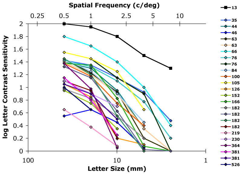
The human visual system is responsive over a range of spatial frequencies. A normally sighted person can see gratings of spatial frequencies up to about 25 or 30 c/deg. Higher levels of contrast are required for the gratings to be seen as the spatial frequencies increase [3, 4]. The inverse of the contrast detection threshold is defined as the contrast sensitivity. Individuals with visual impairment require higher levels of contrast than normally sighted persons to see most stimuli and the differences are greater as spatial frequencies increase. This loss in contrast sensitivity can be thought of as having a low-pass filtering effect, although it is actually a nonlinear, threshold effect. At some critical frequency, no level of presented contrast is sufficient for the stimuli to be seen. This critical frequency and the overall contrast sensitivity vary with the severity of impairment. Figure 1 shows the contrast sensitivity of a normally sighted subject compared with that of subjects with impaired vision who participated in this study, illustrating the nature and range of the loss in sensitivity. Most subjects with impaired vision could not recognize low contrast letter stimuli of fundamental spatial frequencies above 4 c/deg at any contrast.
Figure 1.
Contrast sensitivity function measured for letters of varying sizes. As the fundamental spatial frequency (in cycles per degree) of the letters (based on 2.5 cycles per letter) increases, the contrast sensitivity decreases. The sensitivity of all the people with visual impairments is substantially lower than the sensitivity of a normally sighted subject, (13), and decreases more rapidly with frequency, illustrating the low-pass nature of the impairments. The legend shows the denominator of Snellen acuity at 20ft (20/X). Contrast sensitivity is defined as the inverse of the contrast threshold needed to recognize the letter.
People with vision impairment watch television regularly and to a similar extent as normally sighted viewers [5]. When asked what they miss in the TV picture, low-vision patients (including our participants) report that they cannot see the details and expressions on people’s faces - sometimes to the point of failing to identify who is who in the scene - nor can they read text on the screen. Some report difficulty following a storyline when they fail to identify a character.
Patients with poor central vision frequently use magnification to increase the size of the image on the retina, which shifts the higher-frequency details into lower frequencies and, therefore, greater contrast sensitivity. Magnification of a TV picture can be achieved with optical devices such as spectacles and head born telescopes. Magnification may also be achieved by sitting closer to the TV, increasing the size of the TV screen, or via electronic zooming of the displayed images [6].
Increasing image contrast using image processing, as an alternative or supplement to magnification to make video images more visible to patients with visual impairment has been proposed [7, 8]. Early applications were inspired by the Adaptive Enhancement algorithm [7, 9], a high frequency enhancement that adapts the level of enhancement to local image characteristics. However, those early implementations did not include the adaptive aspects. Yet the effect of enhancing the higher frequencies’ contrast has been shown to be beneficial to these patients, improving their ability to recognize faces from static black and white images [10, 11]. DigiVision (San Diego, CA) has manufactured a series of devices that perform the same (non-adaptive) enhancement in real time [12]. Several studies using these devices in our lab have shown that patients prefer the enhanced video to the original [13–15] and had a small improvement in reading rate of enhanced text [16]. These devices, however, have not yet been marketed to the vision impaired public, mainly because of their high cost.
A recently developed CMOS chip (DV1000, DigiVision) performs similar real-time processing as the earlier, far larger and expensive systems, with additional functionality. In particular it enables a variety of local adaptations. The DV1000 processes digital video (up to 1080p HDTV resolution), and is designed to be integrated within display devices. The end-product manufacturer may restrict or organize these features in various ways to tune the device for a specific application. One such product, RazorVision (Belkin International, Compton, CA, http://www.pureav.com/razorvision/), was designed to be used as a simple in-line cable TV enhancer. It features 3 preset enhancement levels – ‘Low’, ‘Medium’ and ‘High’. It is supplied only with HDMI/DVI connectors, as it is aimed at the high-definition (HD) -viewer market, and has not been marketed to those with visual impairments. The RazorVision device retails for about $250, making image enhancement cost realistic for the low-vision market for the first time. DigiVision provided us with a computer-controlled implementation of the system, with the default settings being those used in the RazorVision model.
The purpose of this study was to investigate whether people with vision impairments would perceive any benefit from using this consumer product, and whether they could differentiate between the 3 preset enhancement levels provided.
2. Methods
2.1 Experimental Setup
We presented a sequence of six short video clips selected to represent various types of programs seen on TV [17]. The videos were taken from DVDs, and played using the VLC media player (www.videolan.org). Subjects were seated 3′ from and centered between two 27″ adjacent standard-definition TVs (approximately 3′6″ diagonally from the center of either TV, each spanning about 27° horizontally from that distance). The source video was split so that an unprocessed version was available along with the DV1000 processed version. The two versions were presented on the two TVs, in a counterbalanced sequence described below, and the subjects were asked which TV picture they preferred (forced choice). Subjects with visual impairment were asked which picture appeared the most clear to them, whereas normally sighted subjects (to whom the effect of the image processing was often evident) where asked simply for their preference. Subjects were encouraged to move their head appropriately to examine the two TV pictures.
The TVs were identical in so far as they were both of the same model, bought at the same time and from the same store. They did not come from the same batch number, so we could not be sure of equal manufacturing specifications. In the default, factory setting, the picture did not appear to have equal brightness and contrast on both screens, so one TV was adjusted slightly until the picture display appeared subjectively equivalent, using both moving video and static test-pattern generator signals. Only the brightness and contrast controls were adjusted, on the left hand television. The experiment was designed to be tolerant to differences in image display on each television (see below).
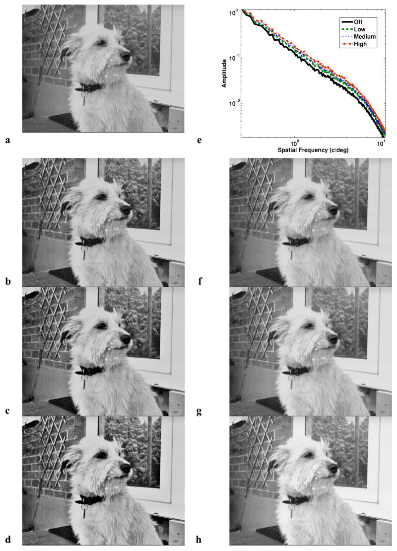
Unlike the RazorVision commercial model, the device we used provided analog S-Video input and output, using analog-to-digital and digital-to-analog circuitry surrounding the DV1000. The device controls allowed selection between an ‘Off’ mode and the three preset levels (‘Low’, ‘Medium’ and ‘High’). The ‘Off’ mode transmitted the video with no enhancement processing (though A/D and D/A processing is still performed). Examples of the effect of these preset enhancement settings are shown in Figure 2. The DV1000 control software allows selection of the active mode. While the software enables changing many of the internal parameters of the system, the default settings matched those of the RazorVision model and were not modified in the current study.
Figure 2.
Illustration of the video processing of the DV 1000 device used in the study. a. No enhancement (‘Off’) b. ‘Low’ c. ‘Medium’ d. ‘High’ enhancement. The enhancement effect varies according to the local spatial frequency content of the image and the local luminance; note, for example, the enhancement of details in the tree reflection in the window, and the enhancement of contrast of the dog’s facial features, but the reduction of visibility of smaller details in and around the eyes. Similarly the high contrast of the collar against the light fur results in masking of finer details on the collar itself. The device settings (Figure 3) can be tuned to achieve optimal results for an intended application/image. e. The spatial frequency amplitude spectra of the image results of all four settings, given in cycles/degree assuming the seating setup used in the study (showing radial-averaged FFT amplitude). Frequencies between 0.3 and 8c/d are shown, as this is where patients’ losses are greatest (see Figure 1). f–h. For comparison, the gray levels of the image shown in (a) were shifted and expanded such that it’s minimum, maximum and midpoint ((Lmax + Lmin) × 0.5) luminance were equal to the DV1000-processed images. No local enhancement of features is apparent, and smaller details are not affected by increased local contrast elsewhere. DV1000 images were recorded using the luminance component only of the S-Video output into a Canon ZR85 camera and captured using Adobe Premiere 6.5 (Adobe Systems Inc., San Jose, CA). The color component is omitted for clarity; the bulk of the effective enhancement is done in the luminance channel only.
Sixteen times during the video presentation, the enhancement level in use by the device and the television (right or left) to show the processed video were selected by the experiment control software. Two sets of the 4 orders of enhancement level (‘Off’, ‘Low’, ‘Medium’ and ‘High’) were randomized, one for each television. Of the 70 possible orders of 4 in 8 television selections, 24 were selected at random. The sequence was repeated for a total of 16 presentations. This randomization ensured that 4 presentations of each enhancement level were made per subject, with each presented on both televisions twice. Equal presentations of conditions on both screens ensured no false bias of preference for any enhancement level due to display differences or subject bias, though such effects could mask a real preference (see Section 2.4). The 12 normally sighted control subjects (see Section 2.3) viewed 12 of these same 24 presentation sequences, selected at random.
The switching of the processed and unprocessed video presentations between the two TVs was achieved using an Extron MMX 42 SVA RCA video switching unit (Extron Electronics, Anaheim CA). This device accepts up to 4 S-Video and audio inputs, and can switch any of these to one or both of two S-Video and audio outputs. The device can be controlled via an RS-232 serial cable. The custom software program started video playback at the correct video, set the video switch to output the processed and unprocessed videos to the appropriate TVs, controlled the DV1000 software to select the correct enhancement level,1 and recorded the subject’s choice of preferred image.
The sequence of videos was started at a video determined by subject number; e.g., the sequences for subjects 1 and 7 began at video number 1. Not all subjects saw all of the available videos, as the sequence length varied depending on individual response time, often not exceeding the length of the first video presented. Audio was provided to the subjects at a low level.
2.2 Image enhancement
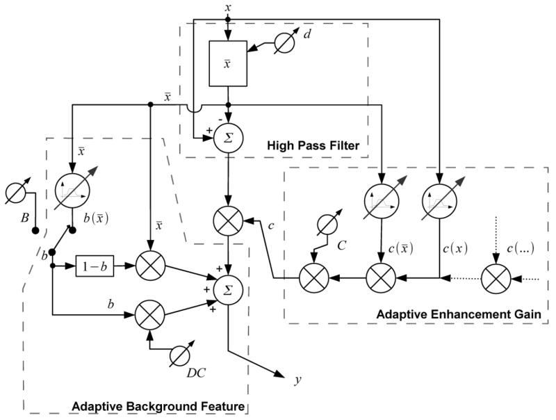
The adaptive enhancement algorithm, as applied in the DV1000, is illustrated in Figure 3. The system first applies a low-pass filter (averaging over a Gaussian kernel window) to the image, x, to produce a mean local luminance image, x̄. Subtracting x̄ from the original image provides an unsharp (high-pass filtered) version [8, 19]. For NTSC resolution video (used here), the size of the averaging kernel used in this operation (which determines the spatial frequencies being enhanced) can be varied from 3.4% to 19.6% of horizontal screen size. The Gaussian kernel size of 9.8% of screen width (full width at half maximum) used in the RazorVision product was applied here. The high-pass filtered version is then amplified by an enhancement gain control, c. The value of c applied at every pixel is the product of a number of multiplicative gain functions, including those based on x̄ and x at the corresponding pixel. Typically higher gain is applied at moderate values of both x and x̄ than near the extremes of the luminance range. This is done to avoid image distortions due to saturation and cut-off at the bright and dark ends of the luminance range, respectively [20], and represents the first adaptation aspect of the algorithm. Other gain functions are available in the DV1000 (Figure 3), but were not used here. The enhanced high-pass version of the image is then added to a modified version of the local luminance mean, x̄. The modified x̄, called the background, can also be adjusted adaptively (as a function of x̄), although this was not used in this study. Typically the background luminance is increased at areas were x̄ is below a set threshold, and decreased at areas where x̄ is above another specified threshold. This additional adaptation allows dynamic range to be cleared, enabling some level of enhancement of the high-pass version to be applied near the extreme luminance range without causing image saturation [20]. Thus, the adaptation can both limit the enhancement and permit some enhancement at bright and dark areas that otherwise would not be enhanced without severe distortions.
Figure 3.
A simplified partial diagram of the DV1000 adaptive enhancement functionality. Subtraction of the mean luminance x̄ (computed locally over a window of adjustable size d), from the original image pixel x, results in a high-pass filtered residual. This is multiplied by a c gain value, obtained by multiplying a number of adaptive gain functions. Two such functions, based on pixel value and local mean luminance are shown; others are available. The value of c can be lowered at the extremes of the luminance range to avoid saturation. An external ‘Clarity’ value (C) mediates these functions. The value of b controls the ratio of original local image background level (x̄) to preset mean luminance (DC) that will be restored/added to the image. The amount of original image background can be set directly (B) or locally adaptively (b(x̄)). The combined background level components are added to the high-pass component, forming output image pixel y.
The settings used in our experiment, per the RazorVision implementation, allow the device to enhance image spatial frequencies between 0.8 and 13.3 c/deg at the observation distance. The increase in enhancement from ‘Low’ to ‘Medium’ to ‘High’ settings is achieved by widening the range of luminance values to which the enhancement is applied and increasing the gain at the more extreme luminance ranges. Many other approaches to increasing the gain and establishing the adaptation are possible within the system, but were not applied here.
2.3 Subjects
24 subjects with impaired central vision (median age 59; range 23 to 87 years) having median visual acuity 0.84 logMAR (20/138; range 20/35 to 20/526) took part in the experiment. Acuity was measured using TestChart2000 Pro (Thompson Software Solutions, Herts. UK). All had reduced contrast sensitivity (Figure 1), measured using custom letter-contrast software substantially modified from a program provided by Bailey et al. [21]. Vision loss was due to diseases affecting their central vision (retinal degenerations or dystrophies and optic nerve head diseases); i.e. age-related macula degeneration (5 subjects), juvenile macula degeneration (e.g. Stargardt’s, cone and rod dystrophy) (5), diabetic retinopathy (2), retinopathy of prematurity (2), myopic retinopathy (2), North Carolina dystrophy (1), unclassified retinopathies (3), Leber’s optic neuropathy (1), unclassified optic atrophy (3). Subjects with vision impairment were recruited from our database of participants from previous studies, and from ophthalmology clinics in the Boston area. Their median estimated hours of TV watched per day was 2.5 hours (range 1 to 5.5), using a median home sitting distance of 3′ 9″ (range 7″ to 12.5′), and TVs of median size 27″ (range 16.2 to 65″). Assuming a 4:3 aspect ratio television, this shows a wide range of visual angles (median 33°; range 9 to 112°) for viewing television at home in the subjects who participated. Values were obtained per-subject (taking account of multiple viewing locations and habits). Twelve control subjects with normal vision (median age 40; range 22 to 74), having median visual acuity −0.12 logMAR (20/15; range 20/14 to 20/20) also participated in the experiment.
2.4 Analysis
Low-vision patients, and particularly those with central field loss, may have a bias towards one side of their field of view [22]. The preferred retinal locus is on one side of the central blind area in about 60% of patients [23]. The patient may have one eye that is much better. At this short viewing distance, such conditions could result in favoring images on one side. A difference in appearance of the two displays (see Section 2.1) could result in subject bias in many or all subjects. Such a side bias could mask the effect of the enhancement we are investigating. Equal numbers of presentations of all conditions on both screens would make a biased subject tend toward a preference of 50% at all levels of enhancement shown. This prevents the bias from causing a ‘false’ result (i.e. showing a false appreciation or rejection of any particular level). However, it does not prevent a masking of the effect of the enhancement, as limiting one subject’s result to 50% would inflate or deflate the result across all subjects. In order to reduce this masking (systemic noise) effect, we identified the subjects with strong side preferences using a one-tailed binomial test [24]. This examines the difference in an observed proportion in a small number of responses from that expected. As equal numbers of presentations of each level are shown on each screen, our expected proportion of positive responses to the left or right is 50% (8 out of 16 responses). We found 5 subjects (4 vision impaired subjects and 1 normally sighted subject) with a significant preference for one side over the other (≥75% in all cases where p ≤ 0.038). The data from these subjects were removed from subsequent analyses. The vision impaired subjects’ blind spot and better eye status agreed with the TV bias side (two to the left and two to the right). Of the remaining 20 subjects with vision impairment, 7 had no left/right bias, 5 had an insignificant left bias and 8 had an insignificant right bias. A number of the normally sighted subjects noted slightly higher levels of red in the right hand television, and the one discarded normally sighted subject data had a bias to the right. Of the remaining 11 subjects without vision impairment, 2 had no left/right bias, 6 had an insignificant left bias and 3 had an insignificant right bias. We confirmed this (small) color difference was present using a photometer characterization of the displays used (this showed a slightly higher red luminance in the right television (by 0.73 cd/m2 on average), and a slightly higher green (by 0.22 cd/m2 on average) and blue (by 0.22 cd/m2 on average) luminance in the left hand television). Data without significant side bias was pooled across presentation side for each subject so that their preference for each level of enhancement (% of 4 responses) was known. In this preliminary, and in all subsequent analyses, an alpha level of 0.05 was used. Non-directional (two-tailed) p values are given for the remaining tests.
Due to the discrete nature and non-normal distribution of the data, non-parametric tests were used for the subsequent analyses, and exact rather than asymptotic significance values were calculated. Kendall’s Tau-b (τ) was used as a measure of correlation between visual impairment and number of presentations preferred at each enhancement level. The low number of responses per enhancement level in our data led to many ‘ties’, where rank order increases in one variable but is tied (static) in the other. The Kendall’s Tau-b (τ) test corrects for such ties in the data. The Friedman test (χ2) gives a measure of how much the use of different ‘treatments’ (enhancement levels in our case) affects an outcome (number of preference responses) from the same subjects. As a large change in response was expected between the ‘Off’ and other (real) levels of enhancement, the test was also applied across the 3 levels of enhancement only. When this test shows significance, it is typically followed by a test that compares pairs of treatments; here the Wilcoxon Signed Rank test (T) was used. This allowed each level to be compared against the preference for the ‘Off’ mode, thus examining the preference for the effect of the algorithm rather than any potential side effect of the A/D and D/A processing. It also allowed differences in preference within the three levels to be tested for significance. In addition, the total proportion of ‘Off’ responses was compared against the expected value (50%) using a binomial test [24]. To compare the differences in preference between the vision impaired and normally sighted groups, the two-sample Kolmogorov-Smirnov test (D) was performed. This gives a measure of how different the population distributions are, in any way. For the two measures were a significant difference between populations was found, a permutation test was performed to ascertain if there was a significant change in the mean value (rather than simply a change in the distribution). The test establishes the proportion of permutations of the data that have group means greater than or equal to the overall mean observed [24]. All tests were carried out using SPSS version 14 (SPSS Inc., Chicago, IL) except for the permutation test [25], which was implemented using Matlab (The Mathworks, Natick, MA). Power calculations were performed using StudySize version 2.0 (CreoStat HB, V. Frolunda, Sweden). Data from this study is available on our web site (http://www.eri.harvard.edu/faculty/peli/).
3. Results
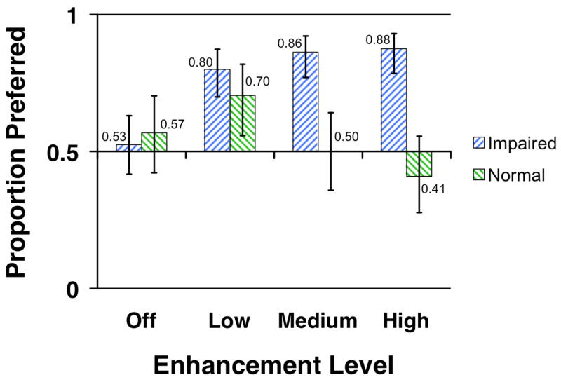
Figure 4 shows that the subjects with impaired vision had a clear preference for the enhanced video at all levels of enhancement. The Friedman test shows a significant effect on preference of the change in enhancement level only when the ‘Off’ mode was included in the analysis (χ23, 20 = 22.3, p < 0.0001) but not among the 3 levels of enhancement (χ22, 20 = 2.84, p = 0.272). This finding was confirmed by the Wilcoxon Signed Ranks test where only the differences between each level of enhancement preference and the ‘Off’ mode were significant (T19 ≥ 97.0, p ≤ 0.004). There was a small preference for the ‘Off’ level over the unprocessed image (53%), which was shown on the other screen without passing through the system (see Section 2.1). This preference was not found to be significant (N = 80, p = 0.738).
Figure 4.
Proportions of video presentations preferred at the various enhancement settings in comparing the enhanced to the unprocessed video for the impaired and normal vision groups. 0.5 represents an overall level of no group preference either way. Levels above this represent a preference for the enhancement; levels below represent an overall rejection of the enhancement relative to the original. The error bars represent the 95% confidence intervals.
Figure 4 also shows the results for the normally sighted subjects. The preferences for enhancement clearly differed between the two groups. Here the effect of a change in enhancement level was also significant (χ23, 11 = 8.59, p = 0.030). While the normally sighted controls showed a preference for the ‘Low’ levels of enhancement, they rejected the device more frequently as enhancement increased, showing a significant change within the 3 levels of enhancement (χ22, 11 = 9.83, p = 0.005), unlike the vision impaired group, and a significant paired difference between the ‘Low’ and ‘High’ levels (T10 = 0.0, p = 0.004). A test of the difference in the distribution of enhancement preferences between the two groups shows significance at the ‘Medium’ and ‘High’ enhancement levels (D20, 11 ≥ 0.445, p ≤ 0.018). Permutation tests reveal a significant change in the mean at the Medium (M20 = 3.45, M11 = 2.00, 1×10⁶ −1 permutations, p = 0.002) and High (M20 = 3.50, M11 = 1.64, 1×10⁶ −1 permutations, p = 0.001) enhancement levels.
For the vision-impaired subjects, the preference for the ‘Low’ enhancement level was significantly negatively correlated with contrast sensitivity at 2 and 8 c/deg (τ19 ≤ −0.405, p = 0.027) and approached significance at 1 c/deg (τ19 = −0.355, p = 0.053). Acuity (in logMAR, where higher values indicate poorer vision) was positively correlated with the preference for the ‘Low’ enhancement level (τ19 = −0.440, p = 0.017).
4. Discussion
We found that subjects with a wide range of central retinal vision impairment were able to see the effect of the DV1000 device from the distance they typically use for TV viewing (shorter than that used by normal viewers). They showed a clear preference for the enhancement, even at the modest enhancement levels designed for normally sighted people. In contrast, normally sighted subjects preferred the enhanced images only for the ‘Low’ level, had no preference for the ‘Medium’ level and rejected the ‘High’ level enhancement (this latter effect was significant when compared to the level of preference for the ‘Low’ level). This rejection (or lack of preference) by the normally sighted subjects may be due to the close viewing distance used in our study. At the shorter distance than normally used by these subjects, the enhancement was applied at lower spatial frequencies (already visible to the normally sighted). Evaluation of the preference by normally sighted users of the system should be carried at the normal viewing distance. Higher spatial frequencies present in HDTV pictures (for which the commercial version of the product was designed) were not present here. We also noted a degradation of the picture sharpness and some luminance saturation with the inclusion of the video splitting equipment used. This may have reduced some of the higher frequencies available for enhancement, making normally sighted subjects more likely to reject the enhancement of a sub-standard picture. Future studies of this enhancement with normally sighted viewers should utilize higher fidelity equipment.
The preference among the vision impaired subjects for the enhancement increased with increasing levels of enhancement (Figure 4), although a significant change was not found within the range of enhancement levels (‘Low’ to ‘High’). The number of subjects and number of tied differences within subjects (due to the low number of responses per enhancement level collected per subject) limited the power of the Wilcoxon Signed Ranks test to at best 15% when examining the small differences between preference for different enhancement levels in the vision impaired population shown in Figure 4. To examine more rigorously the change in preference at different levels of enhancement would require more responses, giving more levels and hence fewer ties.
We would expect that further enhancement would eventually lead to image distortions that will be rejected by the viewers (vision impaired subjects). The finding of such a rejection effect in the normally sighted group supports this expectation. However, it is likely that even higher levels of enhancement could be beneficial for people with impaired vision, as the peak of the function may not have been reached. A similar device could easily be developed for this population, providing higher levels of enhancement – and possibly enhancing different (lower) spatial frequencies. Distortions caused by this method of enhancement may be reached at a relatively low level due to local image saturations [26]. The more fully locally-adaptive capabilities of devices such as the DV1000, combined with newer high dynamic range displays [27] may reduce this limitation by providing higher dynamic range for these higher levels of enhancement (within the light and dark areas of the image prone to saturation). We are planning follow-up studies that will explore the value of the wider range of parameters available, with standard displays as well as high dynamic range displays, and will attempt to identify the useful range of enhancement.
We found significant correlation between the preference for level of enhancement and the vision impaired subjects’ visual acuity. There was also significant correlation with contrast sensitivity. These correlations were only at the ‘Low’ level of enhancement. The lack of a relationship at the two higher enhancement levels suggests that the lowest level of enhancement did not always benefit those with the lowest levels of impairment, but as the level rose, all patients tended to benefit more Tracking the preferred parameters along a scale using a new method for evaluating perceived quality of video that we recently introduced [28] is more likely to further probe the relationship between preferred enhancement and visual impairment. In that study [28], we also found correlation of level of enhancement with contrast sensitivity at 4 c/deg (significant) and at 0.4 c/deg (approaching significance). In that study (and also here) frequencies in the order of 1 to 10 c/deg were enhanced. The enhancement in the current study was limited to a high range of frequencies as the settings were designed for normally sighted observers. Studies with lower spatial frequencies enhanced may be easily conducted with the current system and the available software control.
The number of responses collected from each subject for each condition was deliberately limited in this initial study, giving only a 60% chance of detecting a side bias at the p < 0.05 level in any one subject, and giving many tied ranks for statistical analyses (see above). A future study using the same paradigm should include many more responses, allowing differences in preference to be more clearly explored.
Acknowledgments
We thank Rick Hier of DigiVision for providing the device and his assistance with the control software, Miguel Garcia-Perez, Alex Bowers, Russell Woods, Henry Apfelbaum and Matt Bronstad for advice on data analysis, Fuensanta Vera Diaz for assisting with data collection, Long To for assistance in display characterization and Doris Apfelbaum for recruitment of participants.
Neither author has any financial interest in the DigiVision or the Belkin devices.
Supported in part by NIH grants EY05957 and EY12890.
Footnotes
The data presented here were obtained using a modified version of the system/software that prevented errors we believe might have added noise to data previously reported [18].
References
- 1.Congdon N, O’Colmain B, Klaver CC, Klein R, Munoz B, Friedman DS, Kempen J, Taylor HR, Mitchell P. Causes and prevalence of visual impairment among adults in the United States. Archives of Ophthalmology. 2004;122(4):477–485. doi: 10.1001/archopht.122.4.477. [DOI] [PubMed] [Google Scholar]
- 2.Timberlake GT, Mainster MA, Peli E, Augliere RA, Essock EA, Arend LE. Reading with a macular scotoma I. Retinal location of scotoma and fixation area. Investigative Ophthalmology & Visual Science. 1986;27(7):1137–1147. [PubMed] [Google Scholar]
- 3.Peli E, Arend L, Young G, Goldstein R. Contrast sensitivity to patch stimuli: effects of spatial bandwidth and temporal presentation. Spatial Vision. 1993;7(1):1–14. doi: 10.1163/156856893x00018. [DOI] [PubMed] [Google Scholar]
- 4.Peli E. Contrast sensitivity function and image discrimination. Journal of the Optical Society of America A: Optics, Image Science and Vision. 2001;18(2):283–293. doi: 10.1364/josaa.18.000283. [DOI] [PubMed] [Google Scholar]
- 5.Packer J, Kirchner C. Who’s watching? A profile of the blind and visually impaired audience for television and video. 1997 American Foundation for the Blind http://www.afb.org/section.asp?SectionID=3&TopicID=140&DocumentID=1232.
- 6.Goldstein R, Apfelbaum HL, Luo G, Peli E. Society for Information Display International Symposium. Vol. 34. San Jose, CA: Society for Information Display; 2003. Dynamic magnification of video for people with visual impairment; pp. 1152–1155. [Google Scholar]
- 7.Peli E, Peli T. Image enhancement for the visually impaired. Optical Engineering. 1984;23(1):47–51. [Google Scholar]
- 8.Peli E, Arend LE, Jr, Timberlake GT. Computerized image enhancement for low vision: New technology, new possibilities. Journal of Visual Impairment & Blindness. 1986;80:849–854. [Google Scholar]
- 9.Peli T, Lim JS. Adaptive filtering for image enhancement. Optical Engineering. 1982;21:108–112. [Google Scholar]
- 10.Peli E, Goldstein RB, Young GM, Trempe CL, Buzney SM. Image enhancement for the visually impaired: Simulations and experimental results. Investigative Ophthalmology & Visual Science. 1991;32(8):2337–2350. [PubMed] [Google Scholar]
- 11.Peli E, Lee E, Trempe CL, Buzney S. Image enhancement for the visually impaired: the effects of enhancement on face recognition. Journal of the Optical Society of America A: Optics, Image Science and Vision. 1994;11(7):1929–1939. doi: 10.1364/josaa.11.001929. [DOI] [PubMed] [Google Scholar]
- 12.Hier RG, Schmidt GW, Miller RS, DeForest SE. Real-time locally adaptive contrast enhancement: A practical key to overcoming display and human-visual-system limitations 1993. SID International Symposium Digest of Technical Papers; Seattle, WA: Palisades Institute for Research Services, Inc; 1993. pp. 491–494. [Google Scholar]
- 13.Peli E, Fine EM, Pisano K. Video enhancement of text and movies for the visually impaired. In: Kooijman AC, et al., editors. Low Vision: Research and New Developments in Rehabilitation. IOS Press; Amsterdam: 1994. pp. 191–198. [Google Scholar]
- 14.Fine E, Peli E, Brady N. Video enhancement improves performance of persons with moderate visual loss. Proceedings of the International Conference on Low Vision, “Vision ‘96”; Madrid, Spain Book. 1997. pp. 85–92. [Google Scholar]
- 15.Peli E. Recognition performance and perceived quality of video enhanced for the visually impaired. Ophthalmic and Physiological Optics. 2005;25(6):543–555. doi: 10.1111/j.1475-1313.2005.00340.x. [DOI] [PMC free article] [PubMed] [Google Scholar]
- 16.Fine EM, Peli E. Enhancement of text for the visually impaired. Journal of the Optical Society of America A: Optics, Image Science and Vision. 1995;12(7):1439–1447. doi: 10.1364/josaa.12.001439. [DOI] [PubMed] [Google Scholar]
- 17.Goldstein RB, Woods RL, Peli E. Where people look when watching movies: Do all viewers look at the same place? Computers in biology and medicine. 2007;37(7):957–964. doi: 10.1016/j.compbiomed.2006.08.018. [DOI] [PMC free article] [PubMed] [Google Scholar]
- 18.Fullerton M, Peli E. Society for Information Display International Symposium. Vol. 38. Long Beach, CA: Society for Information Display; 2007. People with visual impairment prefer TV viewing using a contrast enhancement consumer product; pp. 975–978. [Google Scholar]
- 19.Rogowska J, Preston K, Jr, Sashin D. Evaluation of digital unsharp masking and local contrast stretching as applied to chest radiographs. IEEE Transactions Biomedical Engineering. 1988;35(10):817–827. doi: 10.1109/10.7288. [DOI] [PubMed] [Google Scholar]
- 20.Peli E. Limitations of image enhancement for the visually impaired. Optometry and Vision Science. 1992;69(1):15–24. doi: 10.1097/00006324-199201000-00003. [DOI] [PubMed] [Google Scholar]
- 21.Bailey IL, Fitz SN, Akinlabi GA. Contrast sensitivity and efficiency at search and detection tasks (Abstract) Investigative Ophthalmology & Visual Science. 2003;44(5):2771. [Google Scholar]
- 22.Kim J, Vora A, Peli E. MPEG-based image enhancement for the visually impaired. Optical Engineering. 2004;43(6):1318–1328. [Google Scholar]
- 23.Guez JE, Le Gargasson JF, Rigaudiere F, O’Regan JK. Is there a systematic location for the pseudo-fovea in patients with central scotoma? Vision Research. 1993;33:1271–1279. doi: 10.1016/0042-6989(93)90213-g. [DOI] [PubMed] [Google Scholar]
- 24.Siegel S, Castellan NJ., Jr . Nonparametric Statistics for the Behavioral Sciences (2e) McGraw-Hill; New York: 1988. [Google Scholar]
- 25.Good PI. Statistics. Springer; New York: 2000. Permutation tests: A practical guide to resampling methods for testing hypotheses. [Google Scholar]
- 26.Peli E. Limitations of image enhancement for the visually impaired. Optometry and Vision Science. 1992;69(1):15–24. doi: 10.1097/00006324-199201000-00003. [DOI] [PubMed] [Google Scholar]
- 27.Rodney LH, Fairchild MD. Beyond the locus of pure spectral color and the promise of HDR display technology. Information Display. 2007;23(7):22–26. [Google Scholar]
- 28.Fullerton M, Woods RL, Vera-Diaz FA, Peli E. Measuring perceived video quality of MPEG enhancement by people with impaired vision. Journal of the Optical Society of America A. 2007;24(12):B174–B187. doi: 10.1364/josaa.24.00b174. [DOI] [PMC free article] [PubMed] [Google Scholar]


