

Abstract
In this paper, the wire-target technique will be shown to be a useful alternative for beam profile measurements in very high frequency range (30–60 MHz). A 9 cm long tungsten wire with a diameter of 8 μm was used as the pulse-echo target to measure the lateral beam profiles at the focal points of two transducers, a spherically focused 40 MHz Panametrics transducer with an aperture size of 6.35 mm and a geometrical focal length of 12.7 mm and a lense-focused in-house lithium niobate (LiNbO3) 60 MHz transducer with an aperture size of 2 mm and a geometrical focal length around 6.5 mm. For comparison, measurements on the same transducers were performed by three small-aperture hydrophones. The first one is a polyvinylidene fluoride trifluoroethylene (PVDF-TrFE) membrane hydrophone developed by Hewlett-Packard, which has a geometric diameter of 37 μm, a measured effective diameter of less than 100 μm and a −3 dB bandwidth of more than 150 MHz. The second one is a needle-type PVDF hydrophone from Precision Acoustics, which has a 9 μm-thick PVDF element, a 40 μm geometrical aperture and a measured effective diameter of less than 100 μm. The third one is a needle-type PVDF hydrophone from Onda, which has a 150-μm geometrical aperture and an effective diameter of about 180 μm. Experimental results show that the −6 dB two-way beam widths measured by this 8 μm wire-target are in agreement with −3 dB transmitted beam widths measured by small-aperture hydrophones. Compared to small-aperture hydrophones, the wire-target technique is simpler and more cost-effective. Its major advantage however is in the frequency range above 100 MHz in which commercial hydrophones are not yet available.
1.Introduction
Very high frequency ultrasonic imaging with medical applications in dermatology, ophthalmology and intravascular imaging, and small animal imaging has been speculated by many to be the next frontier in ultrasonic imaging. Though the hydrophone method [1,2] is considered the gold standard for the characterization of transducers, conventional hydrophones often suffer severely from the effect of spatial averaging and often cannot meet the requirements of charactering high frequency transducers. Small aperture hydrophones that reduce spatial averaging are expensive and have limitations on the frequency range.
Instead of the hydrophone method, a simple wire-target technique proposed by Raum and O’Brien [3] was used in this research to characterize very high frequency transducers. Using a wire-target as the pulse-echo target, translational scans at the focus of the transducer yield the lateral profile of projections of the beam. For a symmetric beam with relative smallside lobes, the line integral projections closely track the point-to-point measurement. Raum and O’Brien [3] gave results in the frequency range of 3–17 MHz. In this research, we extended the wire-target technique to the frequency range of 35–60 MHz. The results are compared to measurements from small aperture hydrophones.
2.Experiment
The experimental system was comprised of a wire-target, three small aperture hydrophones and a computer-controlled exposimetry system designed in house [6] , which includes a water tank filled with deionized water, a digitizing (500 MS/s) oscilloscope (Model 534, Lecroy Corp., Chestnut Ridge, NY), a three-axis positioning system (Burleigh Inc., Fishers, NY) with a resolution of about 0.5 μm, as shown in Figure 1.
Figure 1.
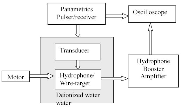
Block diagram of the experimental arrangement.
The wire-target was made from a 9 cm tungsten wire with a diameter of 8 μm (California Fine Wire, Grover Beach, CA).
Also three small-aperture hydrophones were used for comparison. The first one is a PVDF-TrFE membrane hydrophone [4] ]. This hydrophone was fabricated on a 4-μm-thick film of PVDF-TrFE, with a −3 dB bandwidth in excess of 150 MHz. It has a geometric spot diameter of 37 μm, a measured effective spot diameter of less than 100 μm and an on-membrane 7 dB gain buffer amplifier. The second hydrophone is a needle-type PVDF hydrophone (Precision Acoustics, Dorchester, UK). It has a 40 μm geometrical aperture, a 9 μm-thick PVDF element and a 12 dB submersible pre-amplifier. An external 25 dB amplifier with a bandwidth of 125 MHz was used along with those two hydrophones. The third one is a needle-type PVDF hydrophone from Onda (Onda, Sunnyvale, CA), which has a 150-μm geometrical aperture and an effective diameter of about 180 μm. Table 1 lists major characteristics of those small-aperture hydrophones.
Table 1.
Major characteristics of the three small aperture hydrophones
| PA needle hydrophone | HP membrane hydrophone | Onda Needle hydrophone | |
|---|---|---|---|
| Manufacturer | Precision Acoustics Limited | Hewlett-Packard | Onda |
| Piezoelectric material | PVDF | PVDF-TrFE | PVDF |
| Geometrical Diameter | 40 μm | 37 μm | 150 μm |
| Effective Diameter | < 100 μm | < 100 μm | < 180 μm |
| Film thickness | 9 μm | 4 μm | ~ |
| On-membrane Amplifier | 12 dB | 7 dB | ~ |
Two transducers were used in the experiments. A spherically focused 40 MHz Panametrics transducer with an aperture size of 6.35 mm and a geometrical focal length of 12.7 mm and a lens-focused LiNbO3 60 MHz transducer with an aperture size of 2 mm and a geometrical focal length around 6.5 mm fabricated in house. The transducers were excited by the Panametrics pulser/receiver (Model 5900PR and 5910RPP, Panametrics, Waltham, MA). For the pulse-echo signal from wire-target, the echo was amplified and band pass filtered by the pulser/receiver system before displayed on oscilloscope.
During the experiment, wire-target/hydrophones were placed near the focus of the transducers and the wire-target or a hydrophone was scanned by the positioning system perpendicular to acoustic axis of the transducer to obtain a lateral beam profile. The step size was 5 μm for the wire-target in all measurements. For hydrophone measurements, the step size was 25 μm for 40 MHz Panametrics transducer and 15 μm for 60 MHz LiNbO3 transducer.
3.Results and Discussions
The hydrophone-determined lateral beam profiles were compared to the wire-determined beam profile (Figures 2 and 3). In these figures, the normalized transmit lateral beam profiles measured by hydrophones were multiplied by 2 for comparison to the pulse-echo beam profile measured with the wire-target. The two way −3 dB beam widths from hydrophones and −6 dB transmit-receive beam width from the wire-target are given in Table 2. The theoretical value of the beam width is also shown at the bottom row of the table. Table 3 shows the center frequency and −20 dB pulse duration obtained around the focus of the transducers. All acoustic field definitions can be found on [2].
Figure 2.
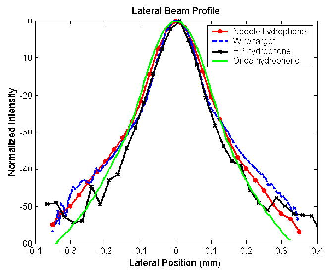
Lateral beam profiles of the 40 MHz Panametrics transducer
Figure 3.
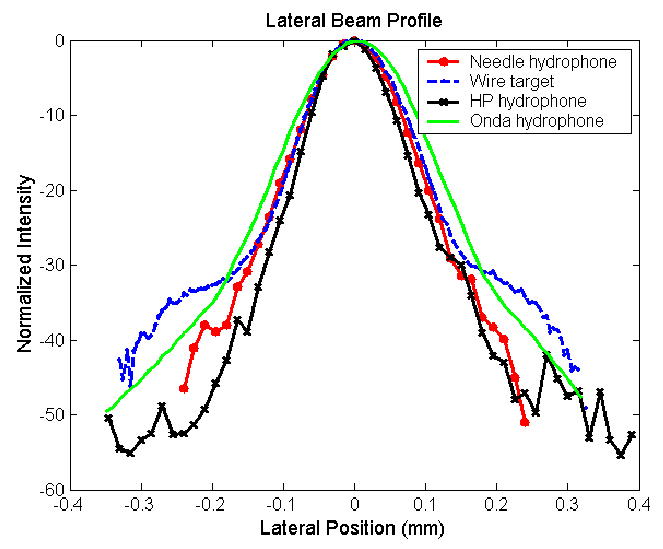
Lateral beam profiles of the 60 MHz LiNbO3 transducer
Table 2.
-6 dB transmit-receive beam widths measured by 8 μm wire target and -3 dB transmit beam widths measured by three small-aperture hydrophones
| Panametrics transducer Beam width (μm) | In-House transducer Beam width (μm) | |
|---|---|---|
| 8 μm wire target | 82.3 | 107.1 |
| HP Hydrophone | 77.4 | 92.4 |
| PA Hydrophone | 89.6 | 103 |
| Onda Hydrophone | 104.7 | 126.4 |
| f-number × wavelength | 74.1 | 80 |
Table 3.
Center frequency and pulse duration measured by wire target and small aperture hydrophones
| Panametrics transducer | In-House transducer | |||
|---|---|---|---|---|
| Center frequency (MHz) | Pulse duration (ns) | Center frequency (MHz) | Pulse duration (ns) | |
| 8 μm wire target | 42.5 | 64.9 | 56.2 | 55.7 |
| HP Hydrophone | 40.6 | 49 | 60.3 | 36.4 |
| PA Hydrophone | 37.5 | 51.3 | 58.4 | 53.2 |
| Onda Hydrophone | 34.7 | 49.2 | 40.7 | 53 |
With its broad bandwidth and smallest aperture size, HP membrane hydrophone suffers less from the spatial averaging. For both transducers, the beam width from HP hydrophone is the smallest and the pulse duration (36.4 ns) for 60 MHz transducer the shortest. However, the signal to noise ratio of HP membrane hydrophone is relatively poor. In Figure 2 and Figure 3, it can be seen that due to noise, there are irregularities in lateral beam profiles measured with the HP hydrophone when the lateral position is away from the center.
The Onda hydrophone has higher signal to noise ratio, but it has the largest spatial averaging effects due to its largest effective diameter (about 180 μm). When measuring the 60 MHz transducer, the center frequency obtained from Onda hydrophone (40.7 MHz) differed substantially from those estimated by other devices.
The wire-target method is the simplest. It has the highest signal to noise ratio though its results may not be as “ideal” as those obtained by HP membrane hydrophone. In fact the utilization of a smaller diameter wire-target may further improve the results because of a smaller spatial averaging effect presented by the area of the wire. It can certainly be extended to the frequency range above 100 MHz. The wire-target method in a sense is analogous to the optical Schlieren method. Tomographic reconstruction algorithms can be used to remove the line integral effect. In order to do so while achieving satisfactory resolution, 180 projections may be needed. One projection will need 5~10 minutes. Thus, for 180 projections 15~30 hours may be needed. In such a case, it is even more time-consuming than the point-to-point hydrophone measurements.
Small aperture hydrophones have directivity. The higher the frequency, the stronger of the directivity. The directivity of wire target is complex, and also depends on the wavelength. More research is needed to further address the effect of this issue on the accuracy of the results.
4.Conclusion
The results reported in this paper demonstrate that the wire-target technique is a useful alternative to small-aperture hydrophones in characterizing ultrasonic transducer radiation patterns. The wire-target technique is simpler and more cost-effective. Its major advantage however is in the frequency range above 100 MHz in which commercial hydrophones are not yet available.
Acknowledgments
This work was supported by NIH grant #P41-EB2182.
References
- 1.IEEE. IEEE guide for medical ultrasound field parameter measurements. IEEE. 19901989:790. [Google Scholar]
- 2.AIUM 1992 Acoustic Output Measurement and Labeling Standard for Diagnostic Ultrasound Equipmenmt (Laurel,MD)
- 3.Kay Raum, O’Brien WD. Pulse-Echo Field Distribution Measurement Technique for High-Frequency Ultrasound Sources. IEEE Trans Ultrason Ferroelec Freq Contr. 1997;44(4):810–815. [Google Scholar]
- 4.Lum P, Greenstein M, Grossman C, Szabo TL. High-freqency membrane hydrophone. IEEE Trans Ultrason Ferroelec Freq Contr. 1996 July;43(4):536–44. [Google Scholar]
- 5.Lum, P.,Greenstein,M.etc. A 150-MHz-Bandwidth Membrane Hydrophone for Acoustic Field Characterization, The Hewlett-Packard Journal, 6–16 August 1988
- 6.Snook KA, Huang B, Smith NB, Shung KK. An exposimetry system for characterization of acoustic fields above 20 MHz. 2000 IEEE Ultrasonics Symposium. 2:1109–12. [Google Scholar]