Abstract
Agrobacterium-mediated transformation for poinsettia (Euphorbia pulcherrima Willd. Ex Klotzsch) is reported here for the first time. Internode stem explants of poinsettia cv. Millenium were transformed by Agrobacterium tumefaciens, strain LBA 4404, harbouring virus-derived hairpin (hp) RNA gene constructs to induce RNA silencing-mediated resistance to Poinsettia mosaic virus (PnMV). Prior to transformation, an efficient somatic embryogenesis system was developed for poinsettia cv. Millenium in which about 75% of the explants produced somatic embryos. In 5 experiments utilizing 868 explants, 18 independent transgenic lines were generated. An average transformation frequency of 2.1% (range 1.2–3.5%) was revealed. Stable integration of transgenes into the poinsettia nuclear genome was confirmed by PCR and Southern blot analysis. Both single- and multiple-copy transgene integration into the poinsettia genome were found among transformants. Transgenic poinsettia plants showing resistance to mechanical inoculation of PnMV were detected by double antibody sandwich enzyme-linked immunosorbent assay (DAS-ELISA). Northern blot analysis of low molecular weight RNA revealed that transgene-derived small interfering (si) RNA molecules were detected among the poinsettia transformants prior to inoculation. The Agrobacterium-mediated transformation methodology developed in the current study should facilitate improvement of this ornamental plant with enhanced disease resistance, quality improvement and desirable colour alteration. Because poinsettia is a non-food, non-feed plant and is not propagated through sexual reproduction, this is likely to be more acceptable even in areas where genetically modified crops are currently not cultivated.
Keywords: Euphorbia pulcherrima, Somatic embryogenesis, Transformation, Agrobacterium tumefaciens, Poinsettia mosaic virus
Introduction
Poinsettia, Euphorbia pulcherrima Willd. Ex Klotzsch, is a contemporary symbol of Christmas in most parts of the world. Since it was introduced to the United States in 1825 from Mexico, poinsettia has become the primary potted flower produced and sold in North America, Europe, Asia and Australia (Ecke et al. 2004; Williams 2005). Today, Europe and North America represent the largest volume of production and sales, but demand is growing quickly in the Australian region as poinsettia becomes more popular each year (Williams 2005). Global production of poinsettia has exceeded hundreds of millions and is still expanding, indicating its economic and market potential for the floral industry.
Genetic engineering is an important tool for breeding ornamental plants with addition of desirable traits such as novel colour, better quality and resistance to pathogens and insects (Mol et al. 1995; Deroles et al. 2002; Hammond 2006; Hammond et al. 2006). This technology has been successfully utilized in the production of a number of important ornamental crops e.g. blue roses (Yoshikazu 2004), novel carnations (http://www.florigene.com), transgenic gladiolus (Kamo et al. 1997) and improvement of chrysanthemums (Teixeira da Silva 2004). To date, transgenic ornamentals of over 30 genera have been produced by different transformation approaches (Hammond 2006; Hammond et al. 2006). However, there are only a few reports describing genetic transformation of poinsettia: one was the US patent 7119262 (Smith et al. 1997) using the biolistic transformation approach, while the other two were electrophoresis-based transformation attempts (Vik et al. 2001; Clarke et al. 2006). Biolistic transformation requires the use of a gene gun device (Sanford et al. 1987) and tends to generate transformants with a high transgene copy number, complex transgene loci and unpredictable silencing of the transgene (Herrera-Estrella et al. 2004). Electrophoresis of DNA into meristems on a living plant was described as a simple method to generate transformants by avoiding tedious tissue culture work and was utilized in producing transgenic orchid (Griesbach 1994). However, no stable transgenic poinsettia was ever produced using electrophoresis, regardless of the strong transient expressions that were detected in both studies (Vik et al. 2001; Clarke et al. 2006). Thus, Agrobacterium-mediated transformation for poinsettia was developed in the present study.
Poinsettia mosaic virus (PnMV) is a single-stranded, positive-sense RNA virus (Bradel et al. 2000) that belongs to the family Tymoviridae (Dreher et al. 2005). Infection of poinsettia plants with PnMV results in mosaic symptoms during parts of the growing season (Fulton and Fulton 1980), which in turn decreases the commercial value of this ornamental plant. Thus, growers are interested in the potential benefits of growing PnMV-free poinsettias. PnMV-free poinsettia plants can be obtained by heat treatment or in vitro culture of apical meristems, which are time-consuming and cost-ineffective methods. An additional problem is that PnMV-free poinsettia tends to be rapidly reinfected, although no vector is known (Blystad and Fløistad 2002; Siepen et al. 2005). There is therefore a need for a new and effective alternative approach, like Agrobacterium-mediated transformation, which can overcome these difficulties.
RNA silencing is a mechanism by which transcription or translation of a gene is suppressed. It is known to occur in plants, fungi and animals (Fire et al. 1998; Waterhouse et al. 1998; Baulcombe 2005). It is triggered by double stranded RNA (dsRNA) molecules (Meister and Tuschl 2004), which are subsequently recognized and cleaved by the host-encoded endoribonuclease dicer (Bernstein et al. 2001) into small interfering RNA (siRNA) molecules of 21–26 nucleotides in length (Hamilton and Baulcombe 1999). These siRNAs, in conjunction with the RNA-induced silencing complex (RISC), target RNA molecules with homologous sequences for sequence-specific degradation (Hammond et al. 2000; Bernstein et al. 2001; Tabara et al. 2002). In plants, RNA silencing can be achieved by genetic transformation with gene constructs that express highly transcribed sense, anti-sense or self complementary hairpin RNA (hpRNA) containing sequences homologous to the target gene (Smith et al. 2000; Wesley et al. 2001; Helliwell and Waterhouse 2003). RNA silencing has been efficiently used to generate resistance against plant viruses in many plant species including ornamentals (Metzlaff et al. 1997; Tenllado et al. 2004; Bucher et al. 2006; Hammond et al. 2006).
In this study, we report the development of an A. tumefaciens-mediated transformation method for poinsettia, which has never previously been described for this plant species. This method could facilitate the improvement of poinsettia by introducing new traits into existing commercial poinsettia cultivars in order to meet market demands. Using this approach, in combination with RNA silencing technology, transgenic PnMV resistant poinsettia plants carrying PnMV-derived hpRNA constructs were produced for the first time.
Materials and methods
Plant materials
Euphorbia pulcherrima, poinsettia cv. Millenium, plants were kindly supplied by the J. Kristiansen nursery, Grimstad, Norway, in the year 2000. The original stock plants were subjected to heat therapy to eliminate PnMV (Fløistad and Blystad, unpublished). PnMV-free cv. Millenium cuttings were grown in the greenhouse under a photoperiod of 16 h light and 8 h dark with a temperature of 22°C. Internode stem explants from 8 to 10-week-old cv. Millenium plants were used for somatic embryogenesis and A. tumefaciens-mediated transformation.
Somatic embryogenesis of poinsettia cv. Millenium
Internode stem explants 5–15 mm long from cv. Millenium plants were excised and used in the establishment of somatic embryogenesis for poinsettia prior to transformation. In Experiments 1 and 2, the explants were surface sterilized for 10 min in 3% NaOCl and then rinsed three times with sterilized deionized and autoclaved H2O for 5, 10 and 15 min according to Preil (1994). Because of a large number of infections encountered in Experiments 1 and 2, the stem explants were sterilized in Experiment 3 using 70% ethanol for 1 min followed by 5 min with 1% NaOCl and 3 rinsings with sterile deionized and autoclaved water for 3, 10 and 20 min, a modification of the protocol of Preil (1994). The modified protocol was repeated twice before being utilized in Experiment 3 and also in subsequent transformation experiments. After sterilization, stem segments of ca. 1–1.5 mm thickness were placed on callus induction (CI) medium [MS medium (Murashige and Skoog 1962) supplemented with 0.2 mg l−1 BAP and 0.2 mg l−1 CPA and 30 g l−1 sucrose, Table 1] for 8–10 days. Embryogenic calli were then transferred to somatic embryo induction (SEI) medium (MS medium contains 0.3 mg l−1 NAA and 0.15 mg l−1 2iP and 30 g l−1 sucrose, Table 1) for somatic embryogenesis. The somatic embryos emerged after an average of 12 weeks were then transferred to somatic embryo maturation (SEM) medium (MS basal medium containing 0.05 mg l−1 BAP and 30 g l−1 sucrose, Table 1). Shoots and plantlets derived from somatic embryos were subsequently cultured on root induction (RI) medium consisting of 1/2 strength MS, 2 mg l−1 IAA and 20 g l−1 sucrose or on hormone free 1/2 strength MS (HFMS) medium supplemented with 20 g l−1 sucrose for root induction (Table 1). Plants with well-developed roots were transferred to soil and grown in the greenhouse at 22°C. The above culture conditions and media compositions for somatic embryogenesis of cv. Millenium were developed according to Preil (1994) with a number of modifications described above. Detailed information regarding media compositions is summarized in Table 1.
Table 1.
Media names and compositions modified according to Preil (1994)
| Medium | MS | Sucrose (%) | CPA (mg l−1) | BAP (mg l−1) | NAA (mg l−1) | 2iP (mg l−1) | IAA (mg l−1) |
|---|---|---|---|---|---|---|---|
| CI | Full strength | 3 | 0.2 | 0.2 | – | – | – |
| SEI | Full strength | 3 | – | – | 0.3 | 0.15 | – |
| SEM | Full strength | 3 | – | 0.05 | – | – | – |
| RI | 1/2 strength | 2 | – | – | – | – | 2 |
| HFMS | 1/2 strength | 2 | – | – | – | – | – |
CI Callus induction medium, SEI somatic embryo induction medium, SEM somatic embryo maturation medium, RI root induction medium, HFMS hormone free MS medium for root induction
Light conditions were 23 μE m−2 s−1 for callus and somatic embryos and 30 μE m−2 s−1 for plantlets in RI and HFMS media with a 16 h photoperiod. Temperature was constant at 24°C. A light microscope connected to a digital camera was used to follow the development of the somatic embryos.
Agrobacterium strain and hairpin (hp) RNA constructs
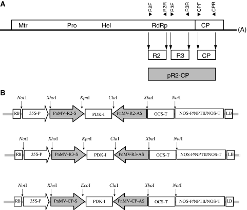
The disarmed A. tumefaciens strain LBA4404 (Hoekema et al. 1983; Invitrogen, California, USA) was utilized throughout the study. Three hpRNA constructs, named as pCP, pR2 and pR3, were generated. Construct pCP targeted the viral coat protein (CP), whereas constructs pR2 and pR3 targeted two distinct regions within the viral RNA-dependant RNA-polymerase (RdRp) (Fig. 1a). Briefly, constructs pCP, pR2 and pR3 were generated by amplifying the corresponding fragments from the viral genome and introducing the appropriate restriction sites. The following restriction sites were introduced: XhoI·KpnI (for sense orientation) and ClaI·XbaI (for antisense orientation) for R2 and R3 fragments, and XhoI·EcoRI (sense) and ClaI·XbaI (antisense) for CP fragment. The primers used are presented in Table 2. Each amplified fragment was 500 bp long. Subsequently, the fragments were inserted into pHANNIBAL vector (kindly provided by CSIRO Plant Industry, Canberra, Australia) in sense and antisense orientations, interrupted by an intron pyruvate orthophosphate dikinase (pdk) gene (Fig. 1b) as described by Helliwell and Waterhouse (2003). The three expression cassettes were cloned into pART27 (Gleave 1992, kindly provided by CSIRO Plant Industry, Canberra, Australia), a binary plasmid vector, at Not1 site under the control of CaMV 35S promoter and with ocs terminator (Helliwell and Waterhouse 2003), generating hpRNA constructs pCP, pR2 and pR3 (Fig. 1b). For selection, the neomycin phosphotransferase II (nptII) gene conferring kanamycin resistance was used under the control of nopaline synthase promoter (Nos-P) and with Nos terminator (NosT, Fig. 1b) in the binary plasmid expression vector pART27. Sequence analysis was utilized to verify the junctions and orientations in the expression vectors pCP, pR2 and pR3. All three plasmid vectors were introduced into A. tumefaciens strain LBA 4404 by electroporation according to the manufacturer’s instructions (Invitrogen, California, USA).
Fig. 1.
Schematic representation of PnMV genome (a) and hairpin (hp) RNA constructs used in the current study (b). The locations of the primers used to generate the constructs as well as for screening purposes are presented over the PnMV genome. Sequences of the primers are presented in Table 2. The location of the probe used to detect siRNA is presented below the PnMV genome (see “Materials and methods”). CP coat protein region, R2 and R3 RNA-dependent RNA-polymerase (RdRp) regions of the PnMV genome
Table 2.
Primers used in this study
| Primer name | Restriction sites | Sequencea | Location |
|---|---|---|---|
| R2F-XhK | XhoI, KpnI | ctcgagggtaccTTTAGCAAAACGCAGCACAAAATCA | 4,371–4,395 |
| R2R-CXb | ClaI, XbaI | catcgattctagaTCTCCAGACACCATGATTGGGTG | 4,848–4,870 |
| R3F-XhK | XhoI, KpnI | ctcgagcggtaccTTCGCTTTAAAACAGAAAGCACCA | 4,939–4,962 |
| R3R-CXb | ClaI, XbaI | catcgatgtctagaGCCTCGTAGCTTGGTTGGGTT | 5,418–5,438 |
| CPF-XhE | XhoI, EcoRI | ctcgaggaattcAACCACGTCGACTCCACTCCAT | 5,478–5,499 |
| CPR-CXb | ClaI, XbaI | atcgattctagaAGCTTGCCGCTCACCAGCAC | 5,958–5,977 |
aPrimer sequences are presented in the 5′–3′ orientation. Sequences complementary to the viral genome are presented in uppercase letters whereas non-complementary restriction sites included in each primer are presented in lowercase letters. The primers used for screening purposes are identical to the ones presented but do not include the non-complementary restriction sites
Transformation of poinsettia stem explants
A. tumefaciens strain LBA4404, harbouring the plasmid pCP, pR2 or pR3, was grown overnight in 15 ml liquid LB medium supplemented with 50 mg l−1 kanamycin (Sigma-Aldrich, St Louis, USA) at 28°C with shaking at 200 rpm until an OD600 of 0.6–0.8 was reached. The bacterium suspension was collected at 2,700 rpm for 10 min, washed twice with MS basal medium supplemented with 2% sucrose (MS-2), and resuspended in 10 ml MS-2.
The internode stem explants taken from 8 to 10-week-old poinsettia plants derived from cuttings were disinfected as described above, excised into stem segments with 1–1.5 mm thickness and inoculated with Agrobacterium suspension for 5 min with gentle shaking. After the infection, stem segments were blotted briefly with sterile filter paper and placed on CI medium at 24°C in the dark for 72 h without selection. After co-cultivation, the explants were blotted gently on sterile filter paper and transferred to the CI medium with selection and light conditions as described above.
Selection and regeneration of transgenic plants
After 8–10 days on CI medium supplemented with 500 mg l−1 claforan (Aventis Pharma Ltd, Norway) and 10 mg l−1 kanamycin, the explants were transferred to SEI medium supplemented with 400 mg l−1 claforan and 25 mg l−1 kanamycin for somatic embryogenesis. The protocol for somatic embryogenesis is detailed above. The somatic embryos obtained after about 12 weeks were then transferred to the SEM medium supplemented with 400 mg l−1 claforan and 10 mg l−1 kanamycin. Shoots and plantlets derived from somatic embryos were subsequently cultured on RI medium or on hormone free 1/2 strength MS (HFMS) medium (Table 1) for root induction and were subsequently transferred to soil and grown in the greenhouse at 22°C.
Screening of transgenic poinsettia plants
Screening of transgenic poinsettia plants was carried out by PCR. The primer pairs used to amplify the CP, R2 and R3 fragments were the same as those for vector constructions, but did not include any additional restriction site (Table 2). HotStarTaq PCR kit purchased from Qiagen (Valencia, California) was used in PCR screening of transformants. Twenty micro litres of reaction mixture containing 2× HotStarTaq Mastermix, 0.4 μM of each primer, 0.1 μg template DNA and H2O were subjected to PCR amplification under the following conditions: 15 min at 95°C (1 cycle), 30 s at 95°C, 30 s at 55°C, 1 min at 72°C (35 cycles) and a final extension 10 min at 72°C (1 cycle) according to the manufacturer’s instructions. All the PCR amplification was performed using Applied Biosystems 96 Thermal Cycler (Applied Biosystems). PCR products were analysed by electrophoresis on 0.8% (W/V) agarose gels.
Southern blot analysis
To confirm the stable integration of transgenes into the poinsettia genome and to evaluate transgene copy number, Southern blot analysis was performed, basically as described by Sambrook et al. (1989). Total genomic DNA was isolated from young leaves of control plant and the putative transformants using a modified CTAB protocol as described by Rogers and Bendich (1988). Ten micrograms of genomic DNA was digested with the restriction enzyme HindIII for 4 h and separated on a 1% (W/V) TBE agarose gel overnight at 37 V followed by transfer onto Gene Screen Transfer membrane (NEN™ Life Science Products Inc., Boston, MA, USA). The HindIII site is not present in the pCP, pR2 and pR3 vectors. The HindIII site from the pHANNIBAL vector was removed during the vector construction. Membranes were hybridized overnight with 32P-labelled probe of 1.5 kb in size targeting the CP, R2 and R3 regions (Fig. 1a). The probe was generated by amplifying a 1.5 kb fragment that included the R2, R3 and CP regions used to generate the hairpin RNA constructs pR2, pR3 and pCP. This allowed us to use one probe instead of three probes for southern blot analyses of pR2, pR3 and pCP transformants.
Northern blot hybridization of low molecular weight RNA
Total RNA was extracted using the Trizol reagent (Invitrogen, USA) as recommended by the manufacturer. Low molecular weight (LMW) RNA was separated from high molecular weight RNA by precipitation with 1 volume of 4 M LiCl4 at 4°C overnight. After centrifugation, the LMW RNA-containing supernatant was transferred to a new tube and precipitated with 1 volume of isopropanol. LMW RNA concentrations were measured using a GeneQuant II spectrophotometer (Amersham Biosciences, UK) and RNA quality was verified by agarose gel electrophoresis. LMW RNA (approximately 30 μg) was mixed with 1 volume of Tris-borate–EDTA–urea sample buffer (Biorad, USA) and heated at 100°C for 5 min. Subsequently, LMW RNA was separated on a 15% polyacrylamid tris-borate–EDTA–urea gel and transferred to a Hybond-N membrane (Amersham Biosciences, UK) overnight. The membranes were hybridized with a digoxigenin (DIG)-labelled (Roche Diagnostics, Germany) RNA probe (Fig. 1a), previously cleaved by alkaline hydrolysis to generated fragments of approximately 50 bp as described by Hamilton and Baulcombe (1999). After hybridization the membranes were washed at room temperature, 2 × 15 min in 5× SSC + 0.5% SDS and 2 × 15 min 1× SSC + 0.5% SDS and then exposed to films.
Virus inoculation and detection
Sap from poinsettia and N. benthamiana plants systemically infected with PnMV was used as an inoculum. Approximately 0.5 g of infected leaf tissue was ground with 4 ml of inoculation buffer [0.03 M sodium phosphate buffer (pH 8)] and mechanically inoculated to leaves of three to four-leaves stage plants lightly dusted with carborundum. Plants were grown in the greenhouse at 22–24°C.
Detection of PnMV infection by double antibody sandwich enzyme-linked immunosorbent assay (DAS-ELISA) was carried out at 6 and 10 weeks post-inoculation basically as described by Clark and Adams (1977). The uppermost fully expanded leaves of each plant were collected in polyvinyl bags (Bioreba, USA). Extraction buffer [100 mM phosphate buffer (pH 7.4), 20 mM Tris, 137 mM NaCl, 3 mM KCl, 2% PVP 24 kD, 0.05% Tween 20, and 0.02% NaN3] was added to the bags (1/10 w/v) and the samples were macerated. A measure of 100 μl of the homogenates were transferred to wells of microtitre plates previously coated with 100 μl of PnMV IgG (Agdia, USA) diluted 1/1,000 in coating buffer [50 mM carbonate–bicarbonate buffer (pH 9.6) and 0.02% NaN3], followed by incubation at +4°C overnight. Microtitre plates were washed with washing buffer [10 mM phosphate buffer (pH 7.4), 140 mM NaCl, 3 mM KCl, and 0.05% Tween 20] and 100 μl of alkaline phosphatase conjugated PnMV IgG (Agdia, USA) diluted 1/1,000 in extraction buffer was added to each well. Microtitre plates were incubated at +4°C overnight. Subsequently, they were washed with washing buffer and 100 μl of substrate buffer [1 M diethanolamine (pH 9.8) and 0.02% NaN3] containing p-nitrophenyl phosphate (0.5 mg ml−1) was added. Absorbance values were recorded at 405 nm with an Expert plus microtitre plate reader using the Kim software (ASYS HITECH, Austria). Values were considered positive when the A450 value was twofold higher than that of the negative control.
Results
Establishment of somatic embryogenesis for poinsettia cv. Millenium
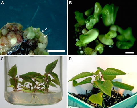
An efficient protocol for somatic embryogenesis of poinsettia cv. Millenium was developed. Stem explants of cv. Millenium responded well to the induction media and a large number of somatic embryos were effectively induced (Table 3; Fig. 2a–d). In Experiments 1 and 2, somatic embryos could only be induced from 21 to 37% of the explants due to infection, whereas more than 75% of the explants produced somatic embryos in Experiment 3 after the modification of the disinfection protocol. The average numbers of somatic embryos on each embryogenic callus varied from 5.3 ± 2.4 and 5.8 ± 3.1 for Experiments 1 and 2, respectively, to 7.2 ± 4.1 for Experiment 3 (Table 3). The highest induction of somatic embryos was found in Experiment 3 with 18 somatic embryos on a single callus. When globular stage somatic embryos (Fig. 2a, arrowed) reached the mature stage (Fig. 2b), they became loosely attached to the calli. Shoots derived from somatic embryos were subsequently cultured on RI medium or HFMS medium (Fig. 2c) for root induction and were transferred into the greenhouse when the roots were well established (Fig. 2d). The three experiments conducted and the efficiency of somatic embryogenesis are summarized in Table 3.
Table 3.
Experiments and efficiency of somatic embryogenesis
| Experiment | No. of explants | % of embryogenic explants | Range of SE/embryogenic explant | Mean SE/embryogenic explanta | No. of regenerated plantletsb |
|---|---|---|---|---|---|
| 1 | 200 | 21 | 2–10 | 5.3 ± 2.4 | 70 |
| 2 | 200 | 37 | 2–14 | 5.8 ± 3.1 | 135 |
| 3 | 350 | 75 | 3–18 | 7.2 ± 4.1 | 768 |
SE somatic embryos
aValues are presented with their corresponding standard deviation
bRegenerated plantlets after 14 weeks
Fig. 2.
Somatic embryogenesis in poinsettia cv. Millenium: a embryogenic structure and globular stage somatic embryos (arrows) that appeared on the callus (bar 1 mm), b cotyledonary stage of somatic embryos (bar 1 mm), c plantlets deriving from somatic embryos on RIM medium; and d regenerated plants established in the greenhouse
The RI and HFMS media used for root induction did not show any clear differences in the percentage (85 and 87% for RI and HFMS, respectively) of plants that developed roots 3 weeks after being transferred to the rooting media.
Development of the A. tumefaciens-mediated transformation method
Prior to the production of transgenic poinsettia plants, a number of optimizations were carried out to reveal the most suitable conditions for inoculation and co-cultivation, concentration of antibiotic for eliminating A. tumefaciens after co-cultivation, and kanamycin selection without inhibiting somatic embryogenesis and regeneration of plants. Results showed that the optimal inoculation time was 5 min with gentle shaking, while the most suitable co-cultivation time was 72 h (Table 4). Using this combination, 26 out of the 80 (33%) explants tested PCR positive after inoculation, co-cultivation and 2 weeks on claforan (Table 4). Those explants were followed and showed normal embryogenesis and regeneration as compared to the control (data not shown). Therefore, these conditions were utilized throughout all the transformation experiments. When inoculation time exceeded 20 min followed by a co-cultivation period of 6 days, only 4% of the explants survived due to the overgrowth of Agrobacterium on the surfaces of the explants (Table 4).
Table 4.
Number of PCR positive explants that survived 2 weeks after Agrobacterium inoculation and co-cultivation treatments. A total of 80 explants were used in each treatment
| Co-cultivation time (days) | Inoculation time | ||
|---|---|---|---|
| 5 min | 10 min | 20 min | |
| 2 | 9 (11%) | 13 (16%) | 21 (26%) |
| 3 | 26 (33%) | 21 (26%) | 20 (25%) |
| 6 | 10 (13%) | 8 (10%) | 3 (4%) |
For elimination of Agrobacterium growth after co-cultivation, the optimized concentration of claforan was 500 mg l−1 for the CI medium, whereas 400 mg l−1 claforan was an adequate concentration for the SEI and SEM media. For selection of transformed cells, 25 mg l−1 kanamycin was found optimal for the SEI medium, while 10 mg l−1 kanamycin was optimal for the CI and SEM media.
Production of transgenic poinsettia plants, molecular analyses and resistance assays
To produce transgenic poinsettia plants, 5 transformation experiments including 868 stem segment explants were carried out using the established inoculation, co-cultivation and selection conditions described above. Of those explants, 228 (26.3%) somatic embryo-derived plants were obtained. The results are summarised in Table 5.
Table 5.
Summary of five Agrobacterium-mediated transformation experiments on poinsettia cv. Millenium with pCP, pR2 and pR3 constructs
| Experimenta | No. of explants | No. of regenerated plants | No. of transformantsb | Transformation efficiency (%) c |
|---|---|---|---|---|
| 1 (CP) | 185 | 54 | 3 | 1.6 |
| 2 (R3) | 132 | 69 | 3 | 2.3 |
| 3 (R3) | 172 | 66 | 6 | 3.5 |
| 4 (R2) | 254 | 21 | 3 | 1.2 |
| 5 (R2) | 125 | 18 | 3 | 2.4 |
aCP, R3, and R2 represent transformation experiments with constructs pCP, pR2 and pR3, respectively
bTransformants verified by PCR and Southern blot analysis
cNumber of transformants/total number of explants transformed
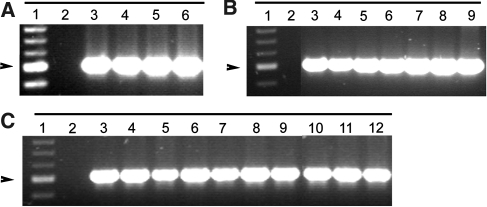
PCR analysis was conducted for screening the putative transformants. All 228 regenerated plants were analysed by PCR and 18 transformants were revealed. Of these, three possessed pCP constructs, while six and nine contained pR2 and pR3 constructs, respectively (Fig. 3a–c). Transformation frequency varied from 1.2 to 3.5% with an average transformation frequency of 2.1% (Table 5).
Fig. 3.
PCR analysis. PCR positive transformants detected with primer pairs for (a) CP, (b) R2 and (c) R3 fragments respectively. Lane A1 1 kb marker; lane A2 non-transformed plant; lanesA3–5 are independent CP-transgenic lines 11-1, 11-2 and 3-1; laneA6 plasmid control. Lane B1 1 kb marker; lane B2 non-transformed plant; lanes B3-8 are R2-transgenic lines 72-1, 72-2, 75-2, 79-1, 84-2, and 84-4; lane B9 plasmid control. Lane C1 1 kb marker; lane C2 non-transformed plant, lanes C3-11 are R3 transgenic lines 18-1, 30A, 38-1, 40A, 40B, 41-2, 41-5, 56-2 and 62-1; lane C12, plasmid control. The 500 bp band of the 100 bp marker is depicted with an arrow
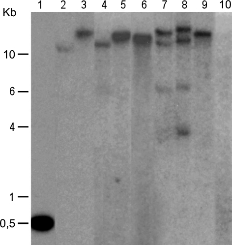
Southern blot analysis was performed on selected PCR positive plants with satisfactory characteristics for studying transgene integration and estimating the transgene copy number. Results of Southern blot analysis confirmed the stable integration of transgene and both single- and multiple-copy transgene integration into the poinsettia genome were detected among the transformants (Fig. 4). Of the eight transformants analysed, six showed single copy integration of transgene (lanes 2–6 and 9 in Fig. 4) with fragment size greater than the length of intact T-DNA (ca 7.5 kb). However, two pR3 transformants (lanes 7–8 in Fig. 4) showed multiple-copy transgene integration with both large (intact T-DNA) and small fragments (partial, non intact T-DNA).
Fig. 4.
Southern blot analysis of selected PCR positive transformants carrying pCP, pR2 and pR3 constructs. The HindIII-digested total genomic DNA was probed with a 1.5 kb probe homologous to the region of CP, R2 and R3 fragments (Fig. 1) allowing us to analyse all the three different types of transformants (CP, R2 and R3 transformants) at the same time. Lane1 plasmid control; lanes2-4 CP transformants 11-1, 11-2 and 3-1; lanes5-6 R2 transformants 72-2, 79-1; lanes 7-9 R3 transformants 38-1, 40B, 18-1; lane10 negative control
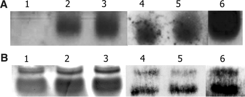
Furthermore, Northern blot analysis of low molecular weight RNA was carried out on the transgenic plants harbouring constructs pCP, pR2 and pR3, prior to inoculation. Transgene-derived siRNA molecules of 21–26 nucleotides length were detected in transgenic lines pCP 11-1, pCP 3-1, pR2 72-1, pR2 72-2 and pR3 18-1 (Fig. 5). This data indicated that the transgene had been transcribed and converted into siRNAs (Fig. 5). Due to limitations in the amount of LMW RNA from transgenic pR3 lines 40B, 41-2, 62-1 and 41-5, these lines were not included in the experiment.
Fig. 5.
Northern blot analysis of low molecular weight (LMW) RNA to detect small interfering RNAs (siRNA) in non-inoculated transgenic plants and non-inoculated control. aLane 1 control; lanes 2–3 CP transgenic lines 11-1, 3-1; lanes 4–5 R2 transgenic lines 72-1,72-2; lane 6 R3 transformant, 18-1. Ribosomal RNA is presented in (b) to indicate presence of LMW RNA in the gel. Lane 1 control; lanes 2 and 3 PCP transformed plants; lanes 4 and 5 PR2 transformed plants; lane 6 PR3 transformed plant
Transgenic plants and non-transformed controls were mechanically inoculated with PnMV at a three to four leaves stage. The upper non-inoculated leaves were tested by DAS-ELISA at 6 and 10 weeks post-inoculation. Results showed that the control plants were systemically infected with PnMV, whereas the tested transgenic lines carrying pCP and pR2 constructs remained virus free (Table 6). However, of the five pR3-containing transgenic lines studied, two lines remained virus free, while the remaining three lines were systemically infected (Table 6).
Table 6.
Immunological detection of PnMV on non-transformed and transformed poinsettia plants inoculated with PnMV
| Construct | Transgenic planta | ELISAb | |
|---|---|---|---|
| 6 wpi | 10 wpi | ||
| R2 | 72-1 | 0.1 | 0.1 |
| 72-2 | 0.1 | 0.2 | |
| R3 | 18-1 | 0.1 | 0.2 |
| 40B | 0.8 | 2.4 | |
| 41-2 | 0.5 | 2.6 | |
| 62-1 | 0.1 | 0.2 | |
| 41-5 | 0.7 | 2.8 | |
| CP | 11-1 | 0.1 | 0.2 |
| 3-1 | 0.1 | 0.1 | |
| Control + | 1 | 0.8 | 2.8 |
| 2 | 0.8 | 2.7 | |
| Control − | 1 | 0.1 | 0.2 |
| 2 | 0.2 | 0.2 | |
| 3 | 0.2 | 0.2 | |
| 4 | 0.1 | 0.1 | |
wpi Weeks post inoculation
aOne plant per transgenic line was challenged in this assay
bELISA absorbance values (405 nm) after 60 min of incubation at room temperature are presented. Samples with twice the value of the highest negative control sample were deemed positive
Discussion
Significance of genetic engineering in control of virus diseases in ornamentals
The application of genetic engineering in the floriculture industry has clearly become instrumental and rewarding in meeting the demand for novel desirable traits. A number of genetic engineering approaches have been developed for ornamental plants (Griesbach 1994; Kamo et al. 1995; Deroles et al. 1997; Zuker et al. 1998; Kamo et al. 2000; Kishimoto et al. 2002; Kim et al. 2004; Teixeira da Silva 2004; Hammond 2006 and the current study) to facilitate the improvement of ornamental crops with better quality and enhanced resistance against diseases and pests. Effective resistance against viruses conferred by transgenes has shown significant potential because no chemical can be applied for control of virus diseases. Regardless of the importance, only a few reports have been published demonstrating the virus resistance obtained in ornamentals through genetic engineering (Kamo et al. 1997; review by Hammond et al. 2006). Furthermore, many of the ornamentals were transformed only with marker genes like gus or selectable antibiotic or herbicide resistant markers (Deroles et al. 2002). Using the Agrobacterium-mediated transformation protocol developed in the current study, we have obtained PnMV resistant transgenic poinsettia lines expressing viral sequences targeting the CP and RdRp regions of the PnMV genome. To our knowledge, no PnMV resistance has previously been obtained using classical or molecular breeding approaches, demonstrating the potential of the transgenic lines produced in the present study for the poinsettia industry.
Somatic embryogenesis and Agrobacterium-mediated transformation in poinsettia cv. Millenium
One of the reasons that transformation of ornamental plants has lagged somewhat behind that of major crops is the lack of efficient regeneration systems, as these are key components leading to the successful development of transformation protocols. Moreover, both transformation and regeneration can be cultivar- or even genotype-dependent. Therefore, we developed an efficient somatic embryogenesis system for poinsettia cv. Millenium prior to transformation, although a number of somatic embryogenesis systems were reported previously for old cultivars such as Angelika, Franzi and Diamond (Preil and Beck 1991; Preil 1994; Osternack et al. 1999). A recent study on somatic embryogenesis of cv. Freedom Red and Freedom White demonstrated the influence of cultivar and even plant genotype on somatic embryogenesis in poinsettia (Castellanos et al. 2006), indicating the necessity of developing an effective regeneration system for the individual cultivar prior to the development of transformation systems.
Poinsettia, like other Euphorbia species, produces latex, a milky solution exuded from cut surfaces. When explants were excised from greenhouse grown poinsettia plants, the latex was released from the cutting points, making surface sterilization difficult. In Experiments 1 and 2, a very low percentage of explants produced somatic embryos due to a severe infection found in the callus induction culture (Table 3). This is likely to have been the result of a combination of latex and insufficient sterilization based on the sterilization procedure described for cv. Angelika, Franzi and Diamond (Preil 1994), which might not be optimal for cv. Millenium (Table 3). A modified protocol was thus used for surface sterilization in Experiment 3 as well as in the five transformation experiments, and the efficiency of somatic embryogenesis was greatly improved (see Table 3), demonstrating the significance of optimization of sterilization and regeneration protocols for the individual cultivar.
In addition to the availability of an efficient regeneration system, successful A. tumefaciens-mediated transformation depends on several factors, e.g. inoculation and co-cultivation times with A. tumefaciens, antibiotic selection and regeneration. The optimized infection conditions and co-cultivation times in our study (Table 4) are consistent with the range of published reports on ornamental plants, where 5–30 min infection time and 1–6 days co-cultivation have been reported (Aida et al. 1999; Kishimoto et al. 2002; Cui and Ezura 2003; Kim et al. 2004; Teixeira da Silva 2004). However, in our study, 6 days co-cultivation led to A. tumefaciens overgrowth and explant death, thus differing from the results for Begonia, in which 6 days co-cultivation is required (Kishimoto et al. 2002). These data indicate that co-cultivation times vary among different plant species.
Antibiotic selection is another key factor influencing the establishment of a transformation protocol. To eliminate Agrobacterium after co-cultivation, several antibiotics such as cefotaxim (claforan), carbenecillin, and timentin have been used in previous studies. The influence of the type of antibiotic and/or concentration on each transformation system varies greatly (Ishida et al. 1996; Nauerbey et al. 1997; Sunikumar and Rathore 2001). In the present study, optimised concentrations of claforan in the CI, SEI and SEM media effectively eliminated Agrobacterium. For selecting transformed cells, the nptII gene was used. Although selection media containing 50 and 100 mg l−1 kanamycin have been documented in the production of transgenic plants (Kishimoto et al. 2002; Kim et al. 2004; Wu et al. 2005), those concentrations severely inhibited the development of embryogenic calli and somatic embryos in poinsettia. In our study the optimal concentrations of kanamycin were 10 and 25 mg l−1. In the case of Agrobacterium-mediated transformation of Platanus acerifolia, kanamycin at 20 mg l−1 was found effective for the inhibition of regeneration in non-transformed shoots (Li et al. 2007), suggesting the variability of antibiotic selection in each transformation system and the necessity of optimization of antibiotic selection for each plant species.
Although transgenic PnMV resistant poinsettias were produced using the Agrobacterium-mediated transformation protocol developed in the current study, the transformation frequency was fairly low (Table 5), suggesting that further improvement is required. This will be carried out in the near future.
Southern blot analysis confirmed the integration of transgenes into the poinsettia genome and detected both single- and multiple-copy integration of the transgenes among the transformants (Fig. 4). The majority (six out of eight) of the transformants showed single copy transgene integration with fragment sizes greater than that of the intact T-DNA (ca 7.5 kb), indicating that the intact T-DNA was integrated into the poinsettia genome. However, the bands with sizes around 4–6 kb, smaller than that of intact T-DNA, were also found in the transgenic lines with multiple insertions (lanes 7 and 8 in Fig. 4).
According to Afolabi et al. (2004) and Zhu et al. (2006), insertion of non-intact T-DNAs were found in more than 70% of the transgenic rice lines causing 14–21% of the loci to contain only part of the T-DNA, due to T-DNA truncation. Moreover, T-DNA rearrangement is well documented in many plant species including both dicots and monocots (Deroles and Gardner 1988; Puchta et al. 1992; Azhakanandam et al. 2000; Yin and Wang 2000; Rai et al. 2007). Almost 50% of the 27 transgenic rice lines studied showed rearrangement of T-DNA inserts according to Rai et al. (2007). Consequently, such T-DNA truncations and/or rearrangements could also possibly occur in our study causing partial T-DNA insertion. Since Agrobacetrium-mediated transformation of poinsettia has never been described before, there is no available information regarding T-DNA transfer and integration into the poinsettia genome.
The hp RNA constructs and PnMV resistance in transgenic poinsettia cv. Millenium
A number of effective approaches have been utilized to control viral diseases (Goldbach et al. 2003). Among these, coat protein-mediated and RNA silencing-based resistance have been shown to be efficient transgenic approaches to engineer virus resistance in plants (Powell-Abel et al. 1986; Barker et al. 1998; Waterhouse et al. 1998; Smith et al. 2000; Domínguez et al. 2002; Lu et al. 2003). Furthermore, according to previous studies, hpRNA-induced RNA silencing is more efficient than sense- or antisense-mediated silencing (Chuang and Meyerowitz 2000).
In the current study, the establishment of Agrobacterium-mediated transformation enabled us to transform poinsettia plants with intron-containing hpRNA constructs homologous to the PnMV genome. PnMV resistant transgenic poinsettia lines showing no systemic infection were identified after mechanical inoculation as determined using the DAS-ELISA assay (Table 6). The presence of transgene-derived siRNA molecules in non-inoculated transgenic poinsettia plants carrying pCP, pR2 and pR3 constructs, and the absence of these in the non-transformed control plants, indicates that the transgenes were expressed and subsequently cleaved. This data indicates that RNA silencing underlies the PnMV resistance observed in our study (Fig. 5). As mentioned previously, LMW Northern analysis included only one pR3 line (i.e.18-1) due to the limitations in the amount of RNA available for the remaining lines. Therefore, the underlying reason for lack of resistance in some of the pR3-containing transformants (Table 6) needs to be clarified by further studies.
Poinsettia is a non-food, non-feed and vegetatively propagated ornamental plant. Pollen mediated transgene outflow is restricted, a significant advantage over sexually propagated plants. The combination of genetic engineering and RNA silencing technologies for the generation of virus-resistant transgenic ornamentals is therefore appealing, especially in an industry in which quality is so important. Transgenic vegetatively propagated ornamentals like PnMV resistant transgenic poinsettia are likely to be more acceptable even in areas where genetically modified crops are currently not cultivated.
In summary, we have developed an A. tumefaciens-mediated transformation method for poinsettia. Using this approach, transgenic PnMV resistant transgenic lines expressing PnMV-derived hpRNA constructs were produced. Phytoplasma, the poinsettia branch-inducing factor for good branching (“free-branching”) and for a compact growth habit (Lee et al. 1997), is currently being reintroduced into the transgenic poinsettias. Candidate transgenic lines with PnMV resistance and desired ornamental values will be selected for further study with the future goal of possible commercialization. The methodology developed here could facilitate the future engineering of poinsettia to meet customers’ expectations for new colours and other desirable traits.
Acknowledgments
Thanks are due to Erling Fløistad, Kari Ørstad and Svein Birkenes for their practical support, CSIRO Plant Industry for providing pHannibal and pART27 vectors and to J. Kristiansen nursery for the poinsettia plants. We are grateful to Dr Sonja Klemsdal, Dr Henry Daniell and Dr Nicholas Clarke for critical reviewing of this manuscript and linguistic correction. This research was supported by the Research Council of Norway, grant: 147147/140.
Abbreviations
- MS
Murashige and Skoog (1962)
- 2iP
2-Isopentenyl adenine
- BAP
6-Benzylaminopurine
- CPA
4-Chlorophenoxy acetic acid
- IAA
Indole-3 acetic acid
- NAA
1-Naphthaleneacetic acid
References
- Afolabi AS, Worland B, Snape JW, Vain P (2004) A large-scale study of rice plants transformed with different T-DNAs provides new insights into locus composition and T-DNA linkage configurations. Theor Appl Genet 109:815–826 [DOI] [PubMed]
- Aida R, Hirose Y, Kishimoto S, Shibata M (1999) Agrobacterium tumefaciens-mediated transformation of Cyclamen persicum Mill. Plant Sci 148:1–7 [DOI]
- Azhakanandam K, Mccabe MS, Power JB, Lowe KC, Cocking EC, Davey MR (2000) T-DNA transfer, integration, expression and inheritance in rice: effects of plant genotype and Agrobacterium super-virulence. J Plant Physiol 157:429–439
- Barker H, Reavy B, McGeachy KD, Dawson S (1998) Transformation of Nicotiana benthamiana with the potato mop-top virus coat protein gene produces a novel resistance phenotype mediated by the coat protein. Mol Plant Microbe Interact 11:626–633 [DOI]
- Baulcombe D (2005) RNA silencing. Trends Biochem Sci 30:290–293 [DOI] [PubMed]
- Bernstein E, Caudy AA, Hammond SM, Hannon GJ (2001) Role for a bidentate ribonuclease in the initiation step of RNA interference. Nature 409:295–296 [DOI] [PubMed]
- Blystad DR, Fløistad E (2002) Transmission of Poinsettia mosaic virus. Acta Hortic 568:225–226
- Bradel GB, Preil W, Jeske H (2000) Sequence analysis and genome organization of poinsettia mosaic virus (PnMV) reveal closer relationship to marafiviruses than to tymoviruses. Virology 271:289–297 [DOI] [PubMed]
- Bucher E, Lohuis D, van Poppel PMJA, Geerts-Dimitriadou C, Goldbach R, Prins M (2006) Multiple virus resistance at a high frequency using a single transgene construct. J Gen Virol 87:3697–3701 [DOI] [PubMed]
- Castellanos M, Power JB, Davey MR (2006) Somatic embryogenesis in red- and white-bract cultivars of poinsettia. Propag Ornam Plants 6:9–14
- Chuang CF, Meyerowitz EM (2000) Specific and heritable genetic interference by double-stranded RNA in Arabidopsis thaliana. Proc Natl Acad Sci USA 97:4985–4990 [DOI] [PMC free article] [PubMed]
- Clark M, Adams A (1977) Characteristics of microplate method of enzyme-linked immunosorbent assay for the detection of plant viruses. J Gen Virol 34:475–483 [DOI] [PubMed]
- Clarke JL, Klemsdal SS, Fløistad E, Hvoslef-Eide AK, Haugslien S, Moe R, Blystad DR (2006) Genetic engineering of poinsettia with the aim of enhancing its resistance to Poinsettia Mosaic Virus. Acta Hort 722:321–325
- Cui ML, Ezura H (2003) Agrobacterium-mediated transformation of Nemesia strumosa Benth, a model plant for asymmetric floral development. Plant Sci 165:863–870 [DOI]
- Deroles SC, Gardner RC (1988) Analysis of the T-DNA structure in a large number of transgenic petunias generated by Agrobacterium-mediated transformation. Plant Mol Biol 11:365–377 [DOI] [PubMed]
- Deroles SC, Boase MR, Konczak I (1997) Transformation protocols for ornamental plants. In: Geneve RL, Preece JE, Merkle SA (eds) Biotechnology of ornamental plants. CAB International, Wallingford, pp 87–119
- Deroles SC, Boase MR, Lee CE, Peters TA (2002) Gene transfer to plants. In: Vainstein A (ed) Breeding for ornamentals: classical and molecular approaches. Kluwer, Dordrecht, pp 156–196
- Domínguez A, Hermoso de Mendoza A, Guerri J, Cambra M, Navarro L, Moreno P, Peña L (2002) Pathogen-derived resistance to Citrus tristeza virus (CTV) in transgenic Mexican lime (Citrus aurantifolia (Christ.) Swing.) plants expressing its p25 coat protein gene. Mol Breed 10:1–10 [DOI]
- Dreher TW, Edwards MC, Gibbs AJ, Haenni AL, Hammon RD, Jupin I, Koenig R, Sabanadzovic S, About Ghanem-Sabanadzovic N, Martelli GP (2005) Family Tymoviridae. In: Fauquet CM, Mayo MA, Maniloff J, Desselberger U, Ball LA (eds) Virus taxonomy. 8th report of the international committee on the taxonomy of viruses. Elsevier Academic Press, London, pp 1067–1076
- Ecke III P, Faust JE, Higgins A, Williams J (2004) The Ecke poinsettia manual. Ball Publishing, Illinois, pp 1–40
- Fire A, Xu S, Montogomery MK, Kostas SA, Driver SE, Mello CC (1998) Potent and specific genetic interference by double stranded RNA in Caenorhabditis elegans. Nature 391:806–811 [DOI] [PubMed]
- Fulton RW, Fulton JL (1980) Characterization of a tymo-like virus common in poinsettia. Phytopathology 70:321–324
- Gleave AP (1992) A versatile binary vector system with a T-DNA organisational structure conducive to efficient integration of cloned DNA into the plant genome. Plant Mol Biol 20:1203–1207 [DOI] [PubMed]
- Goldbach R, Bucher E, Prins M (2003) Resistance mechanisms to plant viruses: an overview. Virus Res 92:207–212 [DOI] [PubMed]
- Griesbach RJ (1994) An improved method for transforming plants through electrophoresis. Plant Sci 102:81–89 [DOI]
- Hamilton JH, Baulcombe DC (1999) A species of small antisense RNA in post-transcriptional gene silencing in plants. Science 286:950–952 [DOI] [PubMed]
- Hammond J (2006) Current status of genetically modified ornamentals. Acta Hortic 722:117–127
- Hammond SM, Bernstein E, Beach D, Hannon GJ (2000) An RNA-directed nuclease mediates post-transcriptional gene silencing in Drosophila cells. Nature 404:293–296 [DOI] [PubMed]
- Hammond J, Hsu HT, Huang Q, Jordan R, Kamo K, Pooler M (2006) Transgenic approaches to disease resistance in ornamental crops. J Crop Improv 17:155–210 [DOI]
- Helliwell CA, Waterhouse PM (2003) Constructs and methods for high-throughput gene silencing in plants. Methods 30:289–295 [DOI] [PubMed]
- Herrera-Estrella L, Simpson J, Martínez-Trujillo M (2004) Transgenic plants. In: Peña L (ed) Transgenic plants—methods and protocols. Humana Press Inc., Totowa, pp 3–31
- Hoekema A, Hirsch PR, Hooykaas PJJ (1983) A binary plant vector strategy based on separation of vir-region and T-region of the Agrobacterium tumefaciens Ti-plasmid. Nature 303:179–180 [DOI]
- Ishida Y, Saito H, Ohta S, Hiei Y, Komari T, Kumashiro T (1996) High efficiency transformation of maize (Zea mays L.) mediated by Agrobacteriumtumefaciens. Nat Biotechnol 14:745–750 [DOI] [PubMed]
- Kamo K, Blowers A, Smith F, van Eck J (1995) Stable transformation of Gladiolus by particle gun bombardment of cornels. Plant Sci 110:105–111 [DOI]
- Kamo K, Hammond J, Roh M (1997) Transformation of Gladiolus for disease resistance. J Korean Soc Hortic Sci 38:188–193
- Kamo K, McElroy D, Chamberlain D (2000) Transforming embryogenic cell lines of Gladiolus with either a bar-uidA fusion or co-bombardment. In Vitro Cell Dev Biol 36:182–187
- Kim CK, Chung JD, Park SH, Burrell AM, Kamo KK, Byrne DH (2004) Agrobacteriumtumefaciens-mediated transformation of Rosa hybrida using the green fluorescent protein (GFP) gene. Plant Cell Tissue Organ Cult 78:107–111 [DOI]
- Kishimoto S, Aida R, Shibata M (2002) Agrobacterium-mediated transformation of Elatior Begonia (Begonia × hiemalis Fotsch). Plant Sci 162:697–703 [DOI]
- Lee IM, Klopmeyer M, Bartoszyk IM, Gundersen-Rindal DE, Chou TS, Thomson KL, Eisenreich R (1997) Phytoplasma induced free-branching in commercial poinsettia cultivars. Nat Biotechnol 15:178–182 [DOI] [PubMed]
- Li ZN, Fang F, Liu GF, Bao MZ (2007) Stable Agrobacterium-mediated genetic transformation of London plane tree (Platanus acerifolia Willd.). Plant Cell Rep 26:641–650 [DOI] [PubMed]
- Lu R, Martin-Hernandez AM, Peart JR, Malcuit I, Baulcombe D (2003) Virus-induced gene silencing in plants. Methods 30:296–303 [DOI] [PubMed]
- Meister G, Tuschl T (2004) Mechanisms of gene silencing by double-stranded RNA. Nature 431:343–349 [DOI] [PubMed]
- Metzlaff M, O’Dell M, Cluster P, Flavell R (1997) RNA-mediated RNA degradation and chalcone synthase A silencing in petunia. Cell 88:845–854 [DOI] [PubMed]
- Mol JNM, Holton TA, Koes RE (1995) Floriculture: genetic engineering of commercial traits. TiBTECH 13:350–355
- Murashige T, Skoog F (1962) A revised medium for rapid growth and bioassays with tobacco tissue cultures. Physiol Plant 15:495–497 [DOI]
- Nauerbey B, Billing K, Wyndaele R (1997) Influence of the antibiotic timentin on plant regeneration compared to carbenicillin and cefotaxime in concentration suitable for elimination of Agrobacteriumtumefaciens. Plant Sci 123:169–177 [DOI]
- Osternack N, Saare-Surminski K, Preil W, Lieberie R (1999) Induction of somatic embryos, adventitious shoots and roots in hypocotyls tissue of Euphorbia pulcherrima Willd. Ex Klotzsch: comparative studies on embryogenic and organogenic competence. J Appl Bot 73:197–201
- Powell-Abel P, Nelson RS, De B, Hoffmann N, Rogers SG, Fraley RT, Beachy RN (1986) Delay of disease development in transgenic plants that express the tobacco mosaic virus coat protein gene. Science 232:738–743 [DOI] [PubMed]
- Preil W (1994) In vitro culture of poinsettia. In Strømme E (ed) The scientific basis of poinsettia production. Agricultural University of Norway, pp 49–55
- Preil W, Beck A (1991) Somatic embryogenesis in bioreactor culture. Acta Hortic 289:179
- Puchta H, Kocher S, Hohn B (1992) Extrachromosomal homologous DNA recombination in plant-cells is fast and is not affected by CpG methylation. Mol Cell Biol 12:3372–3379 [DOI] [PMC free article] [PubMed]
- Rai M, Datta K, Parkhi V, Tan J, Oliva N, Chawla HS, Datta SK (2007) Variable T-DNA linkage configuration affects inheritance of carotenogenic transgenes and carotenoid accumulation in transgenic indica rice. Plant Cell Rep 26:1221–1231 [DOI] [PubMed]
- Rogers SO, Bendich AJ (1988) Extraction of DNA from plant tissues. In: Gelvin SB, Schilperoort RA (eds) Plant molecular biology manual, Kluwer, Dordrecht, pp A6:l–10
- Sambrook J, Fritsch EF, Maniatis T (1989) Molecular cloning: a laboratory manual, 2nd edn. Cold Spring Harbor Laboratory Press, Cold Spring Harbor
- Sanford JC, Klein TM, Wolf ED, Allen N (1987) Delivery of substances into cells and tissues using a particle bombardment process. J Part Sci Technol 5:27–37 [DOI]
- Siepen MAD, Pohl JO, Koo BJ, Wege C, Jeske H (2005) Poinsettia latent virus is not a cryptic virus, but a natural polerovirus-sobemovirus hybrid. Virology 336:240–250 [DOI] [PubMed]
- Smith F, Chou TS, Eisenreich R, Sanford J, Blowers A, Van Eck J (1997) Production of transgenic poinsettia. United States Patent 7119262 pp 1–36
- Smith NA, Singh SP, Wang MB, Stoutjesdijk PA, Green AG, Waterhouse PM (2000) Total silencing by intron-spliced hairpin RNAs. Nature 407:319–320 [DOI] [PubMed]
- Sunikumar G, Rathore KS (2001) Transgenic cotton: factors influencing Agrobacterium-mediated transformation and regeneration. Mol Breed 8:37–52 [DOI]
- Tabara H, Yigit E, Siomi H, Mello CC (2002) The dsRNA binding protein RDE-4 interacts with RDE-1, DCR-1, and a DExH-box helicase to direct RNAi in C. elegans. Cell 109:861–871 [DOI] [PubMed]
- Teixeira da Silva JA (2004) Ornamental chrysanthemum: improvement by biotechnology. Plant Cell Tissue Organ Culture 79:1–18 [DOI]
- Tenllado F, Llave C, Díaz-Ruíz JR (2004) RNA interference as a new biotechnological tool for the control of virus diseases in plants. Virus Res 102:85–96 [DOI] [PubMed]
- Vik NI, Hvoslef-Eide AK, Gjerde H, Bakke K (2001) Stable transformation of Poinsettia via electrophoresis. Acta Hortic 560:101–103
- Waterhouse PM, Graham MW, Wang MB (1998) Virus resistance and gene silencing in plants can be induced by simultaneous expression of sense and antisense RNA. Proc Natl Acad Sci USA 95:13959–13964 [DOI] [PMC free article] [PubMed]
- Wesley SV, Helliwell CA, Smith NA, Wang MB, Rouse DT, Lie Q, Gooding PS, Singh SP, Abbott D, Stoutjesdijk PA, Robinson SP, Gleave AP, Green AG, Waterhouse PM (2001) Construct design for efficient, effective and high-throughput gene silencing in plants. Plant J 27:581–590 [DOI] [PubMed]
- Williams J (2005) Poinsettia production. FlowerTech 8:6–9
- Wu J, Zhang X, Nie Y, Luo X (2005) High-efficiency transformation of Gossypium hirsutum embryogenic calli mediated by Agrobacteriumtumefaciens and regeneration of insect-resistant plants. Plant Breed 124:142–146 [DOI]
- Yin Z, Wang GL (2000) Evidence of multiple complex patterns of TDNA integration into the rice genome. Theor Appl Genet 100:461–470 [DOI]
- Yoshikazu T (2004) Visual biotechnology. Blue rose realized by biotechnology. Biosci Ind 62:789–790
- Zhu QH, Ramm K, Eamens AL, Dennis ES, Upadhyaya NM (2006) Transgene structures suggest that multiple mechanisms are involved in T-DNA integration in plants. Plant Sci 171:308–322 [DOI] [PubMed]
- Zuker A, Tzfira T, Vainstein A (1998) Genetic engineering for cut-flower improvement. Biotechnol Adv 16:33–79 [DOI] [PubMed]


