Abstract
CS1 pili serve as the prototype of a class of filamentous appendages found on the surface of strains of enterotoxigenic Escherichia coli. The four genes needed to synthesize functional CS1 pili in E. coli K12 are: cooA, which encodes the major pilin protein; cooD, which encodes a minor pilin protein found at the tip of the structure; cooC, which encodes a protein found in the outer membrane of piliated bacteria; and cooB. We show here that CooB, which is required for pilus assembly but is not part of the final structure, stabilizes CooA, CooC, and CooD. We previously reported that CooB is complexed with CooA in the periplasm and show here that CooB also is found complexed with CooD in the periplasm. CooB is associated with the membrane fraction only in the presence of CooC, suggesting that these two proteins also interact. This suggests that although it has no homology to known chaperone proteins, CooB serves a chaperone-like role for assembly of CS1.
Diarrheal disease caused by enterotoxigenic Escherichia coli (ETEC) is a major cause of mortality among infants and young children in developing countries and of morbidity in travelers. ETEC produces filamentous cell surface appendages called pili, which are composed of repeating protein subunits. These pili have been implicated in bacterial attachment to eukaryotic cell surface receptors, and because they are believed to be involved in the colonization of the host intestine, these pili are considered to be important virulence factors in the establishment of ETEC infections (1, 2). Because of their involvement in colonization and their location on the exterior of the bacteria, pili are potentially useful targets of vaccines aimed at interfering with bacterial-host cell interactions (2). Furthermore, studies of pili associated with ETEC and other bacteria have been important in elucidating the mechanisms that bacteria use for the targeted and ordered assembly of macromolecular protein structures (3).
Among different ETEC strains isolated from people, several serologically different types of pili have been identified. Of these, the best studied is CS1. The major subunit of this pilus is highly homologous to those of CS2 (4), CFA/I (5), CS4 (EMBL accession no. X97493), CS14 (EMBL accession nos. X97491 and X97492), CS17 (EMBL accession no. X97495), and CS19 (EMBL accession no. X97494) pili, found on other human ETEC strains, and to the cable type II pili of the cystic fibrosis-associated pathogen Burkholderia cepacia (6). For CS1, CS2, and CFA/I, all four of the linked genes required for production of functional pili in E. coli K12 have been cloned and sequenced (4, 5, 7). Sequence analysis shows that the predicted proteins from each system are more than 50% identical to their corresponding homologues. However, because the proteins of this group of pili bear no significant sequence similarity to those of other known pilus systems, this group appears to constitute a distinct class of pili that differs from all other pili previously studied.
Because CS1 represents the prototype for this class of pili, we have been examining the mechanisms for assembly of these structures. The major structural protein that constitutes the CS1 pilus is CooA (8). We recently have found that CooD is located at the tip of the pilus, that CooC is an outer membrane protein, and that CooB is located in the periplasm where it forms intermolecular complexes with CooA in cells producing pili (9). All of these proteins are required for assembly of the CS1 pilus structure (7, 10). This paper examines the role of CooB, which is not present in the assembled pilus, in the process of pilus morphogenesis.
MATERIALS AND METHODS
Bacterial Strains, Plasmids, Media, and Growth Conditions.
The E. coli K12 strain MC4100 is deleted for the lac genes (11). The plasmids used in this study are listed and described in Table 1. Bacteria generally were grown in Luria–Bertani medium (12) with aeration at 37°C. Antibiotics used were 100 μg/ml ampicillin, 50 μg/ml chloramphenicol, and 50 μg/ml kanamycin.
Table 1.
Plasmids
| Plasmid | Replicon | Resistance | Relevant characteristics | Reference |
|---|---|---|---|---|
| pEU476 | pSC101 | Cm | pHSG576 carrying cooACD under control of Plac | (7) |
| pEU479 | pSC101 | Cm | pHSG576 carrying cooC under control of Plac | (7) |
| pEU488 | pSC101 | Cm | pHSG576 carrying cooA under control of Plac | (9) |
| pEU493 | pSC101 | Cm | pHSG576 carrying cooD under control of Plac | (10) |
| pNEBCS1.3 | ColE1 | Ap | pUC19 carrying cooB under control of Plac | (13) |
| pFDX500 | p15A | Km | pACYC177 carrying lacIq | (14) |
| pHSG576 | pSC101 | Cm | Plac | (15) |
| pUC19 | ColE1 | Ap | Plac | (16) |
Cm, chloramphenicol. Ap, ampicillin. Km, kanamycin.
Preparation of Cell Extracts.
When exponential phase broth cultures reached an OD600 of 0.5, they were induced for 5 hr with 1 mM isopropyl β-d-thiogalactoside. Whole-cell and periplasmic extracts were prepared as described (9). Preparation of whole membranes was by freeze/thaw lysis of spheroplasts (17).
Protein Methods and Immunoblotting.
SDS/PAGE was performed as described (18). Native proteins were separated at 4°C by using the continuous Hepes-Imidazole buffer system for native PAGE described by McLellan (19). After PAGE, proteins were electrotransferred to poly(vinylidene difluoride) membranes (20) except that 2% (vol/vol) acetic acid was included in the transfer buffer when proteins were transferred from native gels. Antisera to the Coo proteins (9) were used in immunoblot assays as described (8). For sequence analysis, proteins were electroblotted onto poly(vinylidene difluoride) membranes in 10 mM 3-(cyclohexylamino)-1-propanesulfonic acid (pH 11), 10% (vol/vol) methanol. After staining with Coomassie blue, the protein bands were cut from the membrane, and the amino-terminal sequence was determined directly by using an Applied Biosystems 470 microsequencer. Coomassie blue-stained proteins were isolated from nondenaturing gels by excising them from the gel, macerating the gel slices, and incubating them in SDS/PAGE sample buffer (18) for 2.5 hr at 65°C. The eluted proteins then were separated on SDS/PAGE and subjected to immunoblot analysis.
Hemagglutination and Slide Agglutination Assays.
Hemagglutination assays for the detection of CS1 pili on the bacterial cell surface were performed in the presence of mannose (21). For slide agglutination assays, bacterial cells were suspended in physiological saline and adjusted to an OD600 of 10. Twenty microliters of bacterial suspension was mixed with anti-CS1 antiserum for up to 5 min at room temperature on a glass culture slide. Agglutination of the bacterial suspension indicated the presence of CS1 pilin on the cell surface.
RESULTS
CooB Is Necessary and Sufficient to Stabilize CooA, CooC, and CooD.
Because the periplasmic protein CooB is required for synthesis of CS1 pili but is not part of the final structure, we previously suggested that CooB may serve a chaperone-like function in pilus assembly (9). To investigate whether CooB stabilizes the proteins needed for assembly of CS1 pili against proteolysis in the periplasm, we examined the amount of CooA, CooC, and CooD in isogenic strains expressing and lacking CooB. Whole-cell protein extracts were separated on SDS/PAGE, electroblotted onto poly(vinylidene difluoride) membranes, and analyzed by immunoblotting with antisera specific for each protein in turn.
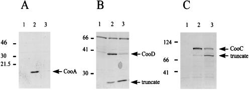
To produce the Coo proteins, we used a system of compatible plasmids in which the genes encoding the major and minor pilins, CooA and CooD, and the outer membrane assembly protein CooC were expressed from a low copy number plasmid under control of the lac promoter (pEU488, pEU493, and pEU479, respectively), and CooB was expressed from a high copy number plasmid under the control of the lac promoter (pNEBCS1.3). A third compatible plasmid containing lacIq was present to repress expression until isopropyl β-d-thiogalactoside was added. The relevant features of these plasmids are described in Table 1. Fig. 1 compares the amount of protein in strains expressing CooA (Fig. 1A), CooC (Fig. 1C), or CooD (Fig. 1B) when they were complemented in trans with CooB (pNEBCS1.3) or a negative control plasmid (pUC19). As expected, the negative control strain did not produce CooA, CooD, or CooC (Fig. 1 A, B, and C, respectively, lane 1). There was a significant amount of CooA when CooB was coexpressed (Fig. 1A, lane 2), but CooA was almost undetectable in the absence of CooB (Fig. 1A, lane 3).
Figure 1.
CooB stabilizes CooA, CooC, and CooD. Whole-cell extracts were separated by SDS/PAGE and probed by immunoblotting with antibodies specific for CooA (A), CooD (B), or CooC (C). (Left) Molecular masses (in kDa) determined by a lane containing markers. (A) Stabilization of CooA. Lane 1, MC4100/pFDX500/pHSG576/pUC19 (Coo−). Lane 2, MC4100/pFDX500/pEU488/pNEBCS1.3 (CooACooB). Lane 3, MC4100/pFDX500/pEU488/pUC19 (CooA). (B) Stabilization of CooD. Lane 1, MC4100/pFDX500/pHSG576/pUC19 (Coo−). Lane 2, MC4100/pFDX500/pEU493/pNEBCS1.3 (CooDCooB). Lane 3, MC4100/pFDX500/pEU493/pUC19 (CooD). (C) Stabilization of CooC. Lane 1, MC4100/pFDX500/pHSG576/pUC19 (Coo−). Lane 2, MC4100/pFDX500/pEU479/pNEBCS1.3 (CooCCooB). Lane 3, MC4100/pFDX500/pEU479/pUC19 (CooC).
The CooD minor pilin exists as two distinct intracellular forms in CS1-piliated bacteria: a full-length form of 38 kDa and a truncated form of approximately 25 kDa (data not shown), although only the high molecular mass form is present in pili (9). When immunoblots were probed with anti-CooD antiserum, the two forms of CooD and a 66-kDa crossreactive protein found in E. coli also were seen in extracts from cells expressing both CooD and CooB (Fig. 1B, compare lanes 1 and 2). The presence of CooB had a positive effect on the production of full-length CooD (Fig. 1B, lane 2), whereas in the absence of CooB, the 25-kDa form of CooD predominated (Fig. 1B, lane 3). When the cell extracts were analyzed with antiserum to the outer membrane protein CooC, the stabilizing effect of CooB was apparent, although it was less dramatic (Fig. 1C, compare lanes 2 and 3). In the absence of CooB, the amount of full-length CooC was reduced, whereas the amount of an approximately 70-kDa truncated form of CooC was enhanced (Fig. 1C, lane 3). From these data we conclude that the expression of CooB is necessary to achieve maximal accumulation of the pilins CooA and CooD and the outer membrane assembly protein CooC.
Inducible Expression System for Functional CS1 Pili.
In each case in the above system, two of the CS1 assembly proteins were always absent and the ratio of CooB to the stabilized protein was much greater than in the wild-type situation in which all four proteins are encoded on the same plasmid and pili are produced. To be sure that functional pili are produced even when there is an excess of CooB, the experiments were repeated with strains in which all three proteins (CooA, CooC, and CooD) are encoded on a low copy plasmid (pEU476, Table 1) with and without the high copy plasmid that encodes CooB (pNEBCS1.3, Table 1). In both plasmids, the coo genes are transcribed from the lac promoter. These strains were unstable in the absence of an additional compatible plasmid containing the lacIq gene (pFDX500, Table 1), which represses their expression in the absence of isopropyl β-d-thiogalactoside (data not shown). To determine whether these strains made functional pili, they were induced with isopropyl β-d-thiogalactoside for different time periods and the presence of pili was assayed by the ability of cells to be agglutinated by antiserum against CS1 pili and to cause hemagglutination of bovine erythrocytes. Strains MC4100/pFDX500/pHSG576/pUC19 (Coo−) and MC4100/pFDX500/pEU476/pUC19 (CooA, CooC, and CooD) served as negative controls, while MC4100/pFDX500/pEU476/pNEBCS1.3 (CooA, CooC, CooD, and CooB) was the test strain. As expected, neither of the negative control strains was able to hemagglutinate bovine erythrocytes or to agglutinate in the presence of anti-CS1 serum, whereas the test strain did both after 2 hr of isopropyl β-d-thiogalactoside induction (data not shown). This indicates that functional CS1 pili are produced even with the high ratio of CooB to the other proteins.
By using this system in which CooA, CooC, and CooD were all present, we compared the amount of each of these proteins by immunoblots with antisera specific for each. In each case, more protein was detected in the presence of CooB than in its absence (data not shown), confirming the previous results.
CooB Interacts Directly with CooD.
In the periplasm of cells producing CS1 pili, we previously found a complex containing the major pilin protein CooA and the assembly protein CooB (9). This suggests that CooB stabilizes CooA by interacting with it directly. To determine whether CooB interacts directly with the other Coo proteins that it stabilizes, periplasmic intermolecular complexes were sought on nondenaturing gels and identified by immunoblot and by N-terminal amino acid sequence analysis.
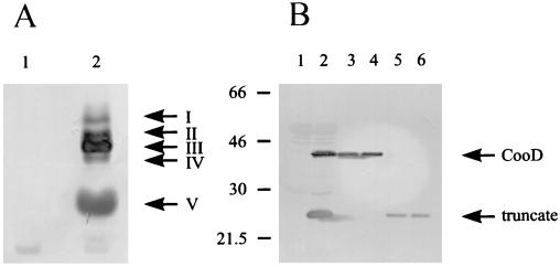
Immunoblot analysis of a periplasmic extract from the strain encoding CooD plus CooB showed five bands, designated I–V, which reacted strongly with anti-CooD antiserum (Fig. 2A, lane 2) and which were not present in the negative control strain (Fig. 2A, lane 1). To characterize the two major forms of CooD (forms III and V), which also were found in piliated cells expressing all of the coo genes (data not shown), the proteins were transferred to poly(vinylidene difluoride) membrane, identified by Coomassie blue staining, and subjected to amino-terminal sequence analysis.
Figure 2.
Interaction between CooB and CooD. (A) Periplasmic extracts were fractionated by native PAGE and probed by immunoblotting with antibodies to CooD. Lane 1, MC4100/pFDX500/pHSG576/pUC19 (Coo−). Lane 2, MC4100/pFDX500/pEU493/pNEBCS1.3 (CooDCooB). (B) Periplasmic extracts (lanes 1 and 2) and different forms of CooD protein eluted from native PAGE (lanes 3–6) were separated by SDS/PAGE and probed by immunoblotting with antibodies to CooD. (Left) Molecular masses (in kDa) determined by a lane containing markers. Lane 1, MC4100/pFDX500/pHSG576/pUC19 (Coo−). Lane 2, MC4100/pFDX500/pEU493/pNEBCS1.3 (CooDCooB). Lane 3, eluted form III. Lane 4, eluted form I. Lane 5, eluted form V. Lane 6, eluted form II.
The protein with the fastest migration (form V) produced a single amino acid sequence corresponding to the amino terminus of the mature form of CooD. Therefore, this form of the protein is not associated with CooB in an intermolecular complex. However, band III, which migrated much more slowly than CooB alone (data not shown), yielded a mixed N-terminal sequence corresponding to the mature forms of both CooD and CooB in a molar ratio of 1:1. This indicates that CooD and CooB are present as an intermolecular complex in the periplasm.
To determine the molecular masses of uncomplexed CooD and the form of CooD that is associated with CooB, bands V and III were excised from Coomassie blue-stained native polyacrylamide gels, separated by SDS/PAGE, and analyzed by immunoblotting with anti-CooD. A periplasmic extract from the positive control strain encoding CooD and CooB contained the full-length 38-kDa form and the truncated 25-kDa form of CooD (Fig. 2B, lane 2), whereas neither form of CooD was present in the extract from the negative control strain with only the vectors (Fig. 2B, lane 1). CooD protein from the CooD-CooB complex (form III) migrated at a size of 38 kDa (Fig. 2B, lane 3), whereas band V contained only the 25-kDa form of CooD (Fig. 2B, lane 5). This is in agreement with the N-terminal sequence analysis of these protein forms. In addition, this showed that because the N-terminal sequences of the full-length and truncated forms of CooD were the same, the 25-kDa form of CooD is truncated at the C terminus.
The molecular masses of the minor CooD forms (bands I, II, and IV) were determined in the same way as described above. Because repeated attempts to isolate form IV of CooD failed, this protein or protein complex is probably unstable. However, forms I and II were both stable and migrated in SDS/PAGE as full-length 38-kDa CooD (Fig. 2b, lane 4) and as a truncated 25-kDa form (Fig. 2B, lane 6), respectively. The full-length form I of CooD is not associated with CooB because it was found in a strain encoding only CooD (data not shown) as well as in the strain encoding CooD and CooB described above. The minor truncated form II of CooD was seen only in the test strain encoding CooD and CooB but not in a strain encoding only CooD (data not shown). Therefore we believe that form II of CooD represents a minor intermolecular complex containing CooB and truncated CooD.
CooB Interacts Directly with CooC.
Because our expression studies showed that CooB is also necessary to achieve high levels of CooC (Fig. 1C), we expect that, as in the case of CooD, CooB may interact directly with CooC to protect it from proteases. CooC is in the insoluble whole membrane fraction after freeze/thaw lysis of spheroplasts (data not shown), so it was not possible to study the CooC–CooB interaction directly by using native PAGE. However, because CooC is located exclusively in the outer membrane (9), if CooB is complexed with it, CooB also should be found in the membrane fraction, but only if CooC is coexpressed. To test this we isolated whole membranes after freeze/thaw lysis of spheroplasts, which, unlike lysis by disruptive methods that use sonication, high pressure, or detergents, does not destroy protein–protein interactions. Whole membrane fractions of various strains then were separated on SDS/PAGE and analyzed with anti-CooB by immunoblots.
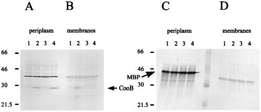
As expected, CooB was found in periplasmic extracts of both strains expressing it (Fig. 3A, lanes 2 and 4) but not in the negative control strains (Fig. 3A, lanes 1 and 3). Examination of the corresponding immunoblot of the whole membrane fractions showed that some fraction of CooB is associated with the membrane when CooC also is expressed (Fig. 3B, lane 2) but that CooB is confined to the periplasm in the absence of CooC (Fig. 3B, lane 4; Fig. 3A, lane 4). To further control for crosscontamination of fractions, the periplasmic maltose binding protein was used as a marker. Immunoblots with antiserum to maltose binding protein showed that it is present exclusively in periplasmic fractions (Fig. 3C) and is not detectable in the whole membrane fractions (Fig. 3D). Therefore, it appears that CooB associates with the membrane in a CooC-dependent manner, indicating that CooB interacts directly with CooC. This interaction also occurs in piliated cells expressing all of the coo genes (data not shown).
Figure 3.
Interaction between CooB and CooC. Periplasmic extracts (A and C, lanes 1–4) and whole membranes (B and D, lanes 1–4) were separated on SDS/PAGE and probed with antibodies to CooB (A and B) or maltose binding protein (C and D). (Left) Molecular masses (in kDa) determined by a lane containing markers. Lane 1, MC4100/pFDX500/pHSG576/pUC19 (Coo−). Lane 2, MC4100/pFDX500/pEU479/pNEBCS1.3 (CooCCooB). Lane 3, MC4100/pFDX500/pEU479/pUC19 (CooC). Lane 4, MC4100/pFDX500/pHSG576/pNEBCS1.3 (CooB).
DISCUSSION
CS1 pili are representatives of a distinct class of pili found in human isolates of enterotoxigenic E. coli. The proteins required for the synthesis of CS1 and related pili have no sequence similarity with those of other pilus systems, including type IV (22) and Pap-related pili (3). In previous studies of CS1 pilus assembly, we found that the assembly protein CooB associates with the major pilin, CooA, in the periplasm, but is not incorporated into the final pilus structure (9). In this respect CooB is similar to the periplasmic chaperones of a number of Pap-related pilus systems even though its sequence has no similarity to them (9). To learn more about the role of CooB in pilus assembly, we have investigated whether CooB has functions that normally are associated with chaperones.
Molecular chaperones are critical in a variety of cellular processes, including the proper folding, secretion, and macromolecular assembly of proteins (3, 23, 24). In general, chaperones prevent the inappropriate intermolecular interaction of misfolded proteins that leads to their aggregation (3, 23, 24) and in most cases to their degradation in the cell (3, 23). Chaperones act either by recognizing and binding to nonnative proteins to promote their proper folding, as in the case of the cytoplasmic chaperone GroEL/ES, or, like SecB, by inhibiting the misfolding of certain secreted proteins so that they are maintained in a secretion-competent configuration (24).
The pilin chaperones, a group of related proteins with conserved motifs, found in piliated Gram-negative bacteria, bind to pilin proteins secreted into the periplasm (3). In these intermolecular complexes, pilins are maintained in a native-like state that allows them to interact productively with other components of the pilus assembly machinery (25).
In this report, we have demonstrated that CooB stabilizes not only the major and minor CS1 pilins CooA and CooD, but also the outer membrane assembly protein CooC. We also demonstrated that CooB associates with CooD in the periplasm, as it does with CooA, (9) and that it associates with CooC in the membrane. This suggests that the interaction of CooB with these proteins is required for their stabilization and therefore that CooB has chaperone-like functions for these proteins.
The degradation of pilins in the absence of a periplasmic chaperone is a common feature of the Pap-related pilus systems (3) and is catalyzed by the DegP protease in the K88 (26) and Pap pilus systems (27). In contrast, the CS1 pilins and CooC are not degraded by the periplasmic protease DegP in the absence of CooB (data not shown). This indicates that at least two distinct proteolytic systems are involved in the removal of unchaperoned pilus proteins in E. coli.
Although the full-length, 38-kDa form of CooD binds to CooB efficiently, the truncated form of CooD binds only poorly. Therefore the interaction of CooD and CooB may be largely dependent on sequences in the C-terminal one-third of the CooD protein. Because the major and minor pilins share a conserved sequence motif near their C termini (9), this sequence may be involved in their binding to CooB. We also had demonstrated previously, in crosscomplementation experiments that used the genes encoding CS1 and CS2 pili, that the CS1 chaperone can interact productively with the CS2 minor pilin and vice versa to form pili (4). This suggests that the sequences involved in chaperone binding are conserved between the minor pilins of both CS1 and CS2 pili. The C-terminal motif described above is the only sequence common to the CS1 major and minor pilins as well as to the CS2 minor pilin, strongly suggesting a role for this sequence in pilin-chaperone binding. However, because this conserved pilin sequence is not present in CooC, if CooB binds to it, CooB must bind differently to CooC.
Although CooB is unrelated to the pilin chaperones of other pilus systems, it appears to have a similar role in pilin stabilization. Why have two unrelated pilus systems evolved a similar mechanism for pilus assembly? The answer may be that both the CS1 and Pap-related pilus systems rely on the Sec-dependent pathway for transport of their pilins across the cytoplasmic membrane and face the same problem of high concentration of pilin proteins in the periplasm (3, 9). Because of this, the pilins have the opportunity to interact prematurely in the periplasm to form aggregates. This may occur through nonspecific hydrophobic interactions between non-native proteins emerging from the cytoplasmic membrane or through the premature periplasmic interaction of the specific sequences normally involved in the polymerization of pilins into pili. The evolution of periplasmic chaperones that prevent such interactions until the pilins are incorporated into growing pili would solve this problem.
In contrast to the CS1 and Pap-related pilus systems, specialized periplasmic pilin chaperones have not been found in type IV pilus systems that do not use the Sec secretion pathway. In Pseudomonas aeruginosa and Vibrio cholerae, no significant pool of Type IV pilins is found in the periplasm (28–30). Instead, in these systems, pilins accumulate almost exclusively in the membrane fraction where they may be prevented by other means from interacting with each other before being transported to the cell surface and assembled (28, 29).
Although CooB plays a role similar to the pilin chaperones in unrelated pilus systems like Pap, it may differ in that in addition to interaction with and stabilization of the pilins, CooB is needed to stabilize the outer membrane protein essential for pilus assembly. In the Pap-related pilus systems, a large outer membrane protein is also required for the assembly of pili (3). Like CooC, the 86-kDa outer membrane PapC protein from the Pap pilus system is degraded to a 70-kDa truncated form (25). However, because experiments testing whether the stability of PapC or its homologues in other systems are chaperone-dependent have not been reported, we do not know whether the CS1 pilus system differs from the Pap-related pili in this respect. The observation that CooB remains associated with CooC even after it has inserted into the outer membrane suggests that CooB may be important for maintaining CooC in an active configuration throughout the pilus assembly process.
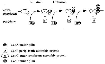
Based on the findings presented in this report, we have refined our model of CS1 pilus assembly (Fig. 4). All of the Coo proteins traverse the cytoplasmic membrane via the Sec pathway because they all possess typical Sec-dependent signal sequences (7, 8, 10). CooB binds to the major and minor pilins as they are secreted into the periplasm, promoting the folding of the pilins and preventing premature polymerization and aggregation in the periplasm. CooB also binds to CooC to maintain it in an active, native conformation in the membrane. The CooB–CooD complex initiates pilus assembly by binding to CooC in the outer membrane, releasing CooB from either the pilin or CooC. In this way, CooB recycles in the periplasm to bind other pilins whereas the interaction between CooB and CooC is maintained to keep CooC in an active conformation throughout the assembly process. After pilus assembly has been initiated, CooA pilin, in the form of a complex with CooB, displaces CooD from CooC and is added to the base of the growing pilus. The pilus then extends by repeated rounds of pilin-chaperone interactions with CooC, leading to the displacement of pilins from CooC to the exterior of the cell. Our studies have shown that although CS1 pili are simpler than the Pap-related pili and there are no sequence similarities between them, they have evolved pathways of assembly that are generally similar.
Figure 4.
Model of CS1 pilus assembly. CooB chaperone binds to CooC in the outer membrane to maintain its conformation and prevent its degradation. CooB also binds to the major and minor pilins, CooA and CooD, respectively, to guide their folding and/or inhibit pilin-pilin interactions leading to aggregation and degradation. A CooB–CooD complex initiates assembly by binding to CooC; CooB is displaced and is recycled into the periplasm where it may interact with other pilins. CooB–CooA complexes displace CooD from CooC, replacing it with CooA. Repeated interactions between CooB–CooA complexes and CooC allow incorporation of CooA at the base of the pilus and extension of the pilus rod.
Acknowledgments
We thank Jan Pohl at the Emory microchemical facility for sequencing proteins, Susan Gottesman for supplying protease-negative E. coli mutants, and the Biomolecular Computing Resource at Emory for facilitating sequence analysis. This work was supported by Public Health Service Grant AI24870 from the National Institutes of Health.
Footnotes
This paper was submitted directly (Track II) to the Proceedings Office.
Abbreviation: ETEC, enterotoxigenic Escherichia coli.
References
- 1.Gaastra W, Svennerholm A-M. Trends Microbiol. 1996;4:444–452. doi: 10.1016/0966-842x(96)10068-8. [DOI] [PubMed] [Google Scholar]
- 2.Levine M M. J Infect Dis. 1987;155:377–389. doi: 10.1093/infdis/155.3.377. [DOI] [PubMed] [Google Scholar]
- 3.Hultgren S J, Normark S, Abraham S N. Annu Rev Microbiol. 1991;45:383–415. doi: 10.1146/annurev.mi.45.100191.002123. [DOI] [PubMed] [Google Scholar]
- 4.Froehlich B J, Karakashian A, Sakellaris H, Scott J R. Infect Immun. 1995;63:4849–4856. doi: 10.1128/iai.63.12.4849-4856.1995. [DOI] [PMC free article] [PubMed] [Google Scholar]
- 5.Jordi B J A M, Willshaw G A, van der Zeist B A M, Gaastra W. DNA Sequence. 1992;2:257–263. doi: 10.3109/10425179209020811. [DOI] [PubMed] [Google Scholar]
- 6.Sajjan U S, Sun L, Goldstein R, Forstner J F. J Bacteriol. 1995;177:1030–1038. doi: 10.1128/jb.177.4.1030-1038.1995. [DOI] [PMC free article] [PubMed] [Google Scholar]
- 7.Froehlich B J, Karakashian A, Melsen L R, Wakefield J C, Scott J R. Mol Microbiol. 1994;12:387–401. doi: 10.1111/j.1365-2958.1994.tb01028.x. [DOI] [PubMed] [Google Scholar]
- 8.Perez-Casal J, Swartley J S, Scott J R. Infect Immun. 1990;58:3594–3600. doi: 10.1128/iai.58.11.3594-3600.1990. [DOI] [PMC free article] [PubMed] [Google Scholar]
- 9.Sakellaris H, Balding D P, Scott J R. Mol Microbiol. 1996;21:529–541. doi: 10.1111/j.1365-2958.1996.tb02562.x. [DOI] [PubMed] [Google Scholar]
- 10.Scott J R, Wakefield J C, Russell P W, Orndorff P E, Froehlich B J. Mol Microbiol. 1992;6:293–300. doi: 10.1111/j.1365-2958.1992.tb01471.x. [DOI] [PubMed] [Google Scholar]
- 11.Casadaban M J. Mol Microbiol. 1976;104:541–555. doi: 10.1016/0022-2836(76)90119-4. [DOI] [PubMed] [Google Scholar]
- 12.Scott J R. Virology. 1974;62:344–349. doi: 10.1016/0042-6822(74)90397-3. [DOI] [PubMed] [Google Scholar]
- 13.Murphree D, Froehlich B, Scott J R. J Bacteriol. 1997;179:5736–5743. doi: 10.1128/jb.179.18.5736-5743.1997. [DOI] [PMC free article] [PubMed] [Google Scholar]
- 14.Schnetz K, Sutrina S L, Saier M H, Rak B. J Biol Chem. 1990;265:13464–13471. [PubMed] [Google Scholar]
- 15.Takeshita S, Sato M, Toba M, Masahashi W, Hashimoto-Gotoh T. Gene. 1987;61:63–74. doi: 10.1016/0378-1119(87)90365-9. [DOI] [PubMed] [Google Scholar]
- 16.Yanisch-Perron C, Vieira C, Messing J. Gene. 1985;33:103–119. doi: 10.1016/0378-1119(85)90120-9. [DOI] [PubMed] [Google Scholar]
- 17.Achtman M, Mercer A, Kusecek B, Pohl A, Heuzenroeder M, Aaronson W, Sutton A, Silver R P. Infect Immun. 1983;39:315–335. doi: 10.1128/iai.39.1.315-335.1983. [DOI] [PMC free article] [PubMed] [Google Scholar]
- 18.Laemmli U K. Nature (London) 1970;227:680–685. doi: 10.1038/227680a0. [DOI] [PubMed] [Google Scholar]
- 19.McLellan T. Anal Biochem. 1982;126:94–99. doi: 10.1016/0003-2697(82)90113-0. [DOI] [PubMed] [Google Scholar]
- 20.Towbin H, Staehelin T, Gordon J. Proc Natl Acad Sci USA. 1979;76:4350–4354. doi: 10.1073/pnas.76.9.4350. [DOI] [PMC free article] [PubMed] [Google Scholar]
- 21.Caron J, Maneval D R, Kaper J B, Scott J R. Infect Immun. 1990;58:3442–3444. doi: 10.1128/iai.58.10.3442-3444.1990. [DOI] [PMC free article] [PubMed] [Google Scholar]
- 22.Strom M S, Lory S. Annu Rev Microbiol. 1993;47:565–596. doi: 10.1146/annurev.mi.47.100193.003025. [DOI] [PubMed] [Google Scholar]
- 23.Hartl F U. Cold Spring Harbor Symp Quant Biol. 1995;110:429–434. doi: 10.1101/sqb.1995.060.01.047. [DOI] [PubMed] [Google Scholar]
- 24.Hardy S J S, Randall L L. Philos Trans R Soc London B. 1993;339:343–354. doi: 10.1098/rstb.1993.0033. [DOI] [PubMed] [Google Scholar]
- 25.Dodson K W, Jacob-Dubuisson F, Striker R T, Hultgren S J. Proc Natl Acad Sci USA. 1993;90:3670–3674. doi: 10.1073/pnas.90.8.3670. [DOI] [PMC free article] [PubMed] [Google Scholar]
- 26.Bakker D, Vader C E M, Roosendaal B, Mooi F R, Oudega B, de Graaf F K. Mol Microbiol. 1991;5:875–886. doi: 10.1111/j.1365-2958.1991.tb00761.x. [DOI] [PubMed] [Google Scholar]
- 27.Striker R, Jacob-Dubuisson F, Frieden C, Hultgren S J. J Biol Chem. 1994;269:12233–12239. [PubMed] [Google Scholar]
- 28.Nunn D, Bergman S, Lory S. J Bacteriol. 1990;172:2911–2919. doi: 10.1128/jb.172.6.2911-2919.1990. [DOI] [PMC free article] [PubMed] [Google Scholar]
- 29.Iredell J R, Manning P A. Trends Microbiol. 1994;2:187–192. doi: 10.1016/0966-842x(94)90109-i. [DOI] [PubMed] [Google Scholar]
- 30.Iredell J R, Manning P A. J Bacteriol. 1997;179:2038–2046. doi: 10.1128/jb.179.6.2038-2046.1997. [DOI] [PMC free article] [PubMed] [Google Scholar]


