Figure 2.—
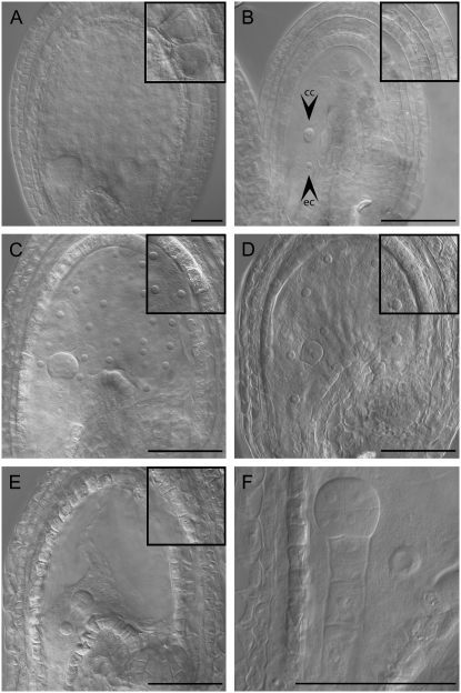
Autonomously proliferating central cell nuclei after single fertilization with cdka;1 mutant pollen. Light micrograph of cleared whole-mount ovules (unfertilized) or seeds originating from the pollination with wild-type pollen (double fertilized) or cdka;1 mutant pollen (only egg cell fertilized). (A) Col-0 seed pollinated with Col-0 wild-type pollen at 3 DAP. (B) Nonfertilized Col-0 ovules at 6 days after emasculation (ec and cc indicate egg cell and central cell, respectively) (C) Col-0 seed pollinated with cdka;1 mutant pollen at 3 DAP; the central cell nucleus has undergone five rounds of division and here 28 nuclei are visible. This represents one of the largest nuclei numbers found upon pollination with cdka;1 mutant pollen and is typical for seeds in the Col genetic background that have initiated autonomous proliferation. (D) Bay-0 seed pollinated with cdka;1 mutant pollen at 3 DAP. The central cell nucleus has undergone three rounds of division and eight nuclei are visible, characteristic for seeds in the Bay genetic background that have initiated autonomous proliferation. (E) Sha seed pollinated with cdka;1 mutant pollen at 3 DAP. The central cell contains only a single nucleus; the predominant number of seeds in the Sha genetic background do not initiate autonomous proliferation. (F) Close-up of a Sha seed pollinated with cdka;1 mutant pollen showing a single central cell nucleus and a 16-cell embryo at 3 DAP. Insets in A–E show the inner integument layers at the same magnification. The integument cells strongly enlarge and vacuolize in double-fertilized seeds (A), whereas in unfertilized ovules (B) no cell expansion is found. Upon single fertilization with cdka;1 mutant pollen (C–E), the integuments slightly expand. All pictures are oriented such that the micropylar pole with the developing embryo points to the left and the chalazal pole of the seed to the right. Bars, 50 μm.

