Abstract
This case report identifies a very rare disease, choriocarcinoma in a male patient, causing another rare entity, an atraumatic rupture of the spleen.
Keywords: Choriocarcinoma, Splenic rupture
Choriocarcinoma is a pure epithelial neoplasm, normally found in females. It is part of the gestational trophoblastic disease group, being derived from the placental trophoblastic tissue. It is a rare entity in men, believed to be derived from the totipotent cells of the testis or the pineal gland.2
Splenic rupture due to choriocarcinoma of the spleen is very rare, with very few cases described in the literature,1,4 and rarely recognised at presentation. A male patient was admitted for investigations of left-sided abdominal pain, which became severe requiring emergency laparotomy. Multiple deposits of chorionic carcinoma were found on the liver and spleen causing the latter to rupture.
Case report
A 71-year-old man with no significant medical history presented to Bronglais Hospital Accident and Emergency Department in April 2006. He was admitted for investigation of left-sided abdominal pain, radiating to the back and left groin, associated with sweating and nausea. There was no history of trauma. His vital signs were stable apart from his pulse, which was 110 per min. Abdominal examination revealed moderate tenderness and an ill-defined, deeply sited mass in the left upper quadrant. Haematological investigations were normal apart from a C-reactive protein (CRP) level of 144 mg/l and an erythrocyte sedimentation rate (ESR) of 51 mm/h. An ultrasound scan revealed a large irregular mass in the left side of the abdomen, anterior and separated from the left kidney. The mass was thought to be related to the splenic flexure of the descending colon.
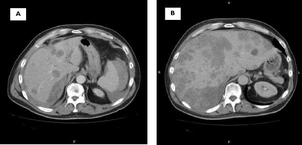
On the third day of his admission, the patient started complaining of severe sudden onset abdominal pain. He was found to be hypotensive and tachycardic. Abdominal examination revealed signs of peritonism. His haemoglobin level had fallen from 12.7 g/dl to 9.9 g/dl and an emergency computer tomography (CT) scan showed a ruptured spleen, multiple liver lesions and a right lung lower lobe lesion (Fig. 1A). Lymphoma was suggested as the cause of the appearances.
Figure 1.
(A) CT of the abdomen 3 days after admission showing the splenic lesion and the multiple liver lesions thought to be lymphoma. (B) CT of the abdomen 6 weeks after the first, showing the enlarged liver almost completely filled with confluent different sized, low-attenuated lesions (metastases).
An emergency laparotomy was performed and 2 l of free blood aspirated from the peritoneum. The cause of the bleeding was the acute spontaneous rupture of the enlarged spleen, full of multiple haemangiomatous type lesions. The liver was similarly affected though intact and the remaining intra-abdominal organs were normal. After splenectomy and peritoneal lavage, the patient's postoperative recovery was uneventful.
The histopathological diagnosis of the specimen was of spleen infiltrated by choriocarcinoma. There were nests of tumour, composed of closely packed cells with clear cytoplasm and fairly uniform nuclei, closely opposed to large cells, with more pleomorphic nuclei and eosinophilic cytoplasm. Tumour islands enclosed cystic spaces and haemorrhagic areas with obvious invasion of the vascular space.
As a result of the histology report, germ cell tumour marker levels were retrospectively assayed on a pre-operative blood sample and the β-human chorionic gonadotrophin (β-HCG) was found to be grossly elevated at 63100 IU/l (normal < 21 IU/l).
Postoperative ultrasound scan of the testes was normal. Because of his poor physical condition, he remained an in-patient. Two weeks after the first operation, he had to undergo another surgical procedure, this time for a strangulated inguinal hernia. After another 2 weeks, he was started on adjuvant chemotherapy (bleomycin, etoposide, platinum). However, his condition steadily deteriorated and soon after the first chemotherapy cycle he developed cardiac arrhythmia and became jaundiced. The β-HCG level rose to 316,000 IU/l and a repeat CT scan showed a massively enlarged liver with multiple metastases (Fig. 1B). It was the patient's wish to go home and he was discharged without follow-up. He died 6 weeks after the initial hospital presentation, 3 days after discharge.
Discussion
Metastatic choriocarcinoma in a man is rare. Surgical presentations have varied from simple abdominal pain to acute abdomen with gastrointestinal haemorrhage.2,3 Choriocarcinoma involving the spleen has only been previously reported on three occasions.1,4,5
The treatment of metastatic choriocarcinoma is surgical excision, if possible, followed by chemotherapy (various chemotherapeutic agents, for example, methotrexate, cisplatin, mitomycin C, fluorouracil, futrafur).3 Even so, the period of survival in male patients is short, usually less than 6 months. There has been one report of a male patient who survived for 4.5 years after radical resection of a gastric choriocarcinoma and subsequent chemotherapy.3
The patient in our case report had multiple metastatic deposits in the liver, lung and spleen at presentation. The subsequent course of the disease was fulminant, as demonstrated by the successive CT scans and the huge rise in β-HCG levels.
The primary site of the choriocarcinoma in our patient is speculative. There are three main hyphotheses for extragonadal choriocarcinoma. Shariat et al.2 enumerated them as: (i) the tumour is derived from the primordial germ cells that migrate abnormally during embryonic development; (ii) the tumour is secondary to differentiation or neometaplasia of non-gonadal tissue; and (iii) the tumour is a metastasis from spontaneously regressed primary gonadal germ tumour. Thus, even though the testicular ultrasound was normal, it is still possible that the tumour originated in the testes.2,5
Conclusion
This case report identifies a very rare disease, chorio-carcinoma in a male patient, causing another rare entity, an atraumatic rupture of the spleen.
References
- 1.Challis DE, Rew KJ, Steigrad SJ. Choriocarcinoma complicated by splenic rupture: an unusual presentation. J Obstet Gynaecol Res. 1996;22:395–400. doi: 10.1111/j.1447-0756.1996.tb00996.x. [DOI] [PubMed] [Google Scholar]
- 2.Shariat SF, Duchene D, Kabbani W, Mucher Z, Lotan Y. Gastrointestinal hemorrhage as first manifestation of metastatic testicular tumor. Urology. 2005;66:13–9. doi: 10.1016/j.urology.2005.06.102. [DOI] [PubMed] [Google Scholar]
- 3.Noguchi T, Takeno S, Sato T, Takahashi Y, Uchida Y, Yokoyama S. A patient with primary gastric choriocarcinoma who received a correct preoperative diagnosis and achieved prolonged survival. Gastric Cancer. 2002;5:112–7. doi: 10.1007/s101200200019. [DOI] [PubMed] [Google Scholar]
- 4.Kristoffersson A, Emdin S, Jarhult J. Acute intestinal obstruction and splenic hemorrhage due to metastatic choriocarcinoma. A case report. Acta Chir Scand. 1985;151:381–4. [PubMed] [Google Scholar]
- 5.Carr AJ, Jacob G, Glanfield PA, Rogers K. Male choriocarcinoma of the spleen: a case report. Eur J Surg Oncol. 1987;13:75–6. [PubMed] [Google Scholar]


