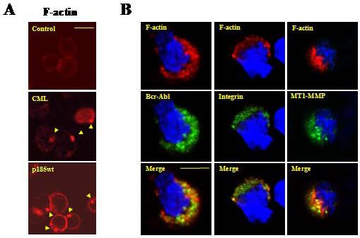
Figure 1. Leukemia cells isolated from CML patient display abnormal F-actin rich structures enriched with Bcr-Abl, β1-integrin, and MT1-MMP.

(A) Mononuclear cells isolated from a Bcr-Abl-positive CML patient (CML) and a Bcr-Abl-negative control human sample (control) were stained with TRITC-conjugated phalloidin and analyzed by confocal microscopy for actin cytoskeleton structure. A murine pro-B cell line Ba/F3 transformed by p185wt (p185wt) was also stained and analyzed. Arrows indicated F-actin rich structures; Bar: 10 μm. (B) Mononuclear cells isolated from a CML patient were probed with the anti-Abl (left panel), anti-β1integrin (middle panel), and anti-MT1-MMP (right panel) antibodies. This was followed by staining with FITC-conjugated secondary antibody. Cells were then counterstained with TRITC-conjugated phalloidin and DAPI to visualize F-actin and nuclei, respectively. The pictures were captured by two-photon confocal microscopy. Bar: 5 μm.
