Abstract
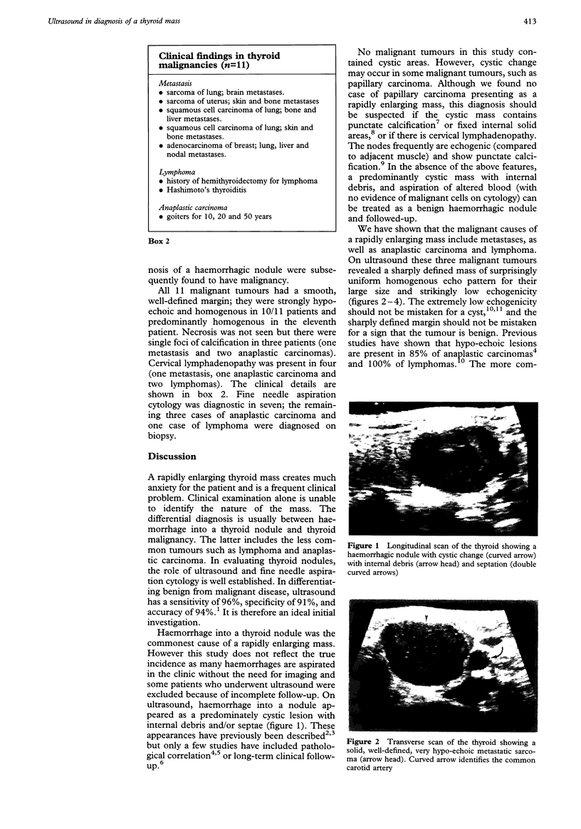
The value of ultrasound in the diagnosis of a large rapidly growing thyroid mass was assessed in a study of 42 patients with a large (> 3 cm) rapidly growing (< two months) solitary mass. Haemorrhage into a thyroid nodule was present in 31 patients and thyroid malignancy in 11. Ultrasound of haemorrhage into a thyroid nodule revealed a large cystic mass in all 31 patients containing internal debris (22), septations (three), or a combination of both (six). The malignant causes of a large rapidly growing mass were lymphoma (two), anaplastic carcinoma (four) and metastasis (five). Ultrasound of these thyroid malignancies revealed a mass with a smooth, well-defined margin and strikingly low homogeneous echogenicity in all cases. Patients with thyroid metastases had evidence of widespread metastatic disease elsewhere. Lymphoma was differentiated from anaplastic carcinoma on fine-needle aspiration cytology or surgical biopsy. Ultrasound was of value in differentiating between a benign haemorrhagic nodule and a malignant tumour. The various malignant tumours had similar appearances, however, and could not be distinguished on ultrasound.
Full text
PDF


Images in this article
Selected References
These references are in PubMed. This may not be the complete list of references from this article.
- Ahuja A. T., Chow L., Chick W., King W., Metreweli C. Metastatic cervical nodes in papillary carcinoma of the thyroid: ultrasound and histological correlation. Clin Radiol. 1995 Apr;50(4):229–231. doi: 10.1016/s0009-9260(05)83475-0. [DOI] [PubMed] [Google Scholar]
- Ahuja A. T., King W., Metreweli C. Role of ultrasonography in thyroid metastases. Clin Radiol. 1994 Sep;49(9):627–629. doi: 10.1016/s0009-9260(05)81880-x. [DOI] [PubMed] [Google Scholar]
- Anscombe A. M., Wright D. H. Primary malignant lymphoma of the thyroid--a tumour of mucosa-associated lymphoid tissue: review of seventy-six cases. Histopathology. 1985 Jan;9(1):81–97. doi: 10.1111/j.1365-2559.1985.tb02972.x. [DOI] [PubMed] [Google Scholar]
- Chang T. C., Hung C. T. Ultrasonographic findings in relation to ease of aspiration and fluid characteristics in thyroid cyst. J Formos Med Assoc. 1990 May;89(5):350–355. [PubMed] [Google Scholar]
- Hatabu H., Kasagi K., Yamamoto K., Iida Y., Misaki T., Hidaka A., Shibata T., Shibata T., Shoji K., Higuchi K. Cystic papillary carcinoma of the thyroid gland: a new sonographic sign. Clin Radiol. 1991 Feb;43(2):121–124. doi: 10.1016/s0009-9260(05)81591-0. [DOI] [PubMed] [Google Scholar]
- Matsuzuka F., Miyauchi A., Katayama S., Narabayashi I., Ikeda H., Kuma K., Sugawara M. Clinical aspects of primary thyroid lymphoma: diagnosis and treatment based on our experience of 119 cases. Thyroid. 1993 Summer;3(2):93–99. doi: 10.1089/thy.1993.3.93. [DOI] [PubMed] [Google Scholar]
- Sackler J. P., Passalaqua A. M., Blum M., Amorocho L. A spectrum of diseases of the thyroid gland as imaged by gray scale water bath sonography. Radiology. 1977 Nov;125(2):467–472. doi: 10.1148/125.2.467. [DOI] [PubMed] [Google Scholar]
- Scheible W., Leopold G. R., Woo V. L., Gosink B. B. High-resolution real-time ultrasonography of thyroid nodules. Radiology. 1979 Nov;133(2):413–417. doi: 10.1148/133.2.413. [DOI] [PubMed] [Google Scholar]
- Simeone J. F., Daniels G. H., Mueller P. R., Maloof F., vanSonnenberg E., Hall D. A., O'Connell R. S., Ferrucci J. T., Jr, Wittenberg J. High-resolution real-time sonography of the thyroid. Radiology. 1982 Nov;145(2):431–435. doi: 10.1148/radiology.145.2.7134448. [DOI] [PubMed] [Google Scholar]
- Solbiati L., Cioffi V., Ballarati E. Ultrasonography of the neck. Radiol Clin North Am. 1992 Sep;30(5):941–954. [PubMed] [Google Scholar]
- Solbiati L., Volterrani L., Rizzatto G., Bazzocchi M., Busilacci P., Candiani F., Ferrari F., Giuseppetti G., Maresca G., Mirk P. The thyroid gland with low uptake lesions: evaluation by ultrasound. Radiology. 1985 Apr;155(1):187–191. doi: 10.1148/radiology.155.1.3883413. [DOI] [PubMed] [Google Scholar]
- Takashima S., Fukuda H., Kobayashi T. Thyroid nodules: clinical effect of ultrasound-guided fine-needle aspiration biopsy. J Clin Ultrasound. 1994 Nov-Dec;22(9):535–542. doi: 10.1002/jcu.1870220904. [DOI] [PubMed] [Google Scholar]
- Takashima S., Morimoto S., Ikezoe J., Arisawa J., Hamada S., Ikeda H., Masaki N., Kozuka T., Matsuzuka F. Primary thyroid lymphoma: comparison of CT and US assessment. Radiology. 1989 May;171(2):439–443. doi: 10.1148/radiology.171.2.2649921. [DOI] [PubMed] [Google Scholar]
- Takashima S., Nomura N., Noguchi Y., Matsuzuka F., Inoue T. Primary thyroid lymphoma: evaluation with US, CT, and MRI. J Comput Assist Tomogr. 1995 Mar-Apr;19(2):282–288. [PubMed] [Google Scholar]


