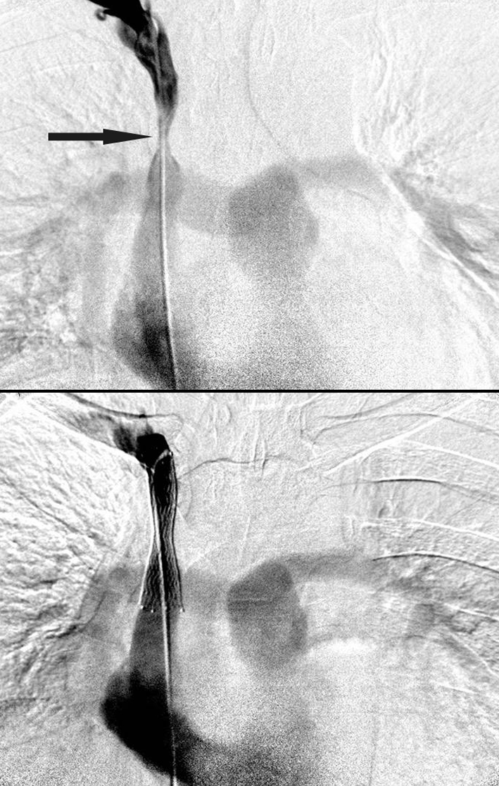
Fig 2 Digital subtraction venogram from a femoral vein approach shows tight focal stenosis of the mid superior vena cava (top; arrow). Digital subtraction venography after placement of a self expanding nitinol 14 mm stent, which was later dilated to 12 mm, shows good flow through the stent at the end of the procedure (bottom)
