

Abstract
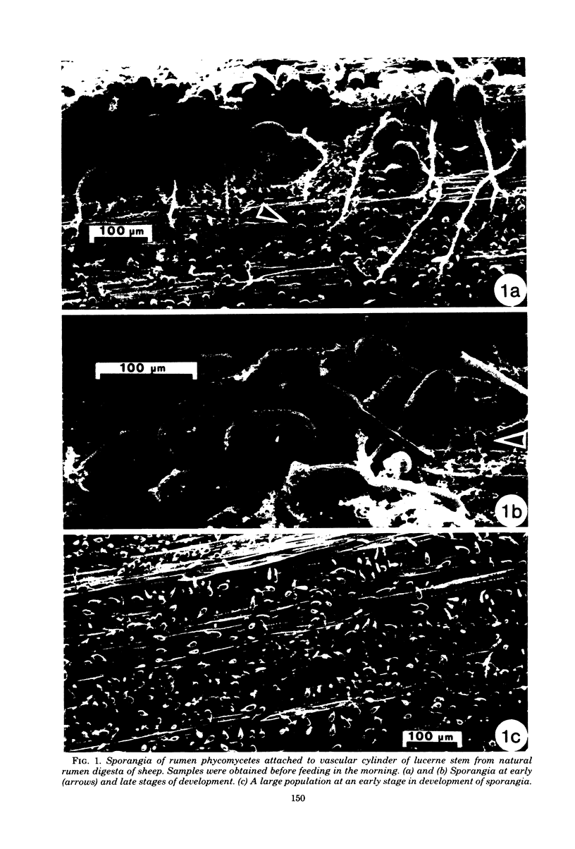
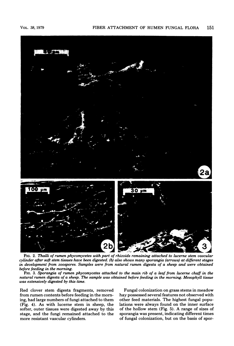
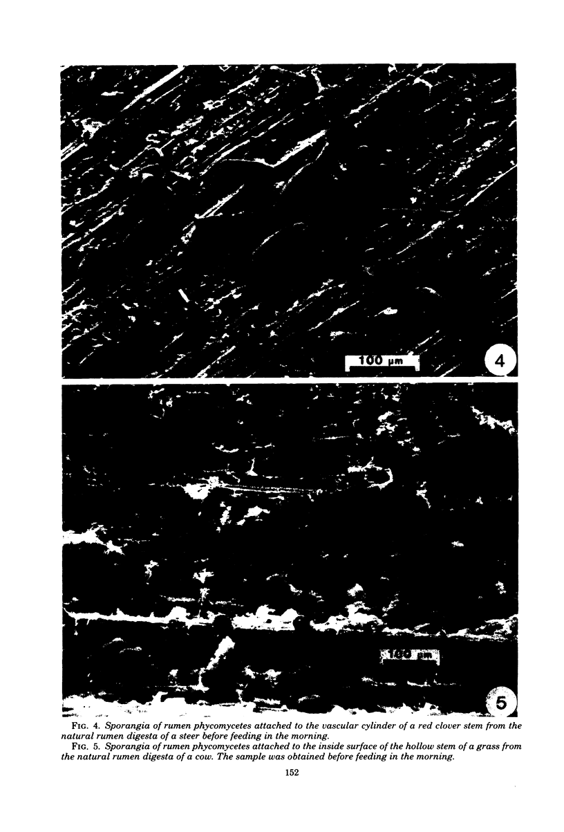
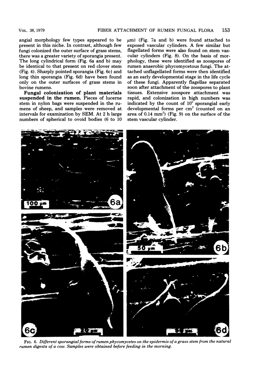
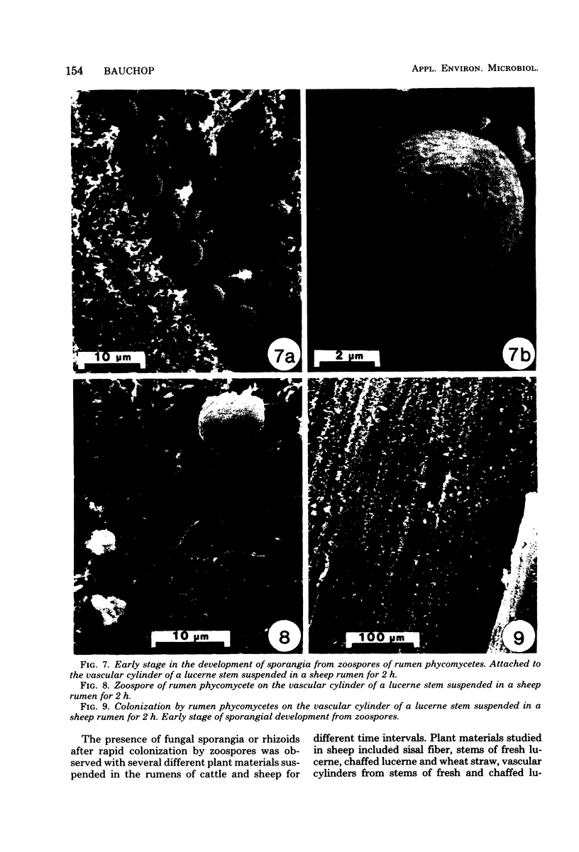
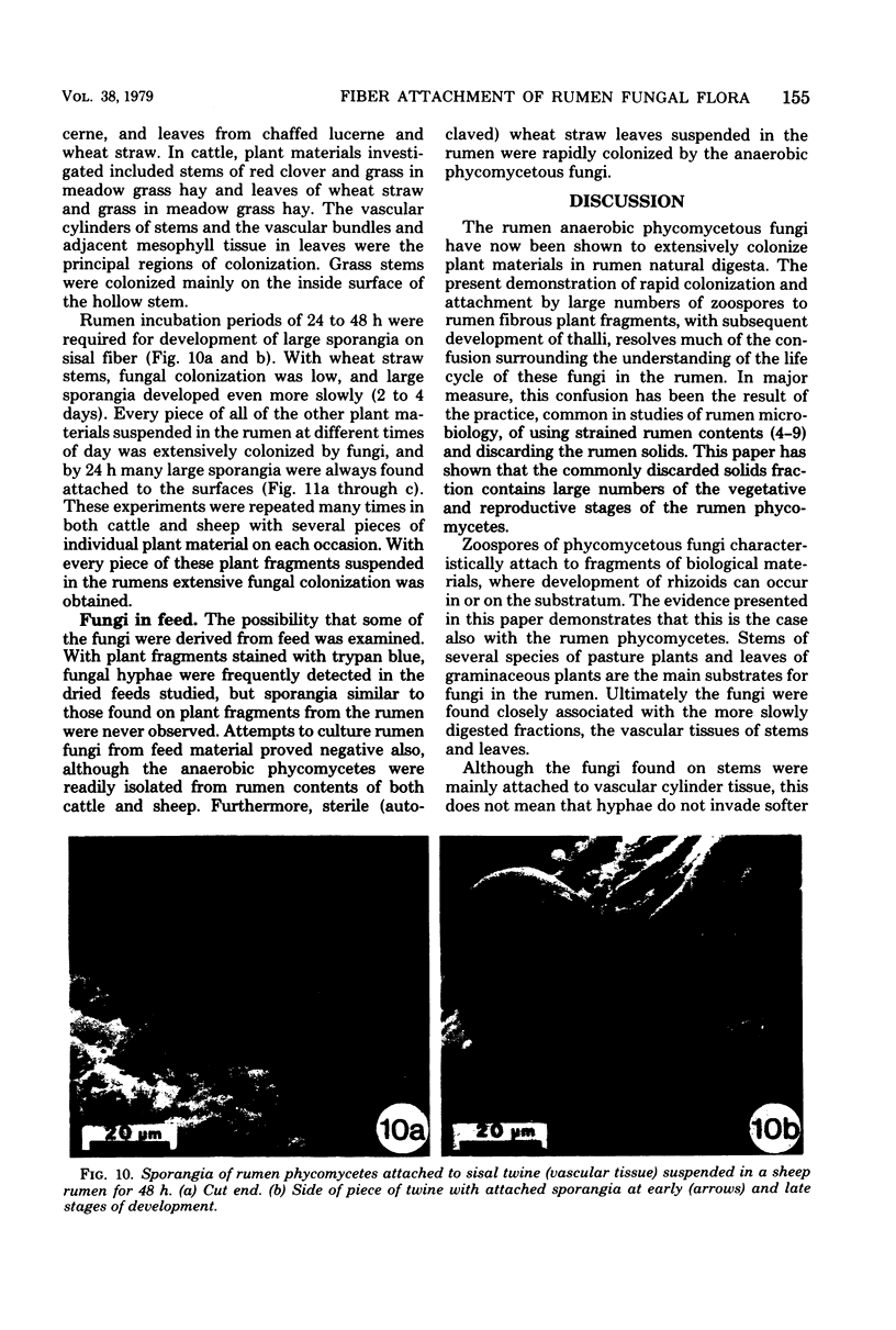
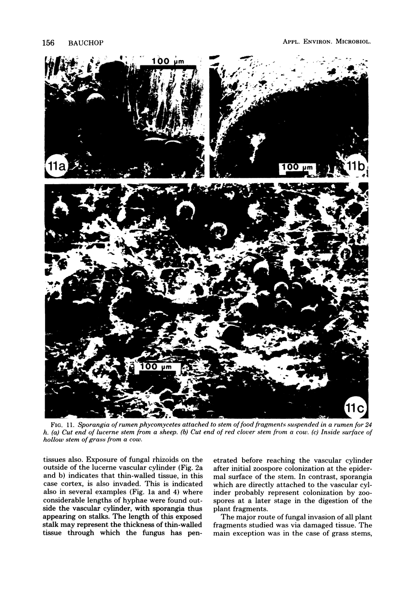
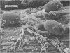
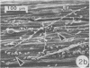
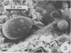
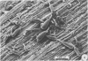
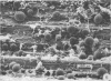
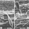
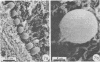
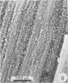
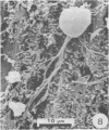
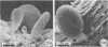
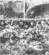
Plant fragments obtained from natural rumen digesta of fistulated cattle and sheep were examined by scanning electron microscopy. Various plant materials suspended in the rumen for different times were examined likewise. By 2 h large numbers of phycomycetous fungal zoospores were found attached to fibrous plant fragments, particularly vascular tissues. The subsequent development of these fungi resulted in production of thalli with extensive rhizoids and with sporangia up to 175 μm long. Scanning electron microscope examination of plant fragments randomly selected from natural rumen contents of both cattle and sheep demonstrated widespread colonization by large populations of these anaerobic fungi. Furthermore, all plant fragments suspended in nylon bags in the rumen were also extensively colonized. These findings demonstrate that plant fragments in the rumen are the sites of colonization and development by the anaerobic phycomycetous fungi. In addition, the results suggest that these fungi may form a significant part of the rumen microbiota in cattle and sheep fed on fibrous diets and suggest that they may be important in fiber digestion.
Full text
PDF
Images in this article
Selected References
These references are in PubMed. This may not be the complete list of references from this article.
- Bauchop T., Clarke R. T. Attachment of the ciliate Epidinium Crawley to plant fragments in the sheep rumen. Appl Environ Microbiol. 1976 Sep;32(3):417–422. doi: 10.1128/aem.32.3.417-422.1976. [DOI] [PMC free article] [PubMed] [Google Scholar]
- Orpin C. G. Invasion of plant tissue in the rumen by the flagellate Neocallimastix frontalis. J Gen Microbiol. 1977 Feb;98(2):423–430. doi: 10.1099/00221287-98-2-423. [DOI] [PubMed] [Google Scholar]
- Orpin C. G. Studies on the rumen flagellate Neocallimastix frontalis. J Gen Microbiol. 1975 Dec;91(2):249–262. doi: 10.1099/00221287-91-2-249. [DOI] [PubMed] [Google Scholar]
- Orpin C. G. Studies on the rumen flagellate Sphaeromonas communis. J Gen Microbiol. 1976 Jun;94(2):270–280. doi: 10.1099/00221287-94-2-270. [DOI] [PubMed] [Google Scholar]
- Orpin C. G. The occurrence of chitin in the cell walls of the rumen organisms Neocallimastix frontalis, Piromonas communis and Sphaeromonas communis. J Gen Microbiol. 1977 Mar;99(1):215–218. doi: 10.1099/00221287-99-1-215. [DOI] [PubMed] [Google Scholar]
- Orpin C. G. The rumen flagellate Piromonas communis: its life-history and invasion of plant material in the rumen. J Gen Microbiol. 1977 Mar;99(1):107–117. doi: 10.1099/00221287-99-1-107. [DOI] [PubMed] [Google Scholar]