

Abstract
We have identified two genes in the genomic database for C. elegans that code for proteins with significant sequence similarity to the mammalian soluble epoxide hydrolase (sEH). The respective transcripts were cloned from a mixed stage cDNA library from C. elegans. The corresponding proteins obtained after recombinant expression in insect cells hydrolyzed standard epoxide hydrolase substrates, including epoxyeicosatrienoic acids (EETs) and leukotoxins (EpOMEs). The enzyme activity was inhibited by urea-based compounds originally designed to inhibit the mammalian sEH. In vivo inhibition of the enzymes using the most potent of these compounds resulted in elevated levels of the EpOMEs in the nematode. These results suggest that the hydrolases are involved in the metabolism of possible lipid signaling molecules in C. elegans.
Keywords: soluble epoxide hydrolase; lipid signaling; epoxyeicosatrienoic acid; leukotoxin; 9,10-epoxy-12-octadecenoate; 12-13-epoxy-9-octadecenoate
1. Introduction
Soluble epoxide hydrolase (sEH), an enzyme first characterized in mammals, hydrolyzes an epoxide moiety through the addition of water.[1, 2] Interestingly, the mammalian soluble epoxide hydrolase possesses two catalytic activities localized to distinct regions of the enzyme.[3] The epoxide hydrolase active site is located on the globular C-terminal region of sEH, while an active site on the N-terminal region has been found to display phosphatase activity.[4] These domains are expressed separately in plants, giving rise to the hypothesis that invertebrates may also express the N- or C-terminal domain separately.[5, 6]
In mammals, the epoxide hydrolase activity of sEH has been implicated in the metabolism of several epoxide-containing lipid signaling molecules.[7, 8] This creates a diol species, which is more water-soluble than the epoxide and more readily eliminated through excretion, thereby depleting the supply of the epoxide signal. Also, in some cases the diols may be biologically active.[9] The epoxyeicosatrienoic acids (EETs) are epoxide-containing compounds produced by the metabolism of arachidonic acid by cytochrome p450 oxygenases.[10] The EETs have anti-inflammatory, as well as vasoconstrictive or vasodilatory properties, depending on the physiological context.[11–13] sEH has also been shown to metabolize 9,10-epoxy-12-octadecenoate (called leukotoxin, coronaric acid, or 9,10-EpOME) and 12,13-epoxy-9-octadecenoate (called isoleukotoxin, vernolic acid or 12,13-EpOME), cytochrome p450 metabolites of linoleic acid.[14–16] These compounds were identified as having a role in the development of acute respiratory distress syndrome (ARDS).[17]
Several recent studies have explored the signaling in C. elegans. C18 and C20 polyunsaturated fatty acids (PUFAs) have been shown to effect directed sperm movement towards the spermatheca and the formation of synaptic vesicles in neuronal tissues.[18, 19] FAT-3 mutants lack the delta6 fatty acid desaturase and so cannot convert linoleic acid into C20 PUFAs because this desaturase activity is a prerequisite to subsequent elongase activity.[20] These mutants show a number of defects which are rescued by dietary supplementation of C20 and C18 PUFAs, including smaller brood size, alterations in the defecation cycle, and defects in chemotaxis.[20, 21] These findings are exciting, because the nematodes amenability to genetic manipulation and the wealth of knowledge concerning its physiology make it an excellent model of lipid signaling. Thus the nematode may have implications for the study of such signaling pathways in mammalian disease models.
There are many similarities between fatty acid synthesis and metabolism in mammals and nematodes. Several desaturases and elongases in the fatty acid synthetic pathway of C. elegans have mammalian counterparts with parallel activities.[22–26] The cytochrome p450 monooxygenases responsible for production of the EETs and EpOMEs are present in the genome, though few have been characterized.[27–29] Finally, C. elegans contain arachidonic acid and linoleic acid, the precursors of these signaling molecules.[30, 31]
The genome of C. elegans contains five enzymes which display significant sequence similarity to the mammalian sEH. Of the five enzymes, two align with the C-terminal region of sEH. This raises the possibility that epoxide hydrolases are involved in the metabolism of epoxide containing lipids in C. elegans. We report the cloning and characterization of both hydrolases which display significant sequence similarity to the C-terminal region of soluble epoxide hydrolase, and their functional disruption in vivo using a small molecule inhibitor.
2. Materials and Methods
Nematode culture
The N2 (Bristol) strain of C. elegans was used. Plated nematodes were cultured on agar plates at 20°C and fed the OP50 strain of E. coli according to standard technique.[32] Liquid cultures of nematodes were grown in S-basal media and fed the NA22 strain of E. coli according to standard technique.[32] For the AUDA-BE liquid culture experiments, the worms were fed OP50.
Rapid Amplification of cDNA ends
Details of worm extract preparation and RNA extraction can be found in supplementary materials. CEEH1 and CEEH2 3′RACE experiments were performed on the total RNA sample with the SMART RACE cDNA Amplification Kit (Clontech, Palo Alto, CA) using the gene specific primers 3CEEH11: 5′ – GGGGAGGTCTTGTTGCGTGGCAATTCGCGG – 3′ and 3CEEH12: 5′ –CTGGGGAACTGCGGACGGAGCATTGGAC – 3′ for CEEH1 and 3CEEH21: 5′ – GGGTCAAAAGCTGGAATCCGGAATTCGG – 3′ and 3CEEH22: 5′ – CAGTCAGCCAGGCGGAACAACTGGTCC – 3′ for CEEH2. CEEH2 5′ RACE experiments were performed on the total RNA sample with the SMART RACE cDNA Amplification Kit (Clontech, Palo Alto, CA) using the gene specific primer 5CEEH21: 5′ – GCTCCCCAATCATGAGCAGCAAGTG – 3′. The 5′UTR of CEEH1 was determined by nested PCRs using the primers 5CEEH1F1: 5′ – CCACTGTCACCTGGTTGGACG – 3′ and 5CEEH1R1: 5′ –CCTTCCAAAACGTTTGGCTTCTCCCGCTGC – 3′, followed by the primers 5CEEH1F2: 5′ – TATAACGCGTTTGGAATCACT – 3′ and 5CEEH1R2: 5′ –CGAACCGAACGCAAGGTCGTGACGGGAGAG – 3′ using cDNA from a ProQuest C. elegans mixed stage library (Invitrogen, Carlsbad, CA).
Cloning
A ProQuest mixed stage cDNA library from C. elegans was purchased (Invitrogen, Carlsbad, CA). Primers for CEEH1 were designed to add BglII endonuclease sites on both ends, and a six histidine tag on the 3′ end of the coding sequence. Primers for CEEH2 were designed to add EcoRI endonuclease sites on both ends, and a six histidine tag on the 3′ end of the coding sequence. The primer pair for CEEH1 was 5′ – AGATCTATGTTGTTTGAAAGTATATACATACAATGT – 3′ and 5′ – AGATCTTTAGTGATGGTGATGGTGATGCTGATACTTATTCAAAAATTTT –3′. The primer pair for CEEH2 was 5′-GGATCCATGGGATTCTTTGCCGACTTGTTCA – 3′ and 5′ –GGATCCCTAGTGATGGTGATGGTGATGCAAGTGGGACTTGAAAGTTTTG – 3′.
Baculovirus Expression
Recombinant baculoviruses harboring the CEEH1 or CEEH2 cDNA sequence were generated by co-transfection of Sf21 cells derived from Spodoptera frugiperda with the recombinant transfer vector plasmid pACUW21 and Bsu36I-cleaved BacPAK6 viral DNA (Clontech, Palo Alto, CA) as previously described.[33]
Protein Purification
Infected High Five cells (250 mL) were pelleted and resuspended in phosphate buffer with 10 mM imidazole. The cells were homogenized with an Ultra-Turax T25 homogenizer (IKA Works, Wilmington, NC) rotating at 17,500 rpm for three 30 s intervals, with 15 s rests on ice between each grinding. The homogenate was centrifuged at 100,000 g for 1 h at 4°C. Ni-NTA HisBind Resin (EMD Biosciences, Inc., Madison, WI) was used to purify the enzymes according to manufacturer’s protocol. Details can be found in supplementary materials.
Protein Analysis
Protein concentration measurements were made using the BCA assay (Pierce, Rockford, IL) with BSA fraction V protein (Sigma-Aldrich, St. Louis, MO) to derive a standard curve. Polyacrylamide gel electrophoresis was performed using Novex gels (Invitrogen, Carlsbad, CA) for both SDS-PAGE analysis and isoelectric focusing.
Radiotracer Based Epoxide Hydrolase Activity Assay
Epoxide hydrolase activity was measured using racemic [3H]-trans-1,3-diphenylpropene oxide (t-DPPO) as substrate as previously described at a final concentration of 50 μM.[34] t-DPPO was previously synthesized and purified [35]. Details of the kinetics and IC50 determination can be found in supplementary materials.
Fluorescent Epoxide Hydrolase Assays
Assays were performed with the fluorescent substrate (3-Phenyl-oxiranyl)-acetic acid cyano-(6-methoxy-naphthalen-2-yl)-methyl ester (PHOME) at a final concentration of 50 μM. Assays were performed as described [36] with the following exception. An enzyme concentration of 0.57 μg/mL for CEEH1 was used in the assay.
Non-radioactive and Non-fluorescent Epoxide Hydrolase Assays
A 5 mM solution of each substrate was made in ethanol for the EETs, 9,10-EpOME and 12,13-EpOME. The substrate solution (1 μL) was added to 100 μL of enzyme preparation in sodium phosphate buffer (0.1 M, pH 7.4) containing 0.1 mg/mL of BSA ([S]final = 50 μM). The enzyme was incubated with the substrate at 30°C for 10 or 30 min, and the reaction quenched by addition of 400 μL of methanol. The EET reaction products were analyzed by HPLC-MS/MS as previously described [37].
Lipid extraction
Pellets of nematodes were resuspended in 1:1 (vol:vol) methanol:water spiked with internal standards and homogenized using an Ultra-Turrax T8 roto-stator grinder rotating at 25,000 rpm for 45 s. Chloroform (2 vol) was then added to the sample. The solution was vortexed 2 min, then centrifuged at 14,000 × g for 5 min at room temperature. The bottom organic layer was removed with a glass pipette and set aside and another two volumes chloroform added to the remaining aqueous phase. This extraction was repeated two more times, and the resulting chloroform fractions combined and evaporated under a nitrogen stream until almost dry then reconstituted in methanol containing 10,11-dihydroxyheptadecanoic acid (10,11-DHHep) and d8 11(12)-EET as internal standards.
In vivo inhibition using AUDA-BE
Worms were staged and 100mL liquid cultures grown at 20ºC. AUDA-BE (0.5mL of 10mM in DMSO) was added to the worms 24h and 36h after adding the staged L1s to the culture to a final concentration of 1% DMSO and 10μM AUDA-BE, and worms collected when the reached the adult stage as judged by examination of aliquots. Control group was treated with vehicle alone, to a final concentration of 1% DMSO.
LC-MS/MS analysis of worm extract
Lipid extracts were analyzed using Shimadzu ASP10 HPLC system (Shimadzu, Pleasanton, CA) and Quattro Ultima tandem mass spectrometer (Waters, Milford, MA). Details can be found in supplementary materials.
3. Results
3.1. Cloning of the hydrolases
A tBLASTx search of the NCBI genomic database of C. elegans employing Gallus gallus (Genbank accession number Q120010) and Xenopus tropicalis (Genbank accession number BC078066) sEH nucleotide and translated amino acid sequences returned five soluble epoxide hydrolase hits. Two of the predicted enzymes, Genbank accession numbers NM_064867 and NM_073261, displayed significant sequence similarity to the C-terminus of mammalian sEH, while the other three, Genbank accession numbers NM_072133, NM_063993 and NM_072107.3 aligned with the N-terminal domain (Table 1). Primers were constructed based on the genomic sequence of enzymes which aligned with the C-terminal domain, and the full length transcripts were verified by 5′ and 3′ RACE experiments. The RACE experiments were performed using both a purchased mixed stage cDNA library, and a cDNA archive prepared in the lab from a mixed stage pellet of Bristol-Meyer worms. The reconstructed full-length mRNA sequences were submitted to Genbank. The enzyme corresponding to the Genbank accession number EU151493 was designated CEEH1, while the enzyme corresponding to the Genbank accession number EU151492 was designated CEEH2. Primers based on these results were designed and the sequences were cloned. (Figures 1 and 2)
TABLE 1.
Protein BLAST results
| Query | Subject | Subject Residues Aligned | Segment identity | Score | E- value |
|---|---|---|---|---|---|
| CEEH1 | sEH from H. sapiens | 241–342 | 27% | 305 | 7e-26 |
| sEH from G. gallus | 248–547 | 26% | 303 | 1e-25 | |
| sEH from X. tropicalis | 249–548 | 28% | 289 | 5e-24 | |
| HLD from X. autotrophicus | 34–148 | 30% | 131 | 1e-05 | |
| CEEH2 | sEH from H. sapiens | 238–548 | 24% | 282 | 3e-23 |
| sEH from G. gallus | 238–547 | 22% | 237 | 5e-18 | |
| sEH from X. tropicalis | 226–556 | 26% | 294 | 1e-24 | |
| HLD from X. autotrophicus | 34–148 | 33% | 156 | 1e-08 | |
| NM_063993 | sEH from H. sapiens | 100–222 | 39% | 225 | 5e-17 |
| NM_072107.3 | sEH from H. sapiens | 1–237 | 27% | 205 | 1e-13 |
| NM_072133 | sEH from H. sapiens | 84–221 | 31% | 170 | 1e-10 |
Figure 1.

Nucleotide sequence and translated protein sequence of the CEEH1 cDNA. This DNA sequence has been assigned Genbank accession no. EU151493.
Figure 2.

Nucleotide sequence and translated protein sequence of the CEEH2 cDNA. This DNA sequence has been assigned Genbank accession no. EU151492.
3.2 Expression and purification of the hydrolases
The constructs encoded for a six histidine tag on the C-terminal end of the enzymes. The recombinant enzymes were expressed in a baculovirus expression system and purified on a nickel chelation column (see supplementary materials). The experimentally determined molecular weight for CEEH1 and CEEH2 were 48 and 44 kDa, respectively, in close agreement with the calculate values of 48.7 and 42.9 kDa. The experimentally determined isoelectric points for CEEH1 and CEEH2 were between the ranges of 8.7–8.8 and 6.7–6.8, respectively, also near the calculated values of 8.8 and 6.8.
3.3 Characterization of recombinant enzyme activity
The epoxide hydrolase activities were initially assayed with t-DPPO, a substrate for the mammalian sEH. The half life for CEEH1 at 37ºC was between 1 and 2 hours, the half life at 24ºC was approximately 8 hours, and the half life at 4ºC was 24 hours. The half life for CEEH2 was approximately 2 hours at 37ºC, 24 hours at 24ºC and over six days at 4ºC.
CEEH1 had a specific activity of 3000 nmol·min−1·mg−1, comparable to the human sEH (Table 2). However, CEEH2 had a specific activity of 14 nmol·min−1·mg−1 (Table 2). Although this was much lower than the activity displayed by CEEH1, insect cells infected with a baculovirus containing the CEEH2 sequence displayed significantly higher activity than insect cells infected with a control virus containing the sequence of an alpha/beta hydrolase fold enzyme which does not possess epoxide hydrolase activity (see supplementary material). This provided evidence that the observed EH activity after partial purification was not due to a co-purified enzyme from the insect cells. Km and kcat were then determined for t-DPPO and CEEH1. The Km was found to be 160 μM, and the kcat 12 s−1 (Table 3).
TABLE 2.
Specific activity with t-DPPO and natural substrates
| Compound name | Structure | Specific activity (nmol·min−1·mg−1) | |
|---|---|---|---|
| Recombinant CEEH1 | Recombinant CEEH2 | ||
| t-DPPO |

|
3,000 ± 230 | 14 ± 2.6 |
| 9,10-EpOME |
|
137 ± 1.77 | 6.77 ± .0033 |
| 12,13-EpOME |

|
132 ± 1.16 | 4.75 ± .085 |
| 14,15-EET |

|
615 ± 10.8 | 7.63 ± .72 |
| 11,12-EET |

|
202 ± 12.3 | 4.10 ± 0.32 |
| 8,9-EET |

|
44.2 ± 1.00 | 2.00 ± 0.14 |
Recombinant CEEH1 and CEEH2 were partially purified as described. Assay conditions are described in the Materials and Methods section. Results are presented as the mean ± standard deviation of two to three separate experiments performed in triplicate. Note: the 5,6 – EET rapidly cyclizes on storage.
TABLE 3.
Km and Kcat with t-DPPO and EpOMEs
| Name Structure | Kinetic parameter | Recombinant CEEH1 | Recombinant CEEH2 | Recombinan t human sEH |
|---|---|---|---|---|

|
Km (μM) | 160 ± 21 | N.D. | 6.2 ± 0.6 |
| kcat (s−1) | 12 ± 0.5 | N.D. | 4.3 ± 0.3 | |
| kcat/Km(μM−1·s−1) | 0.07 | .0004 | 0.7 | |

|
Km (μM) | 7.5 ± 3.9 | 8.4 ± 3.6 | 6.15 ± 1.0 |
| kcat (s−1) | 0.22 ± .035 | 0.01 ± 0.004 | 2.78 | |
| kcat/Km(μM−1·s−1) | 0.029 | 0.0012 | 0.452 | |

|
Km (μM) | 11.4 ± 5.1 | 1.9 ± 0.72 | 5.17 ± 0.56 |
| kcat (s−1) | 0.11 ± 0.022 | .002 ± 0.0004 | 1.93 | |
| kcat/Km(μM−1·s−1) | 0.0096 | 0.001 | 0.37 |
Recombinant CEEH1 and CEEH2 were partially purified as described. Results for t-DPPO are presented as the mean ± standard deviation of two to three separate experiments performed in triplicate. In the case of the EpOMEs, the error is the standard error from a nonlinear regression fitting the results of an experiment performed in triplicate to the Michaelis-Menten equation. The kcat/Km ratio for CEEH2 and t-DPPO was estimated under the assumption of [S]≪ Km. N.D. means not determined. Values for t-DPPO and the human enzyme are from Morisseau (2000).[6] Values for the EpOMEs and the human enzyme are from Greene (2000)[40] and were determined with the methyl ester of these compounds.
Next, natural products were assayed with the recombinant enzymes. The specific activities with the compounds were first determined. 14,15-EET was found to have a value of 615 nmol·min−1·mg−1, 11,12-EET had a value of 202 nmol·min−1·mg−1, and 8,9-EET had the lowest value at 44.2 nmol·min−1·mg−1 (Table 2). The specific activities for 9,10-EpOME and 12,13-EpOME were found to be 137 nmol·min−1·mg−1 and 132 nmol·min−1·mg−1, respectively (Table 2). CEEH2 showed a greater than 20-fold lower activity, compared to CEEH1 (Table 2). Km and kcat were then determined for the EpOMEs (Table 3). The Km values fell in the 1–10 μM range, while the kcat values ranged from approximately 0.2 to 0.002 s−1 (Table 3).
A series of fluorescent substrates has been developed to characterize human sEH epoxide hydrolase activity, and for the use in high-throughput assays.[36] In order to compare the CEEH1 to the human enzyme, a limited structure activity experiment was performed using these substrates. All of the substrates were hydrolyzed by CEEH1 (Table 4). CEEH2 did not display activity when assayed with these substrates.
TABLE 4.
Specific activity with fluorescent substrates
| # |  |
% of highest specific activity | |
|---|---|---|---|
| Recombinant CEEH1 | Recombinant human sEH | ||
| 1 |

|
80 ± 5.9 | 26 |
| 2 |

|
43 ± 2.2 | 15 |
| 3 |
|
52 ± 1.9 | 38 |
| 4 |

|
24 ± 3.2 | < 1 |
| 5 |

|
100 (2300 ± 240 nmole/min/mg) | 100 (2700 ± 44 nmole/min/mg) |
| 6 |

|
41 ± 2.0 | 13 |
| 7 |

|
68 ± 2.8 | 15 |
Recombinant CEEH1 was partially purified as described. Assay conditions are described in the Materials and Methods section. Results are presented as the mean ± standard deviation of three separate experiments performed in triplicate. Values for the recombinant human enzyme are from Jones (2005).[36]
3.4 Analysis of epoxide hydrolase inhibition
A select group of small molecule inhibitors of the mammalian sEH were assayed with recombinant CEEH1 (Table 5). The same inhibitors were used to inhibit t-DPPO activity in crude extract from a mixed stage liquid culture of C. elegans. AUDA was found to be the most potent inhibitor of the five assayed.
Table 5.
IC50s with urea-based inhibitors
| Name | Structure | Mixed stage crude extract IC50 (nM) | Recombinant CEEH1 IC50(nM) | Recombinant Human sEH IC50 (nM) |
|---|---|---|---|---|
| CEU |

|
>50000 | >50000 | 42000 ± 2000 |
| DCU |

|
>50000 | 41000 ±1200 | 160 ±10 |
| CDU |
|
6900 ± 66 | 4300 ± 320 | 100 ± 10 |
| AUDA |
|
650 ± 25 | 530 ± 44 | 100 ± 10 |
IC50 values for the urea based inhibitors N-cyclohexyl-N’-ethylurea (CEU), N,N’-dicyclohexylurea (DCU), N-cyclohexyl-N’-dodecylurea (CDU), and 12-(3-adamantane-1-yl-ureido)-dodecanoic acid (AUDA). Recombinant CEEH1 was partially purified as described. Assay was performed using [3H] t-DPPO as substrate. Conditions are described in the Materials and Methods section. Values for the human enzyme are from Jones (2005), which also used [3H] t-DPPO as substrate.[36] Error bars represent the standard deviation for three separate experiments performed in triplicate.
3.5 Small molecule inhibition
Finally, AUDA-BE was used to inhibit the enzymes in vivo. AUDA and its butyl ester are of similar inhibitory potency. The butyl ester is anticipated to penetrate tissues rapidly and be hydrolyzed to the free AUDA by esterases. Synchronized worms grown in liquid culture were treated with the inhibitor 24 and 36 hours after the first larval stage was transferred from plates to the culture. The worms were harvested at the adult stage and the lipids extracted for LC-MS/MS analysis. It was found that 9,10-EpOME and 12,13-EpOME levels increased by approximately 200% in the treated populations relative to the controls (Figure 3). Changes in 9,10-DiHOME levels were not significantly different from controls. Interestingly, the 12,13-DiHOME levels decreased, rather than increased in the treated populations (Figure 3). The epoxide-to-diol ratio for 9,10-EpOME and 12,13-EpOME both increased. The 9,10-EpOME ratio increased by 174 % while the 12,13-EpOME ratio increased tenfold (Figure 3).
Figure 3.

Inhibition of epoxide hydrolase activity in vivo using the urea-based small molecule inhibitor AUDA-BE. Worms grown in suspension culture were fed the bacterial strain OP50. Inhibited populations were treated twice with AUDA-BE in DMSO to a final inhibitor concentration of 10μM. Controls were treated with vehicle alone. Error bars represent the standard deviation.
4. Discussion
The mammalian sEH is an approximately 60kDa enzyme composed of two globular regions connected by a short proline-rich linker.[2] Mammalian soluble epoxide hydrolase is thought to be a product of the fusion of two ancestral bacterial genes.[5] The C-terminal region contains the epoxide hydrolase active site and is descended from haloalkane dehalogenase (HLD), while the N-terminal region contains a phosphatase active site and is descended from haloacid dehalogenase (HAD).[5, 38] When sequences from the nematode enzymes, vertebrate sEH homologs, and the bacterial gene HLD are compared by BLAST, one sees that CEEH1 and CEEH2 score higher with the vertebrate sEH homologs than with HLD and the aligned segments fall within the C-terminal region, or epoxide hydrolase domain, of the vertebrate sEH homologs (Table 1).
Certain important characteristics of soluble epoxide hydrolases can be identified in the CLUSTALW alignment (Figure 4). The alpha/beta hydrolase fold family catalytic triad is composed of a catalytic nucleophile and a histidine-aspartate pair that activates a water molecule.[3] This triad is shared by all five enzymes, though the spacing between the amino acids is not conserved in the lower organisms. In addition to these residues, the mammalian soluble epoxide hydrolases have two tyrosines which are thought play a role in the orientation and activation of the epoxide moiety.[3] These amino acids are not found in the haloacid dehalogenase, but align in both CEEH1 and CEEH2.
Figure 4.
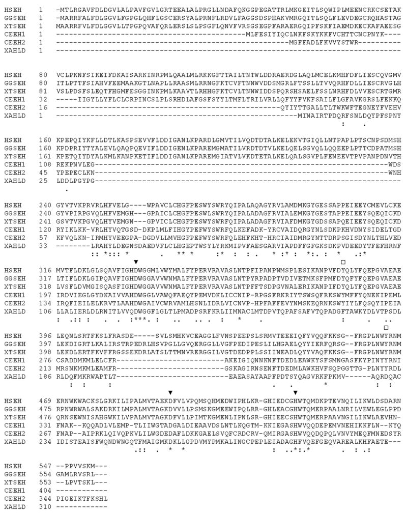
Alignment of the vertebrate sEH homologs with the nematode enzymes and bacterial HLD. The mammalian epoxide hydrolase “catalytic triad” residues are marked by closed triangles. The tyrosines thought to polarize the epoxide moiety of the epoxide hydrolase substrate are marked by open squares. HSEH = sEH from H. sapiens (Genbank accession no. L05779); GGSEH = sEH from G. gallus (DQ120010); XTSEH = sEH from X. tropicalis (BC075370); XAHLD = HLD from X. autotrophicus (M26950).
In addition to the C-terminal homologs CEEH1 and CEEH2, three enzymes were identified that display significant sequence similarity to the N-terminus of vertebrate sEH (Table 1), which may make C. elegans a fascinating case in which both C-terminal and N-terminal precursors of mammalian sEH are expressed individually, as they are in the plants so far examined.[6, 39] The CEEH2 sequence confirmed by RACE agreed with the WormBase predicted mRNA from gene model K06C5.5 (Figure 2). The CEEH1 sequence corresponded to the predicted mRNA from gene model K02F3.6 (Figure 1).
When the enzymes were assayed with t-DPPO, a substrate used to measure mammalian sEH epoxide hydrolase activity, CEEH2 was found to have less than 1/100 the activity of CEEH1 despite the fact that CEEH2 shares the conserved catalytic triad as well as the two orienting tyrosines (Table 2). CEEH2 could, of course, be optimized to hydrolyze very different substrates from CEEH1 or even have quite a different role in the nematode. CEEH1 was found to be less stable than CEEH2. This instability may be due to the dilute state and of the enzyme, which does not reflect the intracellular environment. These data argue against instability accounting for CEEH2′s low activity on the substrates tested. The kcat/Km ratio, or enzyme specificity, of CEEH1 for t-DPPO was lower than the human enzyme, having both a higher kcat and Km (Table 3). The ratio indicated that t-DPPO is a fair substrate for CEEH1 activity. The estimated kcat/Km ratio for CEEH2 shows that t-DPPO is a poor substrate for assaying CEEH2 activity (Table 3).
The structure of the lipid epoxide molecules thought to be endogenous substrates for mammalian sEH differ greatly from t-DPPO. The EETs are 20 carbon fatty acids, in which an epoxide lies near the center of the molecule. Like the human enzyme, CEEH1 hydrolyzed 14,15-EET at the highest rate, which has the epoxide moiety furthest from the carboxylic acid (Table 2). CEEH2 showed twenty-fold less activity when assayed with the molecules, but also hydrolyzed 14,15-EET at the highest rate. 9,10-EpOME and 12,13-EpOME are regioisomeric 18 carbon fatty acid epoxides. The kcat/Km ratios for these compounds reveal that CEEH2 has a ten to twenty-fold lower specificity for the EpOME when compared to CEEH1 (Table 3). CEEH2 has roughly the same or lower Km for the EpOMEs as CEEH1, but a much lower kcat. CEEH2 could influence the metabolism of the EpOMEs in vivo by competing with CEEH1 for substrate binding, resulting in a decrease in the rate of diol production.
To learn more about the substrate selectivity of CEEH1, fluorescent substrates recently developed for use in high-throughput screens of inhibitor libraries were next assayed (Table 4). The left side of the substrates consist of a cyanohydrin paired with a series of right sides, which contain either an ester or carbonate moiety. When released, the cyanohydrin rearranges to yield an intensely fluorescent aromatic aldehyde with a huge Stoke’s shift. These right sides also have a range of steric properties, from a short alkyl chain to substituted phenyl rings, as well as one trisubstituted epoxide. CEEH1 hydrolyzed substrate 5 at the highest rate, like the human enzyme (Table 4), but did not show a marked preference for the aryl carbonate (substrate 5) over the ester (substrate 1). Also, the nematode enzyme showed less of a preference for the aryl carbonate over the alkyl carbonates, compared to the human enzyme. Interestingly, CEEH1 also hydrolyzed substrate 4, the trisubstituted epoxide, which is very slowly turned over by the mammalian enzyme. Substrates 6 and 7, which contain nitro- and chloro-substituted phenyl rings, were also fair substrates for CEEH1, when compared to the human enzyme. This may indicate that the nematode binding site lacks steric constraints found in the mammalian enzyme.
In order to test for inhibition of the enzyme, four inhibitors were chosen that have high, medium and low potency when assayed with the mammalian enzymes. Their structures are based on a urea pharmacophore, which has been shown to bind to the mammalian enzyme in a reversible manner as a putative transition state mimic. These same five inhibitors were also used to characterize the crude extract. The fluorescent probes are attacked by esterases and glutathione S-transferase, and so they cannot easily be used to measure epoxide hydrolase activity in the crude extract. To use these substrates in an epoxide hydrolase “specific assay” one must remove glutathione or inhibit glutathione S-transferase activity and inhibit or remove esterase activity. However, the t-DPPO assay can be made specific for epoxide hydrolase activity after a correction for glutathione transferase activity or elimination of glutathione. In this case, no GST activity was detected. Crude extract from a 20k x g spin of a mixed stage population was prepared. Less than 10% of the total activity was recovered in the pellet, possibly the result of worm carcasses, which trap some soluble material. When the supernatant and recombinant CEEH1 were characterized with the five inhibitors, CEEH1 showed the same pattern and level of response as the crude extract (Table 5) suggesting that CEEH1 is similar or identical to the majority of the enzyme accounting for in vivo activity. The difference in the IC50 values could be due to substrate binding or sequestering, another metabolic pathway for t-DPPO, or the presence of other hydrolases in the crude extract.
When worms were treated with the small molecule inhibitor AUDA-BE, the EpOME levels increased, and their diol levels did not change or increased slightly (Figure 3), though no phenotypic effects were noted. In the simplest model, one might expect that a decrease in epoxide hydrolase activity would result in an increase in epoxides, a decrease in the corresponding diols, resulting in an increased epoxide-to-diol ratio. This assumes one pathway of diol production, which may not be the case in the nematode, and does not consider feedback regulation of other enzymes involved in the formation or elimination of the diols. The EpOMEs are relatively lipophilic compounds that will concentrate in tissues, while the DiHOMEs are dramatically more polar. They are anticipated to rapidly diffuse into the media or be conjugated. It is possible that compensatory mechanisms affected by the decrease in epoxide hydrolase activity, coupled with the rapid clearance of the diols resulted in a relatively unchanged level of diols, regardless of epoxide levels. More work will be required to dissect these metabolic pathways. A second experiment employing RNAi can be found in supplementary materials.
Taken together, the in vitro characterization of the enzymes and the small molecule inhibition results suggest that CEEH1, and possibly CEEH2 to a lesser extent are involved in the metabolism of these C18 lipids in vivo. In mammals, epoxide containing lipids have been found to play numerous signaling roles. The metabolites of 9,10-EpOME and 12,13-EpOME have been shown to promote the onset of ARDS-like symptoms. The EETs have been implicated in a number of physiological processes, including the regulation of inflammation. The role of these epoxide-containing lipids have not been studied in C. elegans.
The demonstration of a nematode epoxide hydrolase with the ability to hydrolyze mammalian soluble epoxide hydrolase substrates offers the possibility of parallel signaling pathways in C. elegans. Their involvement in the metabolism of epoxide containing lipids in vivo has impacts for the study of both lipid metabolism and lipid signaling in C. elegans, which is a model both for parasitic and phytophagus nematodes as well as a more general model for eukaryotic regulatory biology.
Supplementary Material
Acknowledgments
We would like to thank Lesilee Rose and Adam Hayashi for supplying nematodes and bacterial strains and advice on the maintenance of C. elegans. We would also like to thank Kara Schmelzer, Christophe Morisseau and George Kamita for advice at various stages of this project, Katrin Georgi for discussion of oxylipin quantification and Hsing-Ju ‘Cindy’ Tsai for generously sharing instrument time and analytical columns. T.R.H. and P.A.A were supported by NIEHS Advanced Training in Environmental Toxicology Grant T32 ES007059. Funding was also provided by NIEHS Grant R37 ES02710 and NIEHS Superfund Basic Research Program Grant P42 ES004699.
Footnotes
Publisher's Disclaimer: This is a PDF file of an unedited manuscript that has been accepted for publication. As a service to our customers we are providing this early version of the manuscript. The manuscript will undergo copyediting, typesetting, and review of the resulting proof before it is published in its final citable form. Please note that during the production process errors may be discovered which could affect the content, and all legal disclaimers that apply to the journal pertain.
References
- 1.Wixtrom RN, Hammock BD. Membrane-bound and soluble fraction epoxide hydrolases. In: Zakim D, Vessey DA, editors. Biochemical Pharmacology and Toxicology. Wiley; New York: 1985. pp. 1–93. [Google Scholar]
- 2.Newman JW, Morisseau C, Hammock BD. Progress in lipid research. 2005;44:1–51. doi: 10.1016/j.plipres.2004.10.001. [DOI] [PubMed] [Google Scholar]
- 3.Argiriadi MA, Morisseau C, Hammock BD, Christianson DW. Proc Natl Acad Sci U S A. 1999;96:10637–10642. doi: 10.1073/pnas.96.19.10637. [DOI] [PMC free article] [PubMed] [Google Scholar]
- 4.Newman JW, Morisseau C, Harris TR, Hammock BD. Proc Natl Acad Sci U S A. 2003;100:1558–1563. doi: 10.1073/pnas.0437724100. [DOI] [PMC free article] [PubMed] [Google Scholar]
- 5.Beetham JK, Grant D, Arand M, Garbarino J, Kiyosue T, Pinot F, Oesch F, Belknap WR, Shinozaki K, Hammock BD. DNA and cell biology. 1995;14:61–71. doi: 10.1089/dna.1995.14.61. [DOI] [PubMed] [Google Scholar]
- 6.Morisseau C, Beetham JK, Pinot F, Debernard S, Newman JW, Hammock BD. Archives of biochemistry and biophysics. 2000;378:321–332. doi: 10.1006/abbi.2000.1810. [DOI] [PubMed] [Google Scholar]
- 7.Funk CD. Prostaglandins and Leukotrienes: Advances in Eicosanoid Biology. 2001. pp. 1871–1875. [DOI] [PubMed] [Google Scholar]
- 8.Zeilhofer HU. Biochemical Pharmacology. 2007;73:165–174. doi: 10.1016/j.bcp.2006.07.037. [DOI] [PubMed] [Google Scholar]
- 9.Moghaddam MF, Grant DF, Cheek JM, Greene JF, Williamson KC, Hammock BD. Nature medicine. 1997;3:562–566. doi: 10.1038/nm0597-562. [DOI] [PMC free article] [PubMed] [Google Scholar]
- 10.Capdevila JH, Falck JR. Prostaglandins & other lipid mediators. 2002;68–69:325–344. doi: 10.1016/s0090-6980(02)00038-2. [DOI] [PubMed] [Google Scholar]
- 11.Pokreisz P, Fleming I, Kiss L, Barbosa-Sicard E, Fisslthaler B, Falck JR, Hammock BD, Kim IH, Szelid Z, Vermeersch P, Gillijns H, Pellens M, Grimminger F, van Zonneveld AJ, Collen D, Busse R, Janssens S. Hypertension. 2006;47:762–770. doi: 10.1161/01.HYP.0000208299.62535.58. [DOI] [PMC free article] [PubMed] [Google Scholar]
- 12.Node K, Huo Y, Ruan X, Yang B, Spiecker M, Ley K, Zeldin DC, Liao JK. Science. 1999;285:1276–1279. doi: 10.1126/science.285.5431.1276. [DOI] [PMC free article] [PubMed] [Google Scholar]
- 13.Gauthier KM, Edwards EM, Falck JR, Reddy DS, Campbell WB. 14,15-Epoxyeicosatrienoic Acid Represents a Transferable Endothelium-Dependent Relaxing Factor. Bovine Coronary Arteries. 2005:666–671. doi: 10.1161/01.HYP.0000153462.06604.5d. [DOI] [PubMed] [Google Scholar]
- 14.Draper AJ, Hammock BD. Archives of biochemistry and biophysics. 2000;376:199–205. doi: 10.1006/abbi.2000.1705. [DOI] [PubMed] [Google Scholar]
- 15.Ozawa T, Sugiyama S, Hayakawa M, Taki F, Hanaki Y. Biochemical and biophysical research communications. 1988;152:1310–1318. doi: 10.1016/s0006-291x(88)80428-5. [DOI] [PubMed] [Google Scholar]
- 16.Hayakawa M, Sugiyama S, Takamura T, Yokoo K, Iwata M, Suzuki K, Taki F, Takahashi S, Ozawa T. Biochemical and biophysical research communications. 1986;137:424–430. doi: 10.1016/0006-291x(86)91227-1. [DOI] [PubMed] [Google Scholar]
- 17.Ozawa T, Hayakawa M, Kosaka K, Sugiyama S, Ogawa T, Yokoo K, Aoyama H, Izawa Y. Advances in prostaglandin, thromboxane, and leukotriene research. 1991;21B:569–572. [PubMed] [Google Scholar]
- 18.Kubagawa HM, Watts JL, Corrigan C, Edmonds JW, Sztul E, Browse J, Miller MA. Nature cell biology. 2006;8:1143–1148. doi: 10.1038/ncb1476. [DOI] [PubMed] [Google Scholar]
- 19.Lesa GM, Palfreyman M, Hall DH, Clandinin MT, Rudolph C, Jorgensen EM, Schiavo G. Journal of cell science. 2003;116:4965–4975. doi: 10.1242/jcs.00918. [DOI] [PubMed] [Google Scholar]
- 20.Watts JL, Phillips E, Griffing KR, Browse J. Genetics. 2003;163:581–589. doi: 10.1093/genetics/163.2.581. [DOI] [PMC free article] [PubMed] [Google Scholar]
- 21.Kahn-Kirby AH, Dantzker JL, Apicella AJ, Schafer WR, Browse J, Bargmann CI, Watts JL. Cell. 2004;119:889–900. doi: 10.1016/j.cell.2004.11.005. [DOI] [PubMed] [Google Scholar]
- 22.Watts JL, Browse J. Proc Natl Acad Sci U S A. 2002;99:5854–5859. doi: 10.1073/pnas.092064799. [DOI] [PMC free article] [PubMed] [Google Scholar]
- 23.Watts JL, Browse J. Archives of biochemistry and biophysics. 1999;362:175–182. doi: 10.1006/abbi.1998.1024. [DOI] [PubMed] [Google Scholar]
- 24.Oh CS, Toke DA, Mandala S, Martin CE. The Journal of biological chemistry. 1997;272:17376–17384. doi: 10.1074/jbc.272.28.17376. [DOI] [PubMed] [Google Scholar]
- 25.Napier JA, Hey SJ, Lacey DJ, Shewry PR. The Biochemical journal. 1998;330( Pt 2):611–614. doi: 10.1042/bj3300611. [DOI] [PMC free article] [PubMed] [Google Scholar]
- 26.Beaudoin F, Michaelson LV, Hey SJ, Lewis MJ, Shewry PR, Sayanova O, Napier JA. Proc Natl Acad Sci U S A. 2000;97:6421–6426. doi: 10.1073/pnas.110140197. [DOI] [PMC free article] [PubMed] [Google Scholar]
- 27.Gotoh O. Molecular biology and evolution. 1998;15:1447–1459. doi: 10.1093/oxfordjournals.molbev.a025872. [DOI] [PubMed] [Google Scholar]
- 28.Menzel R, Rodel M, Kulas J, Steinberg CE. Archives of biochemistry and biophysics. 2005;438:93–102. doi: 10.1016/j.abb.2005.03.020. [DOI] [PubMed] [Google Scholar]
- 29.Jia K, Albert PS, Riddle DL. Development (Cambridge, England) 2002;129:221–231. doi: 10.1242/dev.129.1.221. [DOI] [PubMed] [Google Scholar]
- 30.Tanaka T, Ikita K, Ashida T, Motoyama Y, Yamaguchi Y, Satouchi K. Lipids. 1996;31:1173–1178. doi: 10.1007/BF02524292. [DOI] [PubMed] [Google Scholar]
- 31.Hutzell PA, Krusberg LR. Comparative biochemistry and physiology. B, Comparative biochemistry. 1982;73B:517–520. [Google Scholar]
- 32.T. Steiernagle, WormBook.
- 33.Merrington CL, King LA, Posse RD. In: Protien Expression: A practical approach. Higgins SJ, Hames BD, editors. Oxford university press; 1999. pp. 101–127. [Google Scholar]
- 34.Borhan B, Mebrahtu T, Nazarian S, Kurth MJ, Hammock BD. Improved radiolabeled substrates for soluble epoxide hydrolase. Analytical biochemistry. 1995:188–200. doi: 10.1006/abio.1995.1520. [DOI] [PubMed] [Google Scholar]
- 35.Borhan B, Mebrahtu T, Nazarian S, Kurth MJ, Hammock BD. Anal Biochem. 1995;231:188–200. doi: 10.1006/abio.1995.1520. [DOI] [PubMed] [Google Scholar]
- 36.Jones PD, Wolf NM, Morisseau C, Whetstone P, Hock B, Hammock BD. Analytical biochemistry. 2005;343:66–75. doi: 10.1016/j.ab.2005.03.041. [DOI] [PMC free article] [PubMed] [Google Scholar]
- 37.Newman JW, Watanabe T, Hammock BD. Journal of lipid research. 2002;43:1563–1578. doi: 10.1194/jlr.d200018-jlr200. [DOI] [PubMed] [Google Scholar]
- 38.Morisseau C, Hammock BD. Annual review of pharmacology and toxicology. 2005;45:311–333. doi: 10.1146/annurev.pharmtox.45.120403.095920. [DOI] [PubMed] [Google Scholar]
- 39.Kiyosue T, Beetham JK, Pinot F, Hammock BD, Yamaguchi-Shinozaki K, Shinozaki K. Plant J. 1994;6:259–269. doi: 10.1046/j.1365-313x.1994.6020259.x. [DOI] [PubMed] [Google Scholar]
- 40.Greene JF, Williamson KC, Newman JW, Morisseau C, Hammock BD. Archives of biochemistry and biophysics. 2000;376:420–432. doi: 10.1006/abbi.2000.1753. [DOI] [PubMed] [Google Scholar]
Associated Data
This section collects any data citations, data availability statements, or supplementary materials included in this article.