Abstract
This study investigated whether endothelin-1 (ET-1), a potent vasoconstrictor, which also stimulates cell proliferation, contributes to endothelial dysfunction and atherosclerosis. Apolipoprotein E (apoE)-deficient mice and C57BL/6 control mice were treated with a Western-type diet to accelerate atherosclerosis with or without ETA receptor antagonist LU135252 (50 mg/kg/d) for 30 wk. Systolic blood pressure, plasma lipid profile, and plasma nitrate levels were determined. In the aorta, NO-mediated endothelium-dependent relaxation, atheroma formation, ET receptor-binding capacity, and vascular ET-1 protein content were assessed. In apoE-deficient but not C57BL/6 mice, severe atherosclerosis developed within 30 wk. Aortic ET-1 protein content (P < 0.0001) and binding capacity for ETA receptors was increased as compared with C57BL/6 mice. In contrast, NO-mediated, endothelium-dependent relaxation to acetylcholine (56 ± 3 vs. 99 ± 2%, P < 0.0001) and plasma nitrate were reduced (57.9 ± 4 vs. 93 ± 10 μmol/liter, P < 0.01). Treatment with the ETA receptor antagonist LU135252 for 30 wk had no effect on the lipid profile or systolic blood pressure in apoE-deficient mice, but increased NO-mediated endothelium-dependent relaxation (from 56 ± 3 to 93 ± 2%, P < 0.0001 vs. untreated) as well as circulating nitrate levels (from 57.9 ± 4 to 80 ± 8.3 μmol/liter, P < 0.05). Chronic ETA receptor blockade reduced elevated tissue ET-1 levels comparable with those found in C57BL/6 mice and inhibited atherosclerosis in the aorta by 31% without affecting plaque morphology or ET receptor-binding capacity. Thus, chronic ETA receptor blockade normalizes NO-mediated endothelial dysfunction and reduces atheroma formation independent of plasma cholesterol and blood pressure in a mouse model of human atherosclerosis. ETA receptor blockade may have therapeutic potential in patients with atherosclerosis.
Diseases related to atherosclerosis such as myocardial infarction and stroke account for the majority of deaths in industrialized countries (1). In patients with cardiovascular risk factors such as hypercholesterolemia, hypertension, or aging (2, 3), endothelial dysfunction precedes the development of atherosclerosis and predisposes to the development of structural vascular changes (1, 4). The endothelium releases vasoactive mediators such as NO and endothelin (ET-1), both of which are importantly involved in the regulation of vascular tone (5, 6) and structure (7, 8). Endothelial NO synthase (9–11) converts l-arginine into NO and l-citrulline (12) and its expression (13), and the release of NO (14) is reduced in atherosclerosis. In experimental atherosclerosis, inhibition of the l-arginine/NO pathway accelerates lesion progression in hypercholesterolemic rabbits (15–17) and low density lipoprotein (LDL) receptor-deficient mice (18). Furthermore, superoxide release in atherosclerosis inactivates NO resulting in formation of peroxynitrite (19, 20), the production of which is further enhanced by cholesterol (21).
In patients with coronary atherosclerosis, circulating ET-1 and immunoreactive staining for ET-1 is increased in the vasculature (22–24). However, it is unknown whether activation of the endothelin system is a cause or consequence of endothelial dysfunction. ET-1 is produced by vascular endothelial cells (6), smooth muscle cells (22, 25), and macrophages (26, 27) and acts through activation of Gi protein-coupled ETA and ETB receptors (28, 29). The production of ET-1 is regulated by endothelium-derived vasoactive substances, components of the coagulation cascade, and growth factors implemented in the atherosclerotic process (30, 31). Interestingly, the density of ETA receptors, which mediate ET-1-induced contraction and cell proliferation, correlates with the proliferative responses of smooth muscle cells (32).
This study investigated the role of the endothelin system in endothelial dysfunction and structural changes in an animal model of human atherosclerosis (33–35). Our results suggest that increased vascular ET-1 production inhibits endothelial NO release thereby impairing endothelium-dependent relaxation and promoting atheroma formation. Chronic ETA receptor blockade normalized NO-mediated endothelium-dependent relaxation and vascular ET-1 levels and reduced atherosclerotic lesions independent of blood pressure and plasma cholesterol.
MATERIALS AND METHODS
Animals and Blood Pressure Measurements.
Male apolipoprotein (apo)E-deficient (33, 34) and C57BL/6 control mice (4- to 5-wk-old) were obtained from The Jackson Laboratory. To accelerate lesion formation in apoE-deficient mice, all animals were treated with a Western type diet (adjusted calories diet; Harlan Teklad TD 88137; 42% from milk fat, 0.15% cholesterol) with or without the non-peptide ETA receptor antagonist LU135252 (36) (50 mg/kg−1/day −1) for 30 wk administered with chow. Animals had free access to water, were maintained at 24°C, and kept at a 12 hr light/dark cycle. Food and drug intake was monitored during the entire study. Systolic arterial blood pressure was measured in conscious animals by using the tail-cuff method with a custom-made pulse pressure transducer (37). An average from at least five independent readings was taken. Animals were anaesthetized with pentobarbital (50 mg/kg i.p.), and a blood sample was collected through puncture of the right ventricle. The study design and the experimental protocols were approved by the institutional animal care committee (Kommission für Tierversuche des Kantons Zürich, Switzerland) and in accordance with the American Heart Association guidelines for research animal use.
Plasma Lipids.
Blood samples were taken in chilled EDTA tubes, kept at 4°C, centrifuged at 5,000 rpm for 10 min, frozen in 100 μl aliquots, and kept at −70°C until further determination. Plasma lipoproteins were determined enzymatically from 100 μl of plasma by using a Cobas Mira Plus autoanalyzer (Roche Diagnostics) as described (38).
Plasma Nitrate.
One hundred microliters of plasma was diluted 1:4 in sterile distilled water and deproteinized (Millipore 10 ultrafiltration membranes). Nitrate, the stable end product of NO oxidation (39), was quantified by RP-HPLC on an ECE250/4.5 Supersil 100 RP column (Machery & Nagel) by using ion-pairing chromatography with photodiode array detection at 210, 215, and 220 nm and related to standard curves in the 0–100 μM range generated in the same sample matrix. Injection volumes were 40 μl with a flow rate of 1.0 ml/min.
Quantification of Atherosclerosis.
The entire aorta from the heart to the iliac bifurcation was removed, placed into cold (4°C) Krebs bicarbonate solution, dissected free from fat and adhering perivascular tissue and photographed against a black background in a standardized fashion allowing plaques and even minor fatty streaks to appear whitish in contrast to the darker, opaquely transparent normal tissue (40, 41). From digitized gross photographs, intimal plaque area was detected by setting a calibrated gray value threshold with image-analysis software image-1 (42) and expressed as a percentage of the total vascular area (40). A second, independent measurement of the extent of plaque involvement was taken from histological cross-sections of the ascending thoracic aorta, the aortic arch including the origins of the carotid arteries (where the relatively largest lesions were found in both groups), and the proximal descending thoracic aorta. Tissue was fixed with 4% formalin and processed for light microscopy, using hematoxilin and eosin, Elastin/vanGieson, and modified trichrome staining (40). Intimal and medial areas were traced manually for all sections from a combined tissue block that contained three different segments, irrespective of orientation using image-1 software. The results were expressed as the intimal area as percentage of the whole cross-sectional area of the vascular wall.
Radioligand-Binding Studies.
Four 10-μm sections were obtained from frozen tissue of the thoracic aorta. One section was stained with hematoxilin and eosin for anatomic orientation of the vessel. Another section was used to determine binding of radioactively labeled [125I]ET-1, and binding was calculated as amol ET-1/mm2 surface area. The two remaining sections were used to specifically assess ETA and ETB receptor-binding capacity by use of 125I-labeled ET receptor antagonists PD 151242 and BQ 3020, respectively, as previously described (43).
Aortic Endothelin Protein Content.
Aortic tissue was snap-frozen in liquid nitrogen and kept at −80°C until assayed. ET-1 was extracted was extracted as previously described (8, 44). Briefly, vascular tissue was minced, weighed, and homogenized by using a polytron (model PT 1200, Kinematica AG, Littau, Switzerland) for 60 sec in 2 ml of ice-cold chloroform:methanol 2:1 containing 1 mmol/l N-ethylmaleimide and 0.1% trifluoroacetic acid. Homogenates were left overnight at 4°C and then 0.8 ml of sterile distilled water was added. The mixture was vortexed and centrifuged at 4,000 rpm for 15 min and the supernatant was removed. One milliliter aliquots of the extract were diluted with 9 ml of 4% acetic acid and then extracted. Eluates were dried in a speed-vac and reconstituted in working assay buffer for the radioimmunoassay. Measurements of ET-1 were verified by RP-HPLC and related to tissue weight (pg⋅g−1).
Vascular Function.
The aorta was placed into cold (4°C) Krebs Ringer bicarbonate solution (mmol/liter: NaCl 118.6/KCl 4.7/CaCl2 2.5/KH2PO4 1.2/MgSO4 1.2/NaHCO3 25.1/edetate calcium disodium 0.026/glucose 11.1), dissected in cold Krebs solution under a microscope (Wild-Heerbrugg, Switzerland), and rinsed with a cannula to remove residual blood cells. Rings of the thoracic aorta (length: 2–3 mm) were suspended to fine tungsten stirr-ups to avoid endothelial damage (diameter: 100 μm) in organ chambers containing Krebs-bicarbonate solution (37°C, pH 7.4, 95% O2/5% CO2) (8). In rings precontracted with norepinephrine in the presence of indomethacin (10 μmol/liter), NO-mediated endothelium-dependent relaxation was investigated by using acetylcholine (0.1–3,000 nmol/liter). In contrast to norepinephrine, ET-1 was only a week agonist in the aorta of apoE-deficient and C57BL/6 mice (not shown). Sodium nitroprusside (0.1–1,000 nmol/liter) was used as an endothelium-independent agonist. Calcium ionophore 23187 did not induce relaxation in aortic rings from apoE-deficient or C57BL/6 mice (unpublished observation). In some experiments, rings were pretreated for 30 min with NO synthase inhibitor NG-l-nitro arginine methyl ester (l-NAME, 0.3 mmol/liter, 30 min of preincubation) or superoxide scavenger superoxide dismutase (150 units/ml, 15 min of preincubation).
Calculations and Statistical Analysis.
Data are given as mean ± SEM, n equals the number of animals. Relaxations to agonists in isolated arteries are given as percent precontraction in rings precontracted to ≈70% of contraction induced by potassium chloride (100 mmol/liter). For multiple comparisons, results were analyzed with ANOVA followed by Bonferroni’s correction; for comparison between two values, the unpaired Student’s t test or the non-parametric Mann–Whitney test were used when appropriate (45). Pearson’s correlation coefficient was calculated by linear regression analysis. A P value <0.05 was considered significant.
RESULTS
Blood Pressure.
Systolic blood pressure was not different between C57BL/6 mice (127 ± 2 mmHg, n = 9, n.s.) and apoE-deficient mice (128 ± 2 mmHg, n = 5, n.s.) and was unaffected by LU135252 treatment in apoE-deficient mice (127 ± 1 mmHg, n = 8).
Plasma Lipid Levels.
Plasma levels of total and LDL-cholesterol were markedly elevated in apoE-deficient mice as compared with C57BL/6 control mice (P < 0.0001, Table 1). In apoE-deficient mice, treatment with LU135252 had no effect on plasma levels of total, VLDL, LDL, or high density lipoprotein (HDL) cholesterol nor on triglycerides levels (Table 1). However, LU135252 treatment increased total cholesterol levels in C57BL/6 mice largely due to an increase in HDL cholesterol (P < 0.0001), which was not seen in apoE-deficient mice (Table 1).
Table 1.
Effect of chronic treatment with ETA receptor antagonist LU135252 on plasma levels of nitrate (NO3−), total cholesterol, VLDL, LDL, and HDL cholesterol and plasma triglycerides in apoE-deficient and C57BL/6 mice
| Group | ApoE deficient
|
C57BL/6
|
||
|---|---|---|---|---|
| Control | LU135252 | Control | LU135252 | |
| Plasma NO3−, μmol/liter | 57.9 ± 4.0 | 80.0 ± 8.3* | 93 ± 10.1† | 92.2 ± 6.0 |
| Total cholesterol, mmol/liter | 16.5 ± 1.4 | 16.2 ± 0.6 | 3.8 ± 0.3† | 6.0 ± 0.3*† |
| VLDL cholesterol, mmol/liter | 1.0 ± 0.23 | 0.69 ± 0.06 | 0.24 ± 0.06† | 0.27 ± 0.15† |
| LDL cholesterol, mmol/liter | 15.1 ± 1.2 | 15.3 ± 0.6 | 0.52 ± 0.17† | 1.0 ± 0.21† |
| HDL cholesterol, mmol/liter | 0.4 ± 0.1 | 0.3 ± 0.03 | 2.9 ± 0.19† | 4.77 ± 0.24*† |
| Triglycerides, mmol/liter | 2.2 ± 0.54 | 1.5 ± 0.14 | 0.65 ± 0.07† | 0.59 ± 0.03† |
Data are means ± SEM.
*P < 0.05 vs. control.
P < 0.05 vs. apoE-deficient.
VLDL, very low density lipoproteins; HDL, high density lipoprotein.
Plasma Nitrate Levels.
Plasma nitrate (NO3−), a marker of NO activity in vivo (39), averaged 93 μmol/liter in C57BL/6 mice (Table 1). In atherosclerotic apoE-deficient mice, NO3− concentrations were 38% lower than in C57BL/6 mice (57.9 ± 4.0 μmol/liter, P = 0.012 vs. C57BL/6). Chronic treatment with LU135252 markedly increased NO3− plasma concentrations in apoE-deficient (80 ± 8.3, P < 0.05 vs. untreated) but not in C57BL/6 mice (n.s., Table 1).
Quantification of Atherosclerosis.
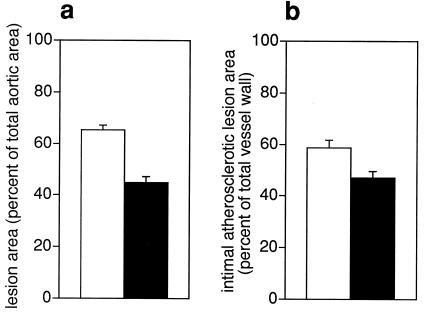
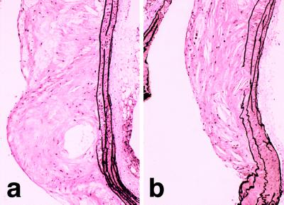
In apoE-deficient mice but not in C57BL/6 mice, severe atherosclerosis developed within 30 wk covering two-thirds of the aortic surface area with atheromatous lesions (Fig. 1a). Treatment with the endothelin ETA receptor antagonist LU135252 reduced the intimal area of the aortic tree occupied with atheromatous lesions by 31% (P < 0.001, Fig. 1a); in histologic sections of the aortic arch (Fig. 2a), lesion size was reduced by 20% (P < 0.05, Figs. 1b and 2b). Histologic analysis of the aortic arch demonstrated advanced atherosclerotic disease with a plaque morphology showing foam cell deposits, calcification, and fibrous caps, which closely resembled human atherosclerosis (35, 46). LU135252 treatment had no effect on plaque morphology.
Figure 1.
Quantification of atherosclerosis in the aorta of control apoE-deficient mice (white) and mice treated with LU135252 for 30 wk (black). Intimal area of the aortic surface area covered with atheromatous plaques (a) and lesion size in histologic sections of the thoracic aorta (b) were reduced by chronic ETA receptor blockade. No macroscopic or microscopic lesions were observed in C57BL/6 mice (unpublished observation and ref. 34). P < 0.001 (a) and P < 0.05 (b) for control vs. LU135252.
Figure 2.
Photomicrographs of atherosclerotic lesion in the aortic arch of apoE-deficient mice. Advanced intimal lesion at the root of the carotid artery in an untreated apoE-deficient mouse exhibiting necrosis, calcification, and cholesterol crystals (a). Lesion at the same location in an apoE-deficient mouse after 30 wk of ETA receptor blockade (b). Cellular composition and morphology of the plaques was similar to that seen in untreated animals. (Elastica van Giesson stain, original magnification X60).
ET Receptor Binding.
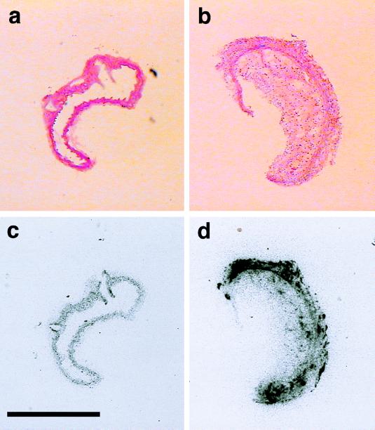
In apoE-deficient mice, the density of total ET receptor-binding sites was higher than in C57BL/6 mice (Fig. 3 c and d). Further evaluation revealed that mainly ETA receptor density was increased in apoE-deficient as compared with C57BL/6 mice (2.73 ± 1.5 vs. 0.47 ± 0.42 amol/μm2). Treatment with LU135252 did not significantly affect this increased density (1.70 ± 0.46 and 0.38 ± 0.25 amol/μm2, respectively). ETB receptor-binding capacity, on the other hand, was not different between apoE-deficient and C57BL/6 mice and was not altered by treatment with LU135252 (data not shown).
Figure 3.
Photomicrographs and autoradiographs of the aorta from C57BL/6 (Left) and apoE-deficient mice (Right). Representative histologic sections from the aorta of a C57BL/6 control mouse (a) and an atherosclerotic apoE-deficient mouse (b). Note the large plaque occluding the lumen in the aorta of the apoE-deficient mouse (b). In these animals, endothelin binding by using [125I]ET-1 was markedly increased as compared with C57BL/6 mice (c) and was localized both in the vascular wall and in the atheromatous plaque (d). (Hematoxilin/eosin stain, original magnification X10. Bar: 1,000 μm).
Vascular Endothelin Protein Content.
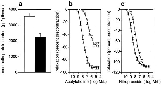
ET-1 protein content in homogenates of aortic rings of atherosclerotic apoE-deficient mice was markedly higher compared with C57BL/6 mice (3668 ± 255 vs. 2415 ± 239 pg/g, P < 0.001, Fig. 4a). LU135252 treatment reduced ET-1 protein content in aortic tissue of the atherosclerosis-prone mice to 2256 ± 214 pg/g, i.e., comparable with levels found in the C57BL/6 controls (P < 0.0001 vs. untreated, Fig. 4a).
Figure 4.
Effects of chronic ETA receptor blockade (black) on aortic ET-1 content and vascular function in isolated aortic rings of apoE-deficient mice compared with untreated animals (white). Endothelin blockade reduced aortic ET-1 protein content comparable to levels of C57BL/6 mice (a). NO-mediated endothelium-dependent relaxation to acetylcholine was normalized by treatment with the ETA antagonist LU135252 (b) and inversely correlated with vascular ET-1 protein content. Sensitivity to exogenous NO as assessed by relaxations to sodium nitroprusside was slightly enhanced (c). Analyses were performed on n = 8–10 (a) and n = 8 (b and c) from each group. P < 0.0001 control vs. untreated.
Vascular Reactivity.
In the aorta of apoE-deficient and C57BL/6 mice, endothelium-dependent relaxation in response to acetylcholine was blocked completely by NO synthase inhibitor l-NAME (0.3 mmol/liter) (refs. 47 and 48 and unpublished observation). In apoE-deficient mice, NO-mediated, endothelium-dependent relaxation to acetylcholine was markedly impaired (Fig. 4b) as compared with C57BL/6 mice (56 ± 3 vs. 99 ± 2%, P < 0.0001). Chronic treatment with the ETA receptor antagonist LU135252 increased relaxations from 56 ± 4 to 93 ± 2% (Fig. 4b) (P < 0.0001 vs. untreated) but had no effect in C57BL/6 mice (data not shown). The extent of the response to acetylcholine was inversely related to tissue endothelin levels (r = −0.695, P < 0.0001). Superoxide dismutase (150 units/ml) had no effect on endothelium-dependent relaxation in apoE-deficient or C57BL/6 mice (data not shown). Maximal relaxations to exogenous NO as assessed by sodium nitroprusside were not affected by LU135252 (109 ± 0.7 vs. 108 ± 1.0%, n.s.); however, the sensitivity to exogenous NO was increased slightly in apoE-deficient (Fig. 4c, P < 0.05) but not in C57BL/6 mice (data not shown).
DISCUSSION
This study demonstrates that in atherosclerotic apoE-deficient mice activation of the endothelin system both at the protein and receptor level is associated with reduced NO release and impaired endothelium-dependent relaxation. Chronic treatment with an orally active ETA receptor antagonist normalized NO-mediated endothelium-dependent relaxation, increased plasma nitrate levels, and reduced atherosclerotic lesions in the aorta. These data suggest that ET-1 contributes to NO-mediated endothelial dysfunction thereby promoting the development of atherosclerosis.
Endothelial dysfunction is a hallmark of cardiovascular risk factors and has been implicated in the pathogenesis of atherosclerosis (1). The vascular endothelium regulates vascular tone (5, 6) and structure (7, 8) through release of vasoactive mediators such as NO and ET-1, and the biosynthesis of ET-1 is regulated by NO in vitro (49). Activation of the endothelin system occurs in human atherosclerosis (22), and indirect evidence suggests that staining intensity for ET-1 in atherectomy specimens of patients with coronary artery disease may be related to clinical symptoms (24). Of note, in these patients endothelium-dependent relaxation is impaired due to impaired release of NO (14, 50) resulting in paradoxical vasoconstriction (51) and increased sensitivity to vasoconstrictors (52). Whether and to what extent the endothelin system contributes to endothelial dysfunction and whether it may modulate the l-arginine/NO pathway in vivo is still unclear. However, indirect evidence suggests a role for ET-1 in the atherosclerotic process. First, ET-1 is chemotactic for macrophages (53), which play an important role in the initiation of lesion formation (1) and are a source of ET-1 (26). Second, ET-1 biosynthesis is regulated by several endothelium-derived vasoactive stimuli, coagulation components, and growth factors implemented in the atherosclerotic process (30, 31), and ET-1 expression is regulated through ETA receptors in an autocrine fashion (44, 54, 55). Third, ET-1 potentiates actions of other vasoactive molecules (56), growth factors (57, 58), and cytokines (59) and stimulates extracellular matrix synthesis (60).
In apoE-deficient mice, an animal model of human atherosclerosis (34, 46), we investigated whether ET-1 is involved in the disease process of atherogenesis. Analysis of ET-1 binding revealed a marked increase in total ET-1-binding density in the vascular wall and atheromatous plaque mainly due to augmented ETA receptor density. These findings indicate that the ETA receptor rather than the ETB receptor is involved in atherosclerotic vascular changes evoked by ET-1. Indeed, ETA receptor density correlates with proliferative responsiveness of vascular smooth muscle cells in vitro (32). In line with this interpretation, the selective ETA receptor antagonist LU135252 reduced atherosclerotic vascular changes in vivo. In the aorta of apoE-deficient mice, tissue ET-1 protein content was increased. In contrast, endothelium-dependent relaxation to acetylcholine and circulating levels of nitrate—a breakdown product of NO (39)—were markedly reduced. As relaxations to acetylcholine were completely blocked by l-NAME and unaffected by superoxide dismutase, these data suggest that atherosclerosis impairs endothelial release of NO, which contributes to the progression of atherosclerosis in hypercholesterolemic rabbits (15, 16) and LDL-receptor deficient mice (16).
To determine whether activation of the endothelin system is causatively related to atherosclerosis and the functional changes associated with it, animals were chronically treated with the selective ETA receptor antagonist LU135252. Chronic ET receptor blockade had no effect on the plasma lipoprotein profile nor on systolic blood pressure, but normalized impaired NO-mediated relaxation and reduced atheromatous lesions in the aorta. These data suggest that, as observed in vitro (61), ET-1 inhibits NO release through ETA receptor activation and that inhibition of NO synthesis as well as the direct vascular effects of the peptide may promote endothelial dysfunction and atherosclerosis. This concept is further supported by the observation that plasma nitrate levels, a marker of NO activity in vivo (39), increased after chronic ETA receptor blockade. As increased activity of the l-arginine/NO pathway inhibits atherosclerosis in other animal models (18), increased sensitivity of endothelium-independent relaxation after chronic ETA receptor blockade in apoE deficient mice as observed in this study may represent a functional correlate of reduced atherosclerotic vascular changes.
Surprisingly, chronic treatment with LU135252 completely prevented the increase in tissue ET-1 levels in the aorta of apoE-deficient mice without affecting the ETA receptor binding density. Similar observations were made previously in the setting of an activated endothelin system in hypertension (8, 44). Thus, it is possible that ETA receptors are also involved in the regulation of ET-1 tissue levels in atherosclerosis. The mechanisms by which endothelin receptor blockade prevents the increase in vascular ET-1 content in vivo are currently unclear, but may involve autocrine, ETA-receptor-mediated regulation of ET-1 mRNA and protein as observed in vascular smooth muscle (54) and cardiac fibroblasts (55). Furthermore, the reduction of ET-1 tissue levels could be related to LU135252-mediated restoration of endothelial release of NO which is a potent inhibitor of ET-1 production (30).
In conclusion, these data suggest that increased vascular ET-1 levels in atherosclerosis, via ETA-receptor activation, promote NO-mediated endothelial dysfunction predisposing to atherosclerotic vascular changes. Chronic treatment with an orally active ETA receptor antagonist markedly augmented NO-mediated endothelium-dependent relaxation and plasma nitrate levels and reduced atheroma formation independent of plasma cholesterol or blood pressure. The beneficial effects of endothelin blockade in an animal model of human atherosclerosis suggest that this approach may also offer therapeutic benefit in humans.
Acknowledgments
We thank P. Albert, J. Boden, Y. Gao, S. Märki, U. Reiter, E. P. Smith, and A. Tjurmin for expert technical assistance, and M. Kirchengast for supplying LU135252. Supported by the Swiss National Research Foundation [Grants 32-51069.97 (to T.F.L.) and 32-49648.96 (to S.S.)], the Deutsche Forschungsgemeinschaft (Ba 1543/1-1), the ADUMED Foundation (to M.B. and L.V.U.), and the National Institutes of Health, Bethesda, MD [Grant RO1-HL-54246 (to C.C.H.)].
ABBREVIATIONS
- ET-1
endothelin-1
- apoE
apolipoprotein E
- LDL
low density lipoprotein
References
- 1.Ross R. Nature (London) 1993;362:801–809. doi: 10.1038/362801a0. [DOI] [PubMed] [Google Scholar]
- 2.Lüscher T F, Tanner F C, Tschudi M R, Noll G. Annu Rev Med. 1993;44:395–418. doi: 10.1146/annurev.me.44.020193.002143. [DOI] [PubMed] [Google Scholar]
- 3.Zeiher A M, Drexler H, Saurbier B, Just H. J Clin Invest. 1993;92:652–662. doi: 10.1172/JCI116634. [DOI] [PMC free article] [PubMed] [Google Scholar]
- 4.Flavahan N A. Circulation. 1992;85:1927–1938. doi: 10.1161/01.cir.85.5.1927. [DOI] [PubMed] [Google Scholar]
- 5.Ignarro L J, Buga G M, Wood K S, Byrns R E, Chaudhuri G. Proc Natl Acad Sci USA. 1987;24:9265–9269. doi: 10.1073/pnas.84.24.9265. [DOI] [PMC free article] [PubMed] [Google Scholar]
- 6.Yanagisawa M, Kurihara H, Kimura S, Tomobe Y, Kobayashi M, Mitsui Y, Yazaki Y, Goto K, Masaki T. Nature (London) 1988;332:411–415. doi: 10.1038/332411a0. [DOI] [PubMed] [Google Scholar]
- 7.Rudic R D, Shesely E G, Maeda N, Smithies O, Segal S S, Sessa W C. J Clin Invest. 1998;101:731–736. doi: 10.1172/JCI1699. [DOI] [PMC free article] [PubMed] [Google Scholar]
- 8.Barton, M., d’Uscio, L., Shaw, S., Meyer, P., Moreau, P. & Lüscher, T. F. (1998) Hypertension 499–504. [DOI] [PubMed]
- 9.Marsden P A, Schappert K T, Chen H S, Flowers M, Sundell C L, Wilcox J N, Lamas S, Michel T. FEBS Lett. 1992;307:287–293. doi: 10.1016/0014-5793(92)80697-f. [DOI] [PubMed] [Google Scholar]
- 10.Nishida K, Harrison D G, Navas J P, Fisher A A, Dockery S P, Uematsu M, Nerem R M, Alexander R W, Murphy T J. J Clin Invest. 1992;90:2092–2096. doi: 10.1172/JCI116092. [DOI] [PMC free article] [PubMed] [Google Scholar]
- 11.Janssens S P, Simouchi A, Quertermous T, Bloch D B, Bloch K D. J Biol Chem. 1992;267:22694. [PubMed] [Google Scholar]
- 12.Palmer R M J, Moncada S. Biochem Biophys Res Commun. 1989;158:384–392. doi: 10.1016/s0006-291x(89)80219-0. [DOI] [PubMed] [Google Scholar]
- 13.Wilcox J N, Subramanian R R, Sundell C L, Tracey W R, Pollock J S, Harrison D G, Marsden P A. Arterioscler Thromb Vasc Biol. 1997;17:2479–2488. doi: 10.1161/01.atv.17.11.2479. [DOI] [PubMed] [Google Scholar]
- 14.Oemar B S, Tschudi M R, Godoay N, Brovkovich V, Malinski T, Lüscher T F. Circulation. 1998;97:2494–2498. doi: 10.1161/01.cir.97.25.2494. [DOI] [PubMed] [Google Scholar]
- 15.Naruse K, Shimizu K, Muramatsu M, Toki Y, Miyazaki Y, Okumura K, Hashimoto H, Ito T. Arterioscler Thromb. 1994;14:746–752. doi: 10.1161/01.atv.14.5.746. [DOI] [PubMed] [Google Scholar]
- 16.Cayatte A J, Palacino J J, Horten K, Cohen R A. Arterioscler Thromb. 1994;14:753–759. doi: 10.1161/01.atv.14.5.753. [DOI] [PubMed] [Google Scholar]
- 17.Wang B, Singer A H, Tsao P, Drexler H, Kosek J, Cooke J P. J Am Coll Cardiol. 1994;23:452–458. doi: 10.1016/0735-1097(94)90433-2. [DOI] [PubMed] [Google Scholar]
- 18.Aji W, Ravalli S, Szabolcs M, Jiang X C, Sciacca R R, Michler R E, Cannon P J. Circulation. 1997;95:430–437. doi: 10.1161/01.cir.95.2.430. [DOI] [PubMed] [Google Scholar]
- 19.White C R, Brock T A, Chang L Y, Crapo J, Briscoe P, Ku D, Bradley W A, Gianturco S H, Gore J, Freeman B A, et al. Proc Natl Acad Sci USA. 1994;91:1044–1048. doi: 10.1073/pnas.91.3.1044. [DOI] [PMC free article] [PubMed] [Google Scholar]
- 20.Beckmann J S, Koppenol W H. Am J Physiol. 1996;271:C1424–C1437. doi: 10.1152/ajpcell.1996.271.5.C1424. [DOI] [PubMed] [Google Scholar]
- 21.Deliconstantinos G, Villiotou V, Stavrides J C. Biochem Pharmacol. 1995;49:1589–1600. doi: 10.1016/0006-2952(95)00094-g. [DOI] [PubMed] [Google Scholar]
- 22.Lerman A, Edwards B S, Hallett J W, Heublein D M, Sandberg S M, Burnett J C J. N Engl J Med. 1991;325:997–1001. doi: 10.1056/NEJM199110033251404. [DOI] [PubMed] [Google Scholar]
- 23.Winkles J A, Alberts G F, Brogi E, Libby P. Biochem Biophys Res Commun. 1993;191:1081–1088. doi: 10.1006/bbrc.1993.1327. [DOI] [PubMed] [Google Scholar]
- 24.Zeiher A M, Ihling C, Pistorius K, Schächinger V, Schaefer H-E. Lancet. 1994;344:1405–1406. doi: 10.1016/s0140-6736(94)90571-1. [DOI] [PubMed] [Google Scholar]
- 25.Hahn A W, Resink T J, Scott-Burden T, Powell J, Dohi Y, Buhler F R. Cell Regul. 1990;1:649–659. doi: 10.1091/mbc.1.9.649. [DOI] [PMC free article] [PubMed] [Google Scholar]
- 26.Ehrenreich H, Anderson R W, Fox C H, Rieckmann P, Hoffman G S, Travis W D, Coligan J E, Kehrl J H, Fauci A S. J Exp Med. 1990;172:1741–1748. doi: 10.1084/jem.172.6.1741. [DOI] [PMC free article] [PubMed] [Google Scholar]
- 27.Zeiher A M, Goebel H, Schachinger V, Ihling C. Circulation. 1995;91:941–947. doi: 10.1161/01.cir.91.4.941. [DOI] [PubMed] [Google Scholar]
- 28.Arai H, Hori S, Aramori I, Ohkubo H, Nakanishi S. Nature (London) 1990;348:730–732. doi: 10.1038/348730a0. [DOI] [PubMed] [Google Scholar]
- 29.Sakurai T, Yanagisawa M, Takuwa Y, Miyazaki H, Kimura S, Goto K, Masaki T. Nature (London) 1990;348:732–735. doi: 10.1038/348732a0. [DOI] [PubMed] [Google Scholar]
- 30.Boulanger C, Lüscher T F. J Clin Invest. 1990;85:587–590. doi: 10.1172/JCI114477. [DOI] [PMC free article] [PubMed] [Google Scholar]
- 31.Boulanger C M, Tanner F C, Bea M L, Hahn A W, Werner A, Lüscher T F. Circ Res. 1992;70:1191–1197. doi: 10.1161/01.res.70.6.1191. [DOI] [PubMed] [Google Scholar]
- 32.Kanse S M, Wijelath E, Kanthou C, Newman P, Kakkar V V. Lab Invest. 1995;72:376–382. [PubMed] [Google Scholar]
- 33.Zhang S, Reddick T L, Piedrahita J A, Maeda N. Science. 1992;258:468–471. doi: 10.1126/science.1411543. [DOI] [PubMed] [Google Scholar]
- 34.Plump A S, Smith J D, Hayek T, Allto-Setälä K, Walsh A, Verstuyft J G, Rubin E M, Breslow J L. Cell. 1992;71:343–353. doi: 10.1016/0092-8674(92)90362-g. [DOI] [PubMed] [Google Scholar]
- 35.Reddick R L, Zhang S H, Maeda N. Arterioscler Thromb. 1994;14:141–147. doi: 10.1161/01.atv.14.1.141. [DOI] [PubMed] [Google Scholar]
- 36.Riechers H, Albrecht H P, Amberg W, Baumann E, Bernard H, Bohm H J, Klinge D, Kling A, Muller S, Raschack M, et al. J Med Chem. 1996;39:2123–2128. doi: 10.1021/jm960274q. [DOI] [PubMed] [Google Scholar]
- 37.Webb R C, Vander A J, Henry J P. Hypertension. 1987;9:268–276. doi: 10.1161/01.hyp.9.3.268. [DOI] [PubMed] [Google Scholar]
- 38.Stein E A, Myers G L. In: Lipoproteins and Apolipoproteins. Burtis C A, Ashwood E R, editors. PA: Philadelphia; 1994. [Google Scholar]
- 39.Forte P, Copland M, Smith L M, Milne E, Sutherland J, Benjamin N. Lancet. 1997;349:837–842. doi: 10.1016/S0140-6736(96)07631-3. [DOI] [PubMed] [Google Scholar]
- 40.Chobanian A V, Lichtenstein A H, Nilakhe V, Haudenschild C C, Drago R, Nickerson C. Hypertension. 1989;14:203–209. doi: 10.1161/01.hyp.14.2.203. [DOI] [PubMed] [Google Scholar]
- 41.Palinski W, Ord V A, Plump A S, Breslow J L, Steinberg D, Witztum J L. Arterioscler Thromb. 1994;14:605–616. doi: 10.1161/01.atv.14.4.605. [DOI] [PubMed] [Google Scholar]
- 42.Han D K M, Haudenschild C C, Hong M K, Tinkle B T, Leon M B, Liau G. Am J Pathol. 1995;147:267–277. [PMC free article] [PubMed] [Google Scholar]
- 43.Dashwood, M. R., Timm, M. & Kaski, J. C. (1995) J. Cardiovasc. Pharmacol. 26, Suppl. 3, S351–S354. [PubMed]
- 44.Barton M, Shaw S, d’Uscio L V, Moreau P, Lüscher T F. Biochem Biophys Res Commun. 1997;238:861–865. doi: 10.1006/bbrc.1997.7394. [DOI] [PubMed] [Google Scholar]
- 45.Wallenstein S, Zucker C L, Fleiss J. Circ Res. 1980;47:1–9. doi: 10.1161/01.res.47.1.1. [DOI] [PubMed] [Google Scholar]
- 46.Breslow J L. Science. 1996;272:685–688. doi: 10.1126/science.272.5262.685. [DOI] [PubMed] [Google Scholar]
- 47.Huang P L, Huang Z, Mashimo H, Bloch K D, Moskowitz M A, Bevan J A, Fishman M C. Nature (London) 1995;377:239–242. doi: 10.1038/377239a0. [DOI] [PubMed] [Google Scholar]
- 48.Bonthu S, Heistad D d, Chappell D A, Lamping K G, Faraci F M. Arterioscler Thromb Vasc Biol. 1997;17:2333–2340. doi: 10.1161/01.atv.17.11.2333. [DOI] [PubMed] [Google Scholar]
- 49.Boulanger C, Luscher T F. J Clin Invest. 1990;85:587–590. doi: 10.1172/JCI114477. [DOI] [PMC free article] [PubMed] [Google Scholar]
- 50.Förstermann U, Mügge A, Alheid U, Haverich A, Frölich J C. Circ Res. 1988;62:185–190. doi: 10.1161/01.res.62.2.185. [DOI] [PubMed] [Google Scholar]
- 51.Ludmer P L, Selwyn A P, Shook T L, Wayne R R, Mudge G H, Alexander R W, Ganz P. N Engl J Med. 1986;315:1046–1051. doi: 10.1056/NEJM198610233151702. [DOI] [PubMed] [Google Scholar]
- 52.Vita J A, Treasure C B, Yeung A C, Vekshtein V I, Fantasia G M, Fish R D, Ganz P, Selwyn A P. Circulation. 1992;85:1390–1397. doi: 10.1161/01.cir.85.4.1390. [DOI] [PubMed] [Google Scholar]
- 53.Haller H, Schaberg T, Lindschau C, Lode H, Distler A. Am J Physiol. 1991;261:L478–L484. doi: 10.1152/ajplung.1991.261.6.L478. [DOI] [PubMed] [Google Scholar]
- 54.Alberts G F, Peifley K A, Johns A, Kleha J F, Winkles J A. J Biol Chem. 1994;269:10112–10118. [PubMed] [Google Scholar]
- 55.Fujisaki H, Ito H, Hirata Y, Tanaka M, Hata W, Lin M, Adachi S, Akimoto H, Marumo F, Hiroe M. J Clin Invest. 1995;96:1059–1065. doi: 10.1172/JCI118092. [DOI] [PMC free article] [PubMed] [Google Scholar]
- 56.Yang Z, Richard V, von Segesser L, Bauer E, Stulz P, Turina M, Lüscher T F. Circulation. 1990;82:188–195. doi: 10.1161/01.cir.82.1.188. [DOI] [PubMed] [Google Scholar]
- 57.Weissberg P L, Witchel C, Davenport A P, Herketh T R, Metcalfe J C. Atherosclerosis. 1990;83:257–262. doi: 10.1016/0021-9150(90)90118-3. [DOI] [PubMed] [Google Scholar]
- 58.Peifley K A, Winkles J A. Biochem Biophys Res Commun. 1998;242:202–208. doi: 10.1006/bbrc.1997.7940. [DOI] [PubMed] [Google Scholar]
- 59.Ruetten H, Thiemermann C. J Physiol Pharmacol. 1997;48:675–688. [PubMed] [Google Scholar]
- 60.Guidry C, Hook M. J Cell Biol. 1991;115:873–880. doi: 10.1083/jcb.115.3.873. [DOI] [PMC free article] [PubMed] [Google Scholar]
- 61.Ikeda U, Yamamoto K, Maeda Y, Shimpo M, Kanbe T, Shimada K. Hypertension. 1997;29:65–69. doi: 10.1161/01.hyp.29.1.65. [DOI] [PubMed] [Google Scholar]


